

Tenascin C Has a Modest Protective Effect on Acute Lung Pathology during Methicillin-Resistant Staphylococcus aureus-Induced Pneumonia in Mice
- PMID: 34319124
- PMCID: PMC8552697
- DOI: 10.1128/Spectrum.00207-21
Tenascin C Has a Modest Protective Effect on Acute Lung Pathology during Methicillin-Resistant Staphylococcus aureus-Induced Pneumonia in Mice
Abstract
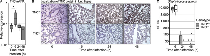
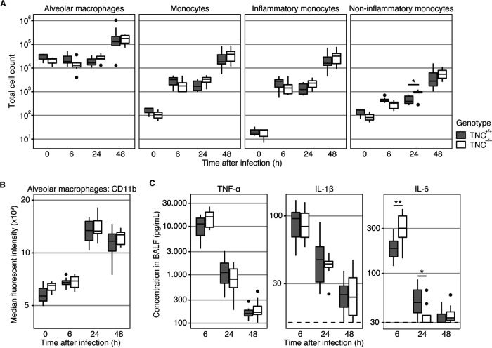
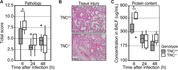
Tenascin C (TNC) is an extracellular matrix protein with immunomodulatory properties that plays a major role during tissue injury and repair. TNC levels are increased in patients with pneumonia and pneumosepsis, and they are associated with worse outcomes. Methicillin-resistant Staphylococcus aureus (MRSA) is a Gram-positive bacterium that is a major causative pathogen in nosocomial pneumonia and a rising cause of community-acquired pneumonia. To study the role of TNC during MRSA-induced pneumonia, TNC sufficient (TNC+/+) and TNC-deficient (TNC-/-) mice were infected with MRSA via the airways and euthanized after 6, 24, and 48 h for analysis. Pulmonary transcription of TNC peaked at 6 h, while immunohistochemistry revealed higher protein levels at later time points. Although TNC deficiency was not associated with changes in bacterial clearance, TNC-/- mice showed increased levels of TNF-α and IL-6 in bronchoalveolar lavage fluid during the acute phase of infection when compared with TNC+/+ mice. In addition, TNC-/- mice showed more severe pulmonary pathology at 6, but not at 24 or 48 h, after infection. Together, these data suggest that TNC plays a moderate protective role against tissue pathology during the acute inflammatory phase, but not during the bacterial clearance phase, of MRSA-induced pneumonia. These results argue against an important role of TNC on disease outcome during MRSA-induced pneumonia. IMPORTANCE Recently, the immunomodulatory properties of TNC have drawn substantial interest. However, to date most studies made use of sterile models of inflammation. In this study, we examine the pathobiology of MRSA-induced pneumonia in a model of TNC-sufficient and TNC-deficient mice. We have studied the immune response and tissue pathology both during the initial insult and also during the resolution phase. We demonstrate that MRSA-induced pneumonia upregulates pulmonary TNC expression at the mRNA and protein levels. However, the immunomodulatory role of TNC during bacterial pneumonia is distinct from models of sterile inflammation, indicating that the function of TNC is context dependent. Contrary to previous descriptions of TNC as a proinflammatory mediator, TNC-deficient mice seem to suffer from enhanced tissue pathology during the acute phase of infection. Nonetheless, besides its role during the acute phase response, TNC does not seem to play a major role in disease outcome during MRSA-induced pneumonia.
Keywords: Gram-positive bacterial infections; Staphylococcus aureus; alarmins; immune system; innate immunity; methicillin-resistant Staphylococcus aureus; mice; pneumonia; tenascin C.
Figures
Similar articles
-
Tenascin-C Deficiency Is Associated With Reduced Bacterial Outgrowth During Klebsiella pneumoniae-Evoked Pneumosepsis in Mice.Front Immunol. 2021 Mar 11;12:600979. doi: 10.3389/fimmu.2021.600979. eCollection 2021. Front Immunol. 2021. PMID: 33776992 Free PMC article.
-
Role of Interleukin-12 in Protection against Pulmonary Infection with Methicillin-Resistant Staphylococcus aureus.Antimicrob Agents Chemother. 2015 Oct;59(10):6308-16. doi: 10.1128/AAC.00968-15. Epub 2015 Jul 27. Antimicrob Agents Chemother. 2015. PMID: 26248370 Free PMC article.
-
Transcription of inflammatory genes in the lung after infection with community-associated methicillin-resistant Staphylococcus aureus: a role for panton-valentine leukocidin?Infect Immun. 2009 May;77(5):2159-67. doi: 10.1128/IAI.00021-09. Epub 2009 Feb 23. Infect Immun. 2009. PMID: 19237525 Free PMC article.
-
Community-acquired necrotizing pneumonia caused by methicillin-resistant Staphylococcus aureus producing Panton-Valentine leukocidin in a Chinese teenager: case report and literature review.Int J Infect Dis. 2014 Sep;26:17-21. doi: 10.1016/j.ijid.2014.02.025. Epub 2014 Jun 26. Int J Infect Dis. 2014. PMID: 24980464 Review.
-
How important is methicillin-resistant Staphylococcus aureus as a cause of community-acquired pneumonia and what is best antimicrobial therapy?Infect Dis Clin North Am. 2013 Mar;27(1):177-88. doi: 10.1016/j.idc.2012.11.006. Epub 2012 Dec 14. Infect Dis Clin North Am. 2013. PMID: 23398873 Review.
Cited by
-
Advances on the roles of tenascin-C in cancer.J Cell Sci. 2022 Sep 15;135(18):jcs260244. doi: 10.1242/jcs.260244. Epub 2022 Sep 14. J Cell Sci. 2022. PMID: 36102918 Free PMC article.
-
Myeloid miR-155 plays a limited role in antibacterial defense during Klebsiella-derived pneumosepsis and is dispensable for lipopolysaccharide- or Klebsiella-induced inflammation in mice.Pathog Dis. 2023 Jan 17;81:ftad031. doi: 10.1093/femspd/ftad031. Pathog Dis. 2023. PMID: 37858304 Free PMC article.
-
Tenascin-C predicts IVIG non-responsiveness and coronary artery lesions in kawasaki disease in a Chinese cohort.Front Pediatr. 2022 Dec 13;10:979026. doi: 10.3389/fped.2022.979026. eCollection 2022. Front Pediatr. 2022. PMID: 36582508 Free PMC article.
-
The Azithromycin Pro-Drug CSY5669 Boosts Bacterial Killing While Attenuating Lung Inflammation Associated with Pneumonia Caused by Methicillin-Resistant Staphylococcus aureus.Antimicrob Agents Chemother. 2022 Sep 20;66(9):e0229821. doi: 10.1128/aac.02298-21. Epub 2022 Aug 16. Antimicrob Agents Chemother. 2022. PMID: 35972289 Free PMC article.
-
Induction of Acute or Disseminating Bacterial Pneumonia in Mice and Sampling of Infected Organs for Studying the Host Response to Bacterial Pneumonia.Bio Protoc. 2022 Jan 5;12(1):e4287. doi: 10.21769/BioProtoc.4287. eCollection 2022 Jan 5. Bio Protoc. 2022. PMID: 35118178 Free PMC article.
References
-
- Midwood K, Sacre S, Piccinini AM, Inglis J, Trebaul A, Chan E, Drexler S, Sofat N, Kashiwagi M, Orend G, Brennan F, Foxwell B. 2009. Tenascin-C is an endogenous activator of Toll-like receptor 4 that is essential for maintaining inflammation in arthritic joint disease. Nat Med 15:774–780. doi:10.1038/nm.1987. - DOI - PubMed
Publication types
MeSH terms
Substances
LinkOut - more resources
Full Text Sources
Medical
Molecular Biology Databases
Miscellaneous
