. 2021 Oct 27;95(22):e0068421.
doi: 10.1128/JVI.00684-21. Epub 2021 Jul 28.

Genome-Wide Patterns of Bracovirus Chromosomal Integration into Multiple Host Tissues during Parasitism

Affiliations

Genome-Wide Patterns of Bracovirus Chromosomal Integration into Multiple Host Tissues during Parasitism

Héloïse Muller et al. J Virol. .

Abstract

Bracoviruses are domesticated viruses found in parasitic wasp genomes. They are composed of genes of nudiviral origin that are involved in particle production and proviral segments containing virulence genes that are necessary for parasitism success. During particle production, proviral segments are amplified and individually packaged as DNA circles in nucleocapsids. These particles are injected by parasitic wasps into host larvae together with their eggs. Bracovirus circles of two wasp species were reported to undergo chromosomal integration in parasitized host hemocytes, through a conserved sequence named the host integration motif (HIM). Here, we used bulk Illumina sequencing to survey integrations of Cotesia typhae bracovirus circles in the DNA of its host, the maize corn borer (Sesamia nonagrioides), 7 days after parasitism. First, assembly and annotation of a high-quality genome for C. typhae enabled us to characterize 27 proviral segments clustered in proviral loci. Using these data, we characterized large numbers of chromosomal integrations (from 12 to 85 events per host haploid genome) for all 16 bracovirus circles containing a HIM. Integrations were found in four S. nonagrioides tissues and in the body of a caterpillar in which parasitism had failed. The 12 remaining circles do not integrate but are maintained at high levels in host tissues. Surprisingly, we found that HIM-mediated chromosomal integration in the wasp germ line has occurred accidentally at least six times during evolution. Overall, our study furthers our understanding of wasp-host genome interactions and supports HIM-mediated chromosomal integration as a possible mechanism of horizontal transfer from wasps to their hosts. IMPORTANCE Bracoviruses are endogenous domesticated viruses of parasitoid wasps that are injected together with wasp eggs into wasp host larvae during parasitism. Several studies have shown that some DNA circles packaged into bracovirus particles become integrated into host somatic genomes during parasitism, but the phenomenon has never been studied using nontargeted approaches. Here, we use bulk Illumina sequencing to systematically characterize and quantify bracovirus circle integrations that occur in four tissues of the Mediterranean corn borer (Sesamia nonagrioides) during parasitism by the Cotesia typhae wasp. Our analysis reveals that all circles containing a HIM integrate at substantial levels (from 12 to 85 integrations per host cell, in total) in all tissues, while other circles do not integrate. In addition to shedding new light on wasp-bracovirus-host interactions, our study supports HIM-mediated chromosomal integration of bracovirus as a possible source of wasp-to-host horizontal transfer, with long-term evolutionary consequences.

Keywords: bracovirus; chromosomal integration; genomics; horizontal transfer; host-parasite relationship; parasitoid wasps; polydnavirus.


Figures

FIG 1
FIG 1
Map of CtBV proviral segments. Proviral segments are represented by filled rectangles. Segments duplicated after circularization are empty. Asterisks indicate HIM-bearing circles found to be integrated into the S. nonagrioides genome, corresponding precisely to all segments originating from the RU2.3 part of the macrolocus and isolated loci (PL3, PL4, PL5, PL6, PL7, and PL8). Each contig or scaffold in which the segments are located is indicated, and lines indicate segments that belong to the same PL. The size of the segments and the spaces between them are shown to scale, unless hash marks are present. The colors represent the quality of the annotation. Green indicates that we delimited both extremities of the segments with confidence (DRJs in proviral segments or J1 and J2 motifs in HIM-mediated duplications). Orange and red indicate that one or both extremities (see Table S5) have to be taken with caution. In the case of the orange ones, the contig was too short to identify the extremity, whereas in the case of red ones, the extremity was long enough but we were not able to find the motif. Although they are shown in green, the DRJs of S37 and S26 are truncated, probably due to sequencing or assembly issues. Blue indicates the segment duplicated after circularization by other means than HIM. In this case, there is no J1 and J2 motifs at the extremities, nor DRJ.
FIG 2
FIG 2
Average sequencing depths in the 5 samples. Green, yellow, and red indicate the average sequencing depths over the whole genome of S. nonagrioides, the whole genome of C. typhae, and the 27 C. typhae proviral segments, respectively.
FIG 3
FIG 3
Map of chimeric reads indicating HIM-mediated chromosomal integration of segment 1. (A) Number of chimeric reads along segment 1 in hemocytes, oriented from the 5′ DRJ to the 3′ DRJ. The white portion represents the HIM (not to scale) near the 3′ DRJ. (B) Magnification of the 121-bp HIM, showing two regions with many chimeric reads, called J2 (left) and J1 (right). (C) Sequence logo of J2 and J1 generated with weblogo.berkeley.edu, using an alignment of the HIMs of the 16 segments that integrated into the S. nonagrioides genome. For J2, we used the 30 bp upstream from the minimum position at which we observed >2 chimeric reads; for J1, we used the 30 bp downstream from the maximum position at which we observed >2 chimeric reads. The highly conserved motif J1 is framed in red and J2 in green.
FIG 4
FIG 4
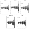
Distribution of microhomology lengths at wasp-host junctions in chimeric reads. Black bars correspond to the numbers of observed chimeric reads for each microhomology length. Red asterisks correspond to the expected numbers of chimeric reads for each microhomology length. (a) Distribution of microhomology lengths for CtBV-host junctions mapped in J1 or J2. (b) Distribution of microhomology lengths for CtBV-host junctions mapped within HIM but outside J1 or J2.
FIG 5
FIG 5
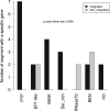
Integration capacity of segments containing ≥1 gene belonging to seven gene families: PTP (protein tyrosine phosphatase), EP1-like (early parasitism-specific protein 1), VANK (viral ankyrin), Ser_rich, RNaseT2, BEN (BEN-domain proteins), and crp (cysteine-rich proteins). Segments containing genes belonging to several gene families are counted for each family. Black bars correspond to segments that integrate into the genome of S. nonagrioides, while white bars correspond to segments that do not integrate.
FIG 6
FIG 6
Number of IEs for each segment and sample. Absolute numbers of IEs and of chimeric reads (in parentheses) are shown at the top of each bar. (a) Barplot comparing the numbers of IEs for each segment. (b) Barplot comparing the total numbers of IEs of all segments in each sample.
FIG 7
FIG 7
Histograms showing the number of chimeric reads and the sequencing depth for each HIM-containing segment. Light gray bars show the number of chimeric reads, while dark gray bars show the sequencing depth. The ratio of sequencing depth to chimeric reads is indicated at the top of each light gray bar. The Spearman rho values indicate the correlation between sequencing depth and the number of chimeric reads for each sample.
FIG 8
FIG 8
Plot of the sequencing depth versus the number of chimeric reads for each of the CtBV segments. Sequencing depths and numbers of chimeric reads were summed for all samples. The same plots are shown for each sample in Fig. S5 in the supplemental material. Blue dots represent proviral segments that do integrate into the S. nonagrioides genome, and red dots represent proviral segments that do not integrate. Green dots represent duplicated segments, i.e., Rdp and Hdp segments. The identification numbers of the segments are shown near each blue dot. For red dots, only the identification numbers S15, S5, and S20/33 are shown; for green dots, only S13_Rdp is indicated. The yellow dashed line shows the average depth on the C. typhae genome when all samples are summed. The Spearman rho value indicates the correlation between sequencing depth and the number of IEs for segments that do integrate into the S. nonagrioides genome.

Similar articles

Cited by

References

    1. Drezen J-M, Leobold M, Bézier A, Huguet E, Volkoff A-N, Herniou EA. 2017. Endogenous viruses of parasitic wasps: variations on a common theme. Curr Opin Virol 25:41–48. 10.1016/j.coviro.2017.07.002. - DOI - PubMed
    1. Strand MR, Burke GR. 2014. Polydnaviruses: nature’s genetic engineers. Annu Rev Virol 1:333–354. 10.1146/annurev-virology-031413-085451. - DOI - PubMed
    1. Gauthier J, Boulain H, van Vugt JJFA, Baudry L, Persyn E, Aury J-M, Noel B, Bretaudeau A, Legeai F, Warris S, Chebbi MA, Dubreuil G, Duvic B, Kremer N, Gayral P, Musset K, Josse T, Bigot D, Bressac C, Moreau S, Periquet G, Harry M, Montagné N, Boulogne I, Sabeti-Azad M, Maïbèche M, Chertemps T, Hilliou F, Siaussat D, Amselem J, Luyten I, Capdevielle-Dulac C, Labadie K, Merlin BL, Barbe V, de Boer JG, Marbouty M, Cônsoli FL, Dupas S, Hua-Van A, Le Goff G, Bézier A, Jacquin-Joly E, Whitfield JB, Vet LEM, Smid HM, Kaiser L, Koszul R, Huguet E, Herniou EA, Drezen JM. 2021. Chromosomal scale assembly of parasitic wasp genome reveals symbiotic virus colonization. Commun Biol 4:104–115. 10.1038/s42003-020-01623-8. - DOI - PMC - PubMed
    1. Strand MR, Burke GR. 2013. Polydnavirus-wasp associations: evolution, genome organization, and function. Curr Opin Virol 3:587–594. 10.1016/j.coviro.2013.06.004. - DOI - PubMed
    1. Sharanowski BJ, Ridenbaugh RD, Piekarski PK, Broad GR, Burke GR, Deans AR, Lemmon AR, Moriarty Lemmon EC, Diehl GJ, Whitfield JB, Hines HM. 2021. Phylogenomics of Ichneumonoidea (Hymenoptera) and implications for evolution of mode of parasitism and viral endogenization. Mol Phylogenet Evol 156:107023. 10.1016/j.ympev.2020.107023. - DOI - PubMed

Publication types

LinkOut - more resources