. 2021 Sep 3;9(1):e0049621.
doi: 10.1128/Spectrum.00496-21. Epub 2021 Jul 28.

Yersinia pseudotuberculosis YopJ Limits Macrophage Response by Downregulating COX-2-Mediated Biosynthesis of PGE2 in a MAPK/ERK-Dependent Manner

Affiliations

Yersinia pseudotuberculosis YopJ Limits Macrophage Response by Downregulating COX-2-Mediated Biosynthesis of PGE2 in a MAPK/ERK-Dependent Manner

Austin E F Sheppe et al. Microbiol Spectr. .

Erratum in

Abstract

Prostaglandin E2 (PGE2) is an essential immunomodulatory lipid released by cells in response to infection with many bacteria, yet its function in macrophage-mediated bacterial clearance is poorly understood. Yersinia overall inhibits the inflammatory circuit, but its effect on PGE2 production is unknown. We hypothesized that one of the Yersinia effector proteins is responsible for the inhibition of PGE2 biosynthesis. We identified that yopB-deficient Y. enterocolitica and Y. pseudotuberculosis deficient in the secretion of virulence proteins via a type 3 secretion system (T3SS) failed to inhibit PGE2 biosynthesis in macrophages. Consistently, COX-2-mediated PGE2 biosynthesis is upregulated in cells treated with heat-killed or T3SS-deficient Y. pseudotuberculosis but diminished in the presence of a MAPK/ERK inhibitor. Mutants expressing catalytically inactive YopJ induce similar levels of PGE2 as heat-killed or ΔyopB Y. pseudotuberculosis, reversed by YopJ complementation. Shotgun proteomics discovered host pathways regulated in a YopJ-mediated manner, including pathways regulating PGE2 synthesis and oxidative phosphorylation. Consequently, this study identified that YopJ-mediated inhibition of MAPK signal transduction serves as a mechanism targeting PGE2, an alternative means of inflammasome inhibition by Yersinia. Finally, we showed that EP4 signaling supports macrophage function in clearing intracellular bacteria. In summary, our unique contribution was to determine a bacterial virulence factor that targets COX-2 transcription, thereby enhancing the intracellular survival of yersiniae. Future studies should investigate whether PGE2 or its stable synthetic derivatives could serve as a potential therapeutic molecule to improve the outcomes of specific bacterial infections. Since other pathogens encode YopJ homologs, this mechanism is expected to be present in other infections. IMPORTANCE PGE2 is a critical immunomodulatory lipid, but its role in bacterial infection and pathogen clearance is poorly understood. We previously demonstrated that PGE2 leads to macrophage polarization toward the M1 phenotype and stimulates inflammasome activation in infected macrophages. Finally, we also discovered that PGE2 improved the clearance of Y. enterocolitica. The fact that Y. enterocolitica hampers PGE2 secretion in a type 3 secretion system (T3SS)-dependent manner and because PGE2 appears to assist macrophage in the clearance of this bacterium indicates that targeting of the eicosanoid pathway by Yersinia might be an adaption used to counteract host defenses. Our study identified a mechanism used by Yersinia that obstructs PGE2 biosynthesis in human macrophages. We showed that Y. pseudotuberculosis interferes with PGE2 biosynthesis by using one of its T3SS effectors, YopJ. Specifically, YopJ targets the host COX-2 enzyme responsible for PGE2 biosynthesis, which happens in a MAPK/ER-dependent manner. Moreover, in a shotgun proteomics study, we also discovered other pathways that catalytically active YopJ targets in the infected macrophages. YopJ was revealed to play a role in limiting host LPS responses, including repression of EGR1 and JUN proteins, which control transcriptional activation of proinflammatory cytokine production such as interleukin-1β. Since YopJ has homologs in other bacterial species, there are likely other pathogens that target and inhibit PGE2 biosynthesis. In summary, our study's unique contribution was to determine a bacterial virulence factor that targets COX-2 transcription. Future studies should investigate whether PGE2 or its stable synthetic derivatives could serve as a potential therapeutic target.

Keywords: Gram-negative infection; Yersinia; eicosanoids; prostaglandin; proteomics.


Figures

FIG 1
FIG 1
Analysis of PGE2 and IL-1β secretion in THP-1 macrophages infected with wild-type, ΔyopB, or heat-killed Y. enterocolitica and Y. pseudotuberculosis. PMA-differentiated THP-1 macrophages were infected at an MOI of 50:1 with wild type, ΔyopB Y. enterocolitica (Ye; A and B) and Y. pseudotuberculosis (Yptb; C and D) or treated with an equivalent number of heat-killed bacteria for 2 h. Uninfected macrophages were used as a control (Ctrl). Bacteria were heat killed at 55°C for 30 min before treating macrophages, and loss of viability was confirmed by plating. Commercial monoclonal ELISAs were used to measure the concentrations of PGE2 (A and C) and IL-1β (B and D) in supernatants from Y. enterocolitica- or Y. pseudotuberculosis-infected THP-1 cells. One-way ANOVA and Tukey’s post hoc test were used to calculate significance (n = 3). P values are indicated (*, P ≤ 0.05; **, P ≤ 0.01; ***, P ≤ 0.001; ****, P ≤ 0.0001). The data are representative of three independent experiments.
FIG 2
FIG 2
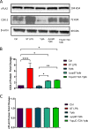
Analysis of COX-2 and cPLA2 transcript and protein expression in THP-1 macrophages after the treatment with viable wild type, heat-killed, ΔyopB, and YopJC172A Y. pseudotuberculosis. PMA-differentiated THP-1 macrophages were infected (or remained uninfected, Ctrl) with live or heat-killed wild type, ΔyopB, and YopJC172A Y. pseudotuberculosis (Yptb) for 2 h. LPS isolated from S. enterica (ST LPS) was used to treat THP-1 macrophages as a positive control. At 2 hpi, cells were collected and lysed, followed by the separation of proteins by SDS-PAGE. (A) Western blotting was performed with specific antibodies to COX-2 and cPLA2. (B and C) Protein fold change was calculated from densitometry analysis using ImageJ, where the protein levels of targets were normalized to β-actin (loading control). Unpaired Student t tests were used to calculate statistical significance from Western blot densitometry (n = 3). (D) The COX-2 mRNA fold change compared to vehicle control-treated cells was calculated using Bio-Rad CFX-manager software and normalized to GAPDH transcript levels (n = 3). *, P ≤ 0.05; **, P ≤ 0.01; ***, P ≤ 0.001; ****, P ≤ 0.0001. The images shown are representative of three independent experiments.
FIG 3
FIG 3
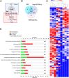
Shotgun proteomics and pathway analysis of THP-1 macrophages infected with wild-type or YopJC172A mutant Y. pseudotuberculosis. PMA-differentiated THP-1 macrophages were infected at an MOI of 50:1 with wild-type or YopJC172A Y. pseudotuberculosis for 2 h. Cell pellets were lysed and separated by SDS-PAGE, and entire lanes were subjected to tryptic digestion, followed by mass spectrometry-based analysis (n = 3). (A) Model of YopJ-mediated inhibition of macrophage response to bacterial LPS. (B) Venn diagram shows a summary of differential protein identifications in wild-type or YopJC172A Y. pseudotuberculosis-infected macrophages. (C) Heat map of augmented proteins in the proteomic analysis analyzed by hierarchical clustering. The grouped samples include identifications in wild-type (WT Yp) or YopJC172A (YopJ C/A) mutant strains of Y. pseudotuberculosis-infected macrophages, as well as uninfected cells (Ctrl). (D and E) Canonical pathway analysis of proteins differentially abundant in panel E. Y. pseudotuberculosis-infected macrophages compared to uninfected cells (D) or YopJC172A mutant of Y. pseudotuberculosis-infected macrophages in comparison to wild-type Y. pseudotuberculosis. The graphs represent downregulated proteins (green), upregulated proteins (red), and proteins with unchanged abundance (gray). (F and G) The top upstream regulator identified in YopJC172A Y. pseudotuberculosis-infected macrophages in comparison to wild-type Y. pseudotuberculosis. The graphs represent downregulated proteins (green), upregulated proteins (red), molecules predicted to be activated (orange; z-score ≥ 2). Orange and blue dashed lines with arrows indicate indirect activation and inhibition, respectively, while solid lines indicate direct effects. Lipopolysaccharide (LPS) signaling (z-score = +2.223) and (C) EGR1 (z-score = +2.162) are predicted to be activated in YopJC172A Y. pseudotuberculosis-infected macrophages compared to wild-type Y. pseudotuberculosis infection. This pathway analysis also indicated the upregulation of proteins involved in the eicosanoid biosynthetic enzymes.
FIG 4
FIG 4
Analysis of PGE2 biosynthesis and MEK function in THP-1 macrophages in response to treatment with ΔyopB Y. enterocolitica, YopJC172A Y. pseudotuberculosis, or the chemical MEK inhibitor PD184161. PMA-differentiated THP-1 macrophages were pretreated with or without 10 μM PD184161 (MEK1/2 inhibitor) 1 h before infection. Macrophages were uninfected (Ctrl) or were infected with wild-type or ΔyopB Y. enterocolitica (Ye) (A) or wild-type or YopJC172A Y. pseudotuberculosis (Yptb) (B and C) for 2 h at an MOI of 50:1. The PGE2 concentration from infected cell supernatants was measured by a commercial monoclonal ELISA recognizing PGE2 (A and B). Western blotting was used to quantify MEK1/2 and phosphorylated MEK/2 (C and D), where the fold change was calculated using densitometry analysis by normalizing to phosphorylated MEK 1/2 to MEK1/2 levels (D). The β-actin was used as a loading control. Two-way ANOVA and Tukey’s post hoc test were used to calculate significance for Western blot densitometry (n = 3). P values are indicated (*, P ≤ 0.05; **, P ≤ 0.01; ***, P ≤ 0.001; ****, P ≤ 0.0001). The images shown are representative of three independent experiments.
FIG 5
FIG 5
Expression of pro-IL-1β from THP-1 macrophages infected with YopJC172A Y. pseudotuberculosis. PMA-differentiated THP-1 macrophages were infected with wild-type, ΔyopB, or YopJC172A mutant Y. pseudotuberculosis (Yptb) at an MOI of 50:1 for 2 h or left uninfected (Ctrl). As a positive control, 10 μg/ml S. enterica LPS was used to treat cells instead for 2 h after PMA differentiation. Cell pellets were collected for Western blotting (A) and qRT-PCR (B and D) analyses. Protein fold changes in IL-1β were calculated using densitometry analysis and were normalized to β-actin (loading control). (C) The IL-1β concentration was measured in cell supernatants at 2 hpi using ELISA. Two-way ANOVA and Tukey’s post hoc test was used to calculate significance for Western blot analysis (n = 3). (D) THP-1 macrophages were infected with wild-type Y. pseudotuberculosis (Yptb), YopJC172A, or YopJC172A mutant expressing YopJ (YopJ-M45) at an MOI of 50:1 for 2 h. YopJC172A+pYopJ-M45 strain was grown in the presence of 0.1 mM IPTG before infection. Alternatively, cells were treated with LPS from Salmonella (ST LPS). The qRT-PCR analysis was used to analyze pro-IL-1β transcripts, where pro-IL-1β mRNA fold change was calculated by normalizing it to GAPDH transcript levels and using a t test to indicate significance (Bio-Rad CFX-manager software; n = 3). P values are indicated (*, P ≤ 0.05; **, P ≤ 0.01; ***, P ≤ 0.001; ****, P ≤ 0.0001). The data are representative of three independent experiments.
FIG 6
FIG 6
Effect of ectopic expression of YopJ on host COX-2 and PGE2 biosynthesis in Y. pseudotuberculosis-infected THP-1 macrophages. (A to C) PMA-differentiated THP-1 macrophages were treated with heat-killed Y. pseudotuberculosis or infected with live wild-type Y. pseudotuberculosis, ΔyopB, YopJC172A, or YopJC172A mutant expressing YopJ (YopJ-M45) at an MOI of 50:1 for 2 h. The YopJC172A-pYopJ-M45 strain contains a plasmid with the wild-type YopJ sequence under the control of an IPTG-inducible promoter. YopJC172A+pYopJ-M45 strain was grown in the presence of 0.01, 0.1, and 0.4 mM IPTG before infection of THP-1 macrophages. The protein fold change was calculated from densitometry analysis (ImageJ) and normalized to β-actin (loading control). Two-way ANOVA and Tukey’s post hoc test were used to calculate significance from Western blot densitometry (n = 3). P values are indicated (*, P ≤ 0.05; **, P ≤ 0.01; ***, P ≤ 0.001; ****, P ≤ 0.0001). (C) PGE2 concentration in cell culture supernatants was measured using monoclonal ELISA. (D) THP-1 cells were treated with 10 μg/ml LPS isolated from Y. pseudotuberculosis grown at 37°C in LB without aeration, a gift kindly provided by Robert Ernst (60). Alternatively, cells were infected with live wild-type Y. pseudotuberculosis, heat-killed ΔyopB, YopJC172A Y. pseudotuberculosis, or the YopJC172A Y. pseudotuberculosis mutant expressing YopJ-M45 at an MOI of 50:1 for 2 h similarly, as described above. The RNA from cells was collected and used to measure mRNA from COX-2 and GAPDH (housekeeping control). The transcripts of COX-2 were analyzed by qRT-PCR, where COX-2 mRNA fold change was normalized to GAPDH mRNA levels. *, P ≤ 0.05; **, P ≤ 0.01; ***, P ≤ 0.001; ****, P ≤ 0.0001. The data are representative of three independent experiments.
FIG 7
FIG 7
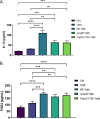
PGE2 and IL-1β secretion from LPS primed primary bone marrow-derived macrophages in response to live or heat-killed wild type, ΔyopB, or YopJC172A Y. pseudotuberculosis infection. Primary BMDMs isolated from wild-type BALB/c mice were primed with 10 μg/ml LPS for 1 h before infection with live or heat-killed wild-type, ΔyopB, or YopJC172A Y. pseudotuberculosis at an MOI of 50:1. At 2 hpi, the supernatant was collected and probed for PGE2 (A) and IL-1β (B) concentrations by commercial ELISA. Student t tests were used to calculate significance (n = 3). P values are indicated (*, P ≤ 0.05; **, P ≤ 0.01; ***, P ≤ 0.001; ****, P ≤ 0.0001).
FIG 8
FIG 8
Survival of intracellular Y. pseudotuberculosis in cells treated with EP4 agonist. (A and B) THP-1 macrophages were treated with EP4 agonist L-902688, JUN inhibitor, or vehicle control (ethanol, 0.01% [vol/vol] DMSO) in RPMI lacking antibiotics for 30 min before infection with wild-type and YopJC172A GFP-Y. pseudotuberculosis (MOI of 15:1). At 1 hpi, media were removed, and cells were washed with PBS to remove extracellular bacteria. RPMI media lacking antibiotics supplemented with gentamicin (100 μg/ml), with or without EP4 agonist and with or without JUN inhibitor, were added, and the cells were incubated further for another hour. At 2 hpi, the media were removed again, and the cells were washed with PBS and resuspended in media containing a lower concentration of gentamicin (10 μg/ml) for the remainder of the infection (total, 2 and 24 hpi). For fluorescence imaging, the cells were stained with Hoechst stain, and the entire well was imaged using Cytation 5 at ×10 magnification. (C) The number of GFP-Yersinia per 1,000 cells was calculated by dividing total GFP cell counts by total host cell counts at 2 and 24 hpi multiplied by 1,000. (D) THP-1 cell count as determined by Hoechst staining and cell counting using GenV software. All data were analyzed by two-way ANOVA, followed by multiple testing correction (Tukey’s; n = 4). P values are indicated (*, P ≤ 0.05; **, P ≤ 0.01; ***, P ≤ 0.001; ****, P ≤ 0.0001).
FIG 9
FIG 9
Model of PGE2 and IL-1β biosynthesis in macrophages during Gram-negative bacterial infection. (Step 1) LPS ligand binding to TLR4 induces rapid phosphorylation of host MAPKs, such as MEK1/2, with adaptor protein assistance, including MyD88. (Step 2) MEK1/2 becomes activated and phosphorylates ERK1/2, leading to the activation and migration of transcription factors to the nucleus to induce proinflammatory gene transcription, including upregulation of COX-2 transcripts. (Step 3) Activated ERK1/2 phosphorylates and activates cPLA2 at Ser 515/505 residue, resulting in the liberation of AA from membrane phospholipids and AA’s conversion into PGH2/PGE2 by COX-2. (Step 4) PGE2 is secreted and binds in an autocrine fashion to specific EP2/4 receptors to activate IL-1β transcription by altering cAMP levels or PI3K-induced migration of transcription factors NF-κB and AP-1. In the presence of Gram-negative bacteria, pro-IL-1β is cleaved to mature IL-1β by caspase-1 due to inflammasome activation. (Step 5) IL-1β forms a positive-feedback loop with PGE2 by binding to IL-1R and increasing COX-2 transcription. Y. pseudotuberculosis and S. flexneri modulate inflammasome activation and PGE2 biosynthesis by manipulating host MAPK/ERK-dependent signaling such as T3SS factors YopJ (Y. pseudotuberculosis) or OspB/OspF (S. flexneri).

Similar articles

Cited by

References

    1. Rainsford KD. 2009. Ibuprofen: pharmacology, efficacy, and safety. Inflammopharmacology 17:275–342. doi:10.1007/s10787-009-0016-x. - DOI - PubMed
    1. Park JY, Pillinger MH, Abramson SB. 2006. Prostaglandin E2 synthesis and secretion: the role of PGE2 synthases. Clin Immunol 119:229–240. doi:10.1016/j.clim.2006.01.016. - DOI - PubMed
    1. Scheiman JM. 2013. The use of proton pump inhibitors in treating and preventing NSAID-induced mucosal damage. Arthritis Res Ther 15(Suppl 3):S5. doi:10.1186/ar4177. - DOI - PMC - PubMed
    1. FitzGerald GA. 2020. Misguided drug advice for COVID-19. Science 367:1434. doi:10.1126/science.abb8034. - DOI - PubMed
    1. Rådmark O, Werz O, Steinhilber D, Samuelsson B. 2015. 5-Lipoxygenase, a key enzyme for leukotriene biosynthesis in health and disease. Biochim Biophys Acta 1851:331–339. doi:10.1016/j.bbalip.2014.08.012. - DOI - PubMed

Publication types

MeSH terms

Substances