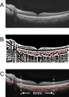
Differences in Retinal and Choroidal Vasculature and Perfusion Related to Axial Length in Pediatric Anisomyopes
- PMID: 34319397
- PMCID: PMC8322721
- DOI: 10.1167/iovs.62.9.40
Differences in Retinal and Choroidal Vasculature and Perfusion Related to Axial Length in Pediatric Anisomyopes
Abstract
Purpose: The purpose of this study was to evaluate the interocular differences in choroidal vasculature, choriocapillaris perfusion, and retinal microvascular network, and to explore their associations with interocular asymmetry in axial lengths (ALs) in children with anisomyopia.
Methods: Refractive error, AL, and other biometric parameters were measured in 70 children with anisomyopia. Using optical coherence tomography (OCT) and OCT-angiography, we measured the submacular choroidal thickness (ChT), total choroidal area (TCA), luminal area (LA), stromal area (SA), choroidal vascularity index (CVI), choriocapillaris flow deficit (CcFD), retinal vessel density (VD), and foveal avascular zone (FAZ) area.
Results: The mean interocular differences in spherical equivalent refraction and AL were -2.26 ± 0.94 diopters and 0.95 ± 0.46 mm, respectively. Submacular ChT, TCA, LA, SA, and CVI were all significantly lower in the more myopic (longer AL) eyes than in the less myopic (shorter AL) fellow eyes. In eyes with longer ALs, both the CcFD and FAZ areas were significantly greater, whereas the superficial and deep retinal VDs were significantly less. After adjusting for corneal power and intraocular pressure, interocular differences in LA (β = -0.774), SA (β = -0.991), and CcFD (β = 0.040) were significantly associated with interocular asymmetry in AL (all P < 0.05).
Conclusions: In pediatric anisomyopes, eyes with longer ALs tended to have lower choroidal vascularity and choriocapillaris perfusion than the contralateral eyes with shorter ALs. Longitudinal investigations would be useful follow-ups to test for a causal role of choroidal circulation in human myopia.
Conflict of interest statement
Disclosure:
Figures


Similar articles
-
The Effect of Axial Length on Macular Vascular Density in Eyes with High Myopia.Rom J Ophthalmol. 2025 Jan-Mar;69(1):88-100. doi: 10.22336/rjo.2025.15. Rom J Ophthalmol. 2025. PMID: 40330963 Free PMC article.
-
Assessment of Choroidal Vascularity and Choriocapillaris Blood Perfusion in Anisomyopic Adults by SS-OCT/OCTA.Invest Ophthalmol Vis Sci. 2021 Jan 4;62(1):8. doi: 10.1167/iovs.62.1.8. Invest Ophthalmol Vis Sci. 2021. PMID: 33393974 Free PMC article.
-
Quantitative evaluation of retinal and choroidal vascularity and retrobulbar blood flow in patients with myopic anisometropia by CDI and OCTA.Br J Ophthalmol. 2023 Aug;107(8):1172-1177. doi: 10.1136/bjophthalmol-2021-320597. Epub 2022 Apr 20. Br J Ophthalmol. 2023. PMID: 35443997
-
Correlation between refractive errors and ocular biometric parameters in children and adolescents: a systematic review and meta-analysis.BMC Ophthalmol. 2023 Nov 21;23(1):472. doi: 10.1186/s12886-023-03222-7. BMC Ophthalmol. 2023. PMID: 37990308 Free PMC article.
-
Choroidal Perfusion Changes After Vitrectomy for Myopic Traction Maculopathy.Semin Ophthalmol. 2024 May;39(4):261-270. doi: 10.1080/08820538.2023.2283029. Epub 2023 Nov 21. Semin Ophthalmol. 2024. PMID: 37990380 Review.
Cited by
-
EFEMP1 is a potential biomarker of choroid thickness change in myopia.Front Neurosci. 2023 Feb 20;17:1144421. doi: 10.3389/fnins.2023.1144421. eCollection 2023. Front Neurosci. 2023. PMID: 36891459 Free PMC article.
-
Analysis of White and Dark without Pressure in a Young Myopic Group Based on Ultra-Wide Swept-Source Optical Coherence Tomography Angiography.J Clin Med. 2022 Aug 18;11(16):4830. doi: 10.3390/jcm11164830. J Clin Med. 2022. PMID: 36013068 Free PMC article.
-
Swept-Source OCT Angiography-Derived Regional Normative Data of Peripapillary Vessel Density in Healthy Populations.Transl Vis Sci Technol. 2025 Aug 1;14(8):5. doi: 10.1167/tvst.14.8.5. Transl Vis Sci Technol. 2025. PMID: 40747988 Free PMC article.
-
Longitudinal Changes in Choroidal Vascularity in Myopic and Non-Myopic Children.Transl Vis Sci Technol. 2024 Aug 1;13(8):38. doi: 10.1167/tvst.13.8.38. Transl Vis Sci Technol. 2024. PMID: 39177994 Free PMC article.
-
Assessment of Choroidal Vascularity and Choriocapillaris Blood Perfusion After Accommodation in Myopia, Emmetropia, and Hyperopia Groups Among Children.Front Physiol. 2022 Mar 17;13:854240. doi: 10.3389/fphys.2022.854240. eCollection 2022. Front Physiol. 2022. PMID: 35370764 Free PMC article.
References
-
- Dolgin E. The myopia boom. Nature . 2015; 519(7543): 276–278. - PubMed
-
- Morgan IG, French AN, Ashby RS, et al. .. The epidemics of myopia: Aetiology and prevention. Prog Retin Eye Res . 2018; 62: 134–149. - PubMed
-
- Baird PN, Saw SM, Lanca C, et al. .. Myopia. Nat Rev Dis Primers . 2020; 6(1): 99. - PubMed
-
- Flitcroft DI. The complex interactions of retinal, optical and environmental factors in myopia aetiology. Prog Retin Eye Res . 2012; 31(6): 622–660. - PubMed
-
- Ohno-Matsui K, Jonas JB.. Posterior staphyloma in pathologic myopia. Prog Retin Eye Res . 2019; 70: 99–109. - PubMed
Publication types
MeSH terms
LinkOut - more resources
Full Text Sources
Miscellaneous

