. 2021 Sep 27;95(20):e0067221.
doi: 10.1128/JVI.00672-21. Epub 2021 Jul 28.

Phosphorylation of JIP4 at S730 Presents Antiviral Properties against Influenza A Virus Infection

Affiliations

Phosphorylation of JIP4 at S730 Presents Antiviral Properties against Influenza A Virus Infection

Juliana Del Sarto et al. J Virol. .

Abstract

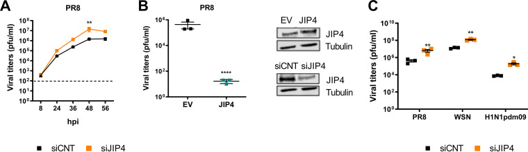
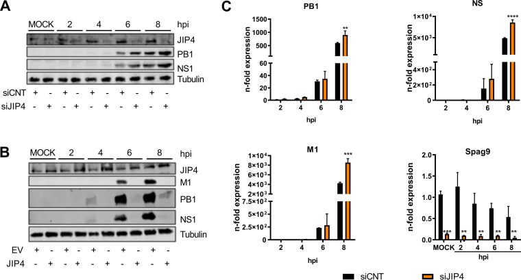
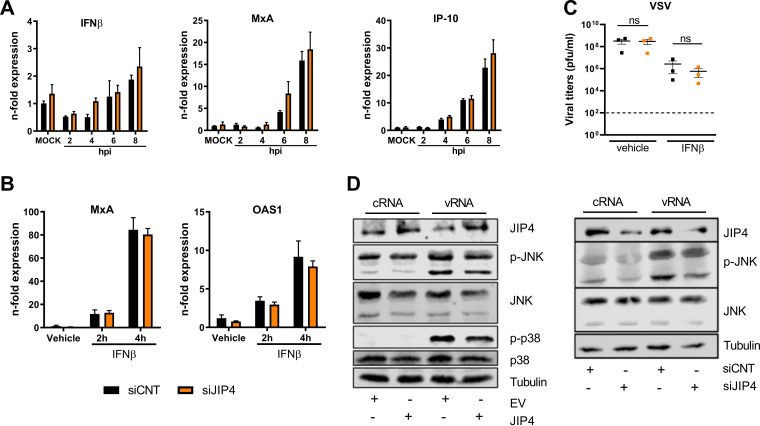
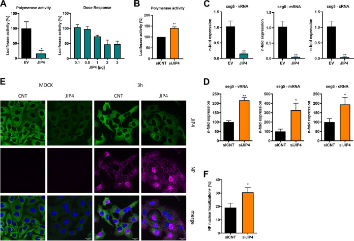
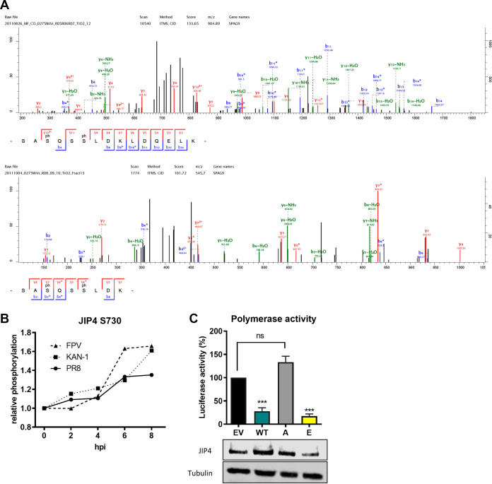
Influenza A virus (IAV) is the causative agent of flu disease that results in annual epidemics and occasional pandemics. IAV alters several signaling pathways of the cellular host response in order to promote its replication. Therefore, some of these pathways can serve as targets for novel antiviral agents. Here, we show that c-Jun NH2-terminal kinase (JNK)-interacting protein 4 (JIP4) is dynamically phosphorylated in IAV infection. The lack of JIP4 resulted in higher virus titers, with significant differences in viral protein and mRNA accumulation as early as within the first replication cycle. In accordance, decreased IAV titers and protein accumulation were observed during the overexpression of JIP4. Strikingly, the antiviral function of JIP4 does not originate from modulation of JNK or p38 mitogen-activated protein kinase (MAPK) pathways or from altered expression of interferons or interferon-stimulated genes but rather originates from a direct reduction of viral polymerase activity. Furthermore, the interference of JIP4 with IAV replication seems to be linked to the phosphorylation of the serine at position 730 that is sufficient to impede the viral polymerase. Collectively, we provide evidence that JIP4, a host protein modulated in IAV infection, exhibits antiviral properties that are dynamically controlled by its phosphorylation at S730. IMPORTANCE Influenza A virus (IAV) infection is a world health concern, and current treatment options encounter high rates of resistance. Our group investigates host pathways modified in IAV infection as promising new targets. The host protein JIP4 is dynamically phosphorylated in IAV infection. JIP4 absence resulted in higher virus titers and viral protein and mRNA accumulation within the first replication cycle. Accordingly, decreased IAV titers and protein accumulation were observed during JIP4 overexpression. Strikingly, the antiviral function of JIP4 does not originate from modulation of JNK or p38 MAPK pathways or from altered expression of interferons or interferon-stimulated genes but rather originates from a reduction in viral polymerase activity. The interference of JIP4 with IAV replication is linked to the phosphorylation of serine 730. We provide evidence that JIP4, a host protein modulated in IAV infection, exhibits antiviral properties that are dynamically controlled by its phosphorylation at S730.

Keywords: JIP4; host pathway; influenza virus; protein phosphorylation.


Figures

FIG 1
FIG 1
JIP4 presents antiviral activity against IAV infection. (A) A549 cells were transfected with siRNAs targeting JIP4 (siJIP4) or control siRNA (siCNT) 2 days prior to infection. Cells were then infected with PR8 IAV (MOI of 0.1). Supernatants were collected at 8, 24, 36, 48, and 56 hpi and analyzed for viral titers by standard plaque assays. Cells were collected for evaluation of the knockdown efficiency via Western blotting; tubulin detection served as a loading control. Results were statistically analyzed by two-way analysis of variance (ANOVA). (B) A549 cells were transfected with either an empty vector (EV) or plasmids containing the JIP4 sequence and infected with PR8 (MOI of 0.1). Supernatants were collected at 48 hpi for analysis of viral titers by standard plaque assays. Cells were collected for evaluation of the overexpression efficiency by Western blotting; tubulin detection served as a loading control. Results were statistically analyzed by a t test. (C) A549 cells were transfected with siJIP4 or siCNT and infected with different IAV strains 2 days post-transfection. PR8, WSN, or H1N1pdm09 was administered at an MOI of 0.1. Supernatants were collected at 48 hpi for determination of viral replication abilities by standard plaque assays. Results were analyzed by independent t tests per strain. *, P < 0.05; **, P < 0.01; ****, P < 0.0001 (compared to the respective controls). All Western blot images are representative of results from three independent experiments. Graphs are a compilation of results from three independent experiments.
FIG 2
FIG 2
JIP4 interferes with IAV replication prior to viral protein expression. (A) A549 cells were transfected with either control siRNA (siCNT) or siRNAs targeting JIP4 (siJIP4) and infected with PR8 (MOI of 5). Cells were lysed every 2 h. Lysates were submitted to Western blot assay and incubated with antibodies against the viral proteins PB1 and NS1 as well as against JIP4 for silencing confirmation. Detection of tubulin served as a loading control. + and − indicate the presence and absence, respectively, of the respective siRNAs. (B) A549 cells were transfected with either an empty vector (EV) or plasmids encoding JIP4 and infected with PR8 (MOI of 5). + and − indicate the presence and absence, respectively, of the respective plasmids. Cells were lysed every 2 h. Lysates were submitted to Western blot assays and incubated with antibodies against the viral proteins PB1, NS1, and M1 as well as against JIP4 for overexpression confirmation. Detection of tubulin was used as a loading control. (C) For analyses of viral mRNA expression, A549 cells were transfected with either siCNT or siJIP4 and infected with PR8 (MOI of 5). Samples were collected every 2 h and processed for qRT-PCR analyses. Viral mRNAs were normalized to the 2 h control group. SPAG9 gene expression was normalized to the mock control. Results were statistically analyzed by two-way ANOVA. **, P < 0.01; ***, P < 0.001; ****, P < 0.0001 (compared to the respective controls). All Western blot images are representative of results from three independent experiments. Graphs are a compilation of results from three different experiments.
FIG 3
FIG 3
The antiviral role of JIP4 does not correlate with changes in IFN responses or JNK/p38 activation. (A) A549 cells were transfected with either control siRNA (siCNT) or siRNAs targeting JIP4 (siJIP4) and infected with PR8 (MOI of 5). Cells were collected every 2 h for qRT-PCR analysis focusing on IFN and ISG mRNA expression. Results were statistically analyzed by two-way ANOVA. (B) A549 cells were transfected with either siCNT or siJIP4 and stimulated with IFN-β for 2 or 4 h. ISG mRNA levels were analyzed by qRT-PCR. Results were statistically analyzed by two-way ANOVA. Results in panels A and B are depicted as n-fold expression over mock/vehicle after normalization to GAPDH expression. (C) A549 cells were transfected with either siCNT or siJIP4, infected with VSV (MOI of 0.01), and treated with the vehicle or 100 U IFN-β. Supernatants were collected at 24 hpi, and virus replication was analyzed by standard plaque assays. Results are expressed as PFU per milliliter and were statistically analyzed by a t test. ns, not significant. (D) For analysis of JNK and p38 activity, A549 cells were transfected with either EV or JIP4-expressing plasmids (left) or siRNAs (right) and incubated for 24 h. Cells were then further transfected with cRNAs/vRNAs to stimulate the activation of JNK and p38 kinases. After 3 h of stimulation, cells were lysed for Western blot analysis, and the activation of MAPKs was determined by using p-JNK and p-p38 antibodies. Detection of p38, JNK, and tubulin served as loading controls; successful overexpression or knockdown was confirmed by JIP4 detection. All Western blot images are representative of results from three independent experiments. + and − indicate the presence and absence, respectively, of the respective siRNAs or plasmids. Graphs are a compilation of results from two independent experiments.
FIG 4
FIG 4
JIP4 overexpression decreases viral polymerase activity. (A) Vero cells were transfected with either an empty vector (EV) or JIP4-expressing plasmids together with all viral polymerase subunits and a reporter gene. At 24 hpt, cells were collected for luciferase assays. The activity in EV-transfected cells was arbitrarily set to 100%. For dose-response determinations, luciferase activities measured in JIP4-transfected cells are presented as percentages over the respective GST-expressing controls. (B) Vero cells were transfected with either control siRNA (siCNT) or siRNAs targeting JIP4 (siJIP4). At 48 hpt, cells were transfected with all viral polymerase subunits and a reporter gene. Twenty-four hours after the second transfection, cells were collected for luciferase assays. The activity in siCNT-transfected cells was arbitrarily set to 100%. (C) A549 cells were transfected with either EV or JIP4-expressing plasmids for 24 h and infected with PR8 (MOI of 5). Total RNA was collected at 4 hpi for strand-specific qRT-PCR. (D) A549 cells were transfected with either siCNT or siJIP4 and infected with PR8 (MOI of 5). Total RNA was collected at 4 hpi for strand-specific qRT-PCR. (E) A549 cells were transfected with either control siRNA or siRNAs targeting JIP4. After 48 h of transfection, cells were infected with PR8 (MOI of 20) and collected for immunofluorescence assays at 3 hpi. The localization of JIP4 and NP was analyzed by using specific antibodies; nuclei were stained with DAPI. (F) Percentage of cells with nuclear NP localization expressed over the total number of cells in the field. **, P < 0.01; *, P < 0.05 (compared to the respective controls). Polymerase activity graphs are a compilation of results from three independent experiments. Strand-specific PCR graphs with overexpression represent data from one experiment with three replicates; JIP4 knockdown strand-specific PCR graphs are compilations of data from two independent experiments. The immunofluorescence image is representative of results from three independent experiments. All results mentioned above were statistically analyzed by a t test (Welch’s t test [B]).
FIG 5
FIG 5
Phosphorylation of JIP4 S730 results in decreased viral polymerase activity. (A) A549 cells were labeled by using SILAC and infected with low-pathogenic IAV strain PR8 (H1N1) or one of two highly pathogenic strains (H5N1 strain KAN-1 and H7N7 strain FPV). Samples were collected at 2, 4, 6, and 8 hpi and analyzed for JIP4 phosphorylation. Representative MS/MS spectra of the tryptic peptides SASQSSLDKLDQELK (top) and SASQSSLDK (bottom) show phosphorylation at S730. ITMS, ion trap mass spectrometry; CID, collision-induced dissociation. (B) Relative phosphorylation of JIP4 at S730 at different time points compared to non-infected cells after normalization to the respective JIP4 protein amounts. (C) Vero cells were transfected with either EV or plasmids expressing WT JIP4 or the JIP4 phospho-mutant S730A or S730E together with all viral polymerase subunits and a reporter gene. At 24 hpt, cells were collected for luciferase assays. The activity in EV-transfected cells was arbitrarily set to 100%, and all groups were statistically compared with EV-transfected cells by one-way ANOVA. Overexpression was confirmed by Western blotting. ***, P < 0.001 (compared to the control group). Mass spectrometry phosphorylation kinetic data are the results from a single experiment. The polymerase activity graph is a compilation of data from three independent experiments.

Similar articles

Cited by

References

    1. Dong G, Peng C, Luo J, Wang C, Han L, Wu B, Ji G, He H. 2015. Adamantane-resistant influenza A viruses in the world (1902-2013): frequency and distribution of M2 gene mutations. PLoS One 10:e0119115. 10.1371/journal.pone.0119115. - DOI - PMC - PubMed
    1. Hayden FG, de Jong MD. 2011. Emerging influenza antiviral resistance threats. J Infect Dis 203:6–10. 10.1093/infdis/jiq012. - DOI - PMC - PubMed
    1. Laure M, Hamza H, Koch-Heier J, Quernheim M, Müller C, Schreiber A, Müller G, Pleschka S, Ludwig S, Planz O. 2020. Antiviral efficacy against influenza virus and pharmacokinetic analysis of a novel MEK-inhibitor, ATR-002, in cell culture and in the mouse model. Antiviral Res 178:104806. 10.1016/j.antiviral.2020.104806. - DOI - PubMed
    1. Nacken W, Ehrhardt C, Ludwig S. 2012. Small molecule inhibitors of the c-Jun N-terminal kinase (JNK) possess antiviral activity against highly pathogenic avian and human pandemic influenza A viruses. Biol Chem 393:525–534. 10.1515/hsz-2011-0270. - DOI - PubMed
    1. Ludwig S, Ehrhardt C, Neumeier ER, Kracht M, Rapp UR, Pleschka S. 2001. Influenza virus-induced AP-1-dependent gene expression requires activation of the JNK signaling pathway. J Biol Chem 276:10990–10998. 10.1074/jbc.M009902200. - DOI - PubMed

Publication types

MeSH terms

Substances