. 2021 Jul 28;16(7):e0254763.
doi: 10.1371/journal.pone.0254763. eCollection 2021.

Computational modelling unveils how epiblast remodelling and positioning rely on trophectoderm morphogenesis during mouse implantation

Affiliations

Computational modelling unveils how epiblast remodelling and positioning rely on trophectoderm morphogenesis during mouse implantation

Joel Dokmegang et al. PLoS One. .

Abstract

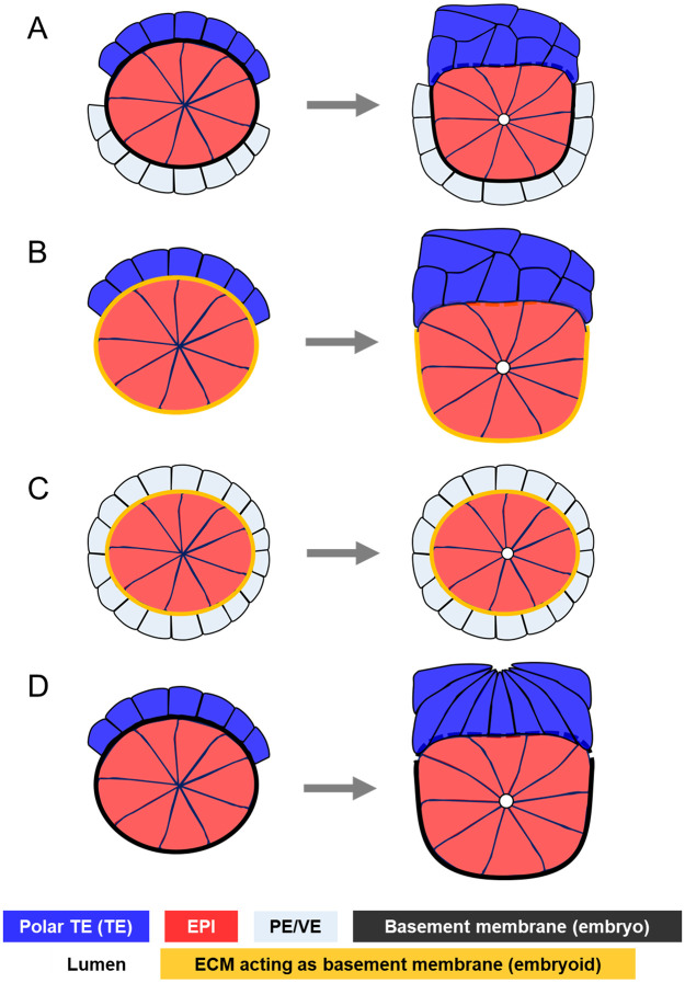
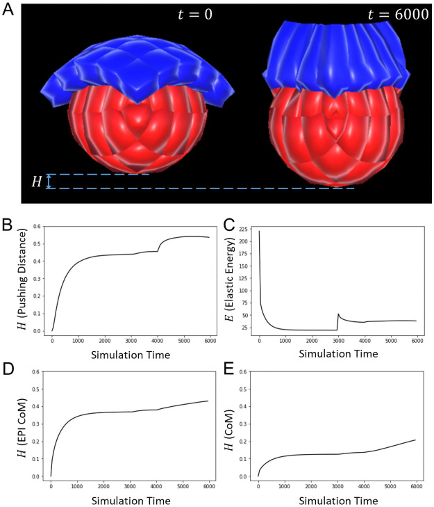
Understanding the processes by which the mammalian embryo implants in the maternal uterus is a long-standing challenge in embryology. New insights into this morphogenetic event could be of great importance in helping, for example, to reduce human infertility. During implantation the blastocyst, composed of epiblast, trophectoderm and primitive endoderm, undergoes significant remodelling from an oval ball to an egg cylinder. A main feature of this transformation is symmetry breaking and reshaping of the epiblast into a "cup". Based on previous studies, we hypothesise that this event is the result of mechanical constraints originating from the trophectoderm, which is also significantly transformed during this process. In order to investigate this hypothesis we propose MG# (MechanoGenetic Sharp), an original computational model of biomechanics able to reproduce key cell shape changes and tissue level behaviours in silico. With this model, we simulate epiblast and trophectoderm morphogenesis during implantation. First, our results uphold experimental findings that repulsion at the apical surface of the epiblast is essential to drive lumenogenesis. Then, we provide new theoretical evidence that trophectoderm morphogenesis indeed can dictate the cup shape of the epiblast and fosters its movement towards the uterine tissue. Our results offer novel mechanical insights into mouse peri-implantation and highlight the usefulness of agent-based modelling methods in the study of embryogenesis.


Conflict of interest statement

The authors have declared that no competing interests exist.

Figures

Fig 1
Fig 1. Review of epiblast symmetry breaking theories.
A. The basement membrane separating the epiblast and the primitive endoderm moulds the epiblast into a cup while it disintegrates between the epiblast and the trophectoderm in mouse embryos [4]. B. Embryoid structures featuring epiblast and trophectoderm stem cells surrounded by an ECM acting as a basement membrane (ETS-embryoids) replicate mouse embryogenesis by forming body structures similar to those observed in normal embryonic development [13]. Here the presence of the trophecdoderm shows that this tissue might be required for symmetry breaking in the epiblast and cup shape acquisition. C. Embryoid structures featuring epiblast and primitive endoderm stem cells surrounded by an ECM acting as a basement membrane (EXE-embryoids) do not break symmetry in the epiblast, but initiate lumenogenesis [14]. This evidences the requirement of the trophectoderm for the remodelling of the epiblast. D. Trophectoderm morphogenesis during mouse implantation. Trophectodermal cells elongate, then undergo apical constriction, resulting in the tissue folding [10]. This suggests that epiblast remodelling into a cup might be a mechanical response to trophectoderm dynamics.
Fig 2
Fig 2. Computational model.
A. 3D representation of a cell: The cell is abstracted by an agglomeration of particles (small white spheres, 34 in the picture), whose triangulation (white edges) forms the membrane, and by an intracellular particle (big white sphere). Interactions between the intracellular and membrane particles (blue lines) mimic the cytoskeleton. B. 3D rendering of a cell without its sub-cellular elements. C. Forces acting within a cell: Fjiint, Fkiint are the forces that membrane particles j, k exert on another membrane particle i. Fiχ is the force that the intracellular particle χ exerts on i. D. External forces acting on a cell via its particles. Here, Fi2ext=Fj2i2ext=(Fj1j2int+Fj3j2int)+Fj2χ. E. Plots of the magnitude of Morse forces under different values of J, with ρ = 1 and req = 0.5. F. Apical constriction of an epithelial cell with original radius R shrinking by d. G. Formulas of the new equilibrium lengths in an apically constricted cell.
Fig 3
Fig 3. Lumenogenesis in the epiblast.
A. A 3D model of a rosette-shaped epiblast. B. A 2D slice of the epiblast in A showing apically constricted cells of the building block of the epiblast rosette. C. Creation of the lumen cavity by repulsion at the apical surface of the epiblast. Green arrows represent the direction of repulsive forces. The snapshots (from left to right) were taken respectively at t = 0, 500 and 2000. D. Lateral view of the sliced epiblast showing the lumen volume. The lumen has been greyed to allow a better view over the black background. E. Evolution over time of the volume of the lumen. Values of the equation parameters: JEPI = 2.5, λ = 2, ρ = 1, Rlum = 0.25.
Fig 4
Fig 4. Trophectoderm morphogenesis regulates epiblast shape.
A-D. 3D snapshots of the simulation of TE and EPI morphogenesis during mouse implantation, and the regulation of EPI shape, taken respectively at t = 0, 3000, 6000 and 9000. E-H. Corresponding 2D slices of the cell population at the same stages. (A,E). The initial stage features a single layered TE with cuboidal cells resting upon the rosette-shaped epiblast. (B,F). TE cells have transited to a columnar shape. (C,G). The TE has folded by apical constriction of single cells. Concomitantly, lumenogenesis was initiated in the epiblast (the process starts at t = 4000). (D,H). After adhesive links were broken between TE and EPI, the EPI bounces back to its near spherical shape. I. Definitions of the metrics used to evaluate the model, involving the curvature θ, TE/EPI interface diameter D, TE/EPI interface length L, and interface ratio L/D. J. Plot of the population’s elastic energy E. Discontinuities mark the start of new morphological events at t = 0, 3000, 4000, and 6000). After removal of the TE, E falls closer to zero than ever before, meaning that cells are closer to their resting stage, hence less externally constrained. K. Plot of the interface curvature θ. During TE morphogenesis, θ rises towards a flat angle, then sharply drops when the TE is removed. L. Plot of the interface ratio L/D. During TE morphogenesis, the interface curvature decreases towards 1, then sharply increases when the TE is removed. Values of the equation parameters: JEPI = JTE = 2.5, λmed = λχ = 2, ρ = 1, d = 0.5.
Fig 5
Fig 5. Trophectoderm fosters epiblast movement towards maternal sites.
A. Snapshots of the simulation of TE and EPI morphogenesis during mouse implantation, and their influence on EPI positioning, taken respectively at t = 0 and 6000. B. Plot of the pushing distance, which increases with time. C. Plot of the elastic energy E. Discontinuities mark the start of new morphological events (t = 0 and 3000). The sudden soar observed at t = 4000 reflects the slight elongation of the tissue due to hollowing-driven lumenogenesis in the epiblast. D. Plot of the pushing distance on the epiblast Centre of Mass (CoM), which also increases with time. E. Plot of the pushing distance on the cell population Centre of Mass (CoM), which also increases with time. Values of the equation parameters: JEPI = JTE = 2.5, λmed = λχ = 2, ρ = 1, d = 0.5, Rlum = 0.25.
Fig 6
Fig 6. Mechanical properties of EPI and TE determine mouse implantation.
A. In“Silico” experimental protocol used to determine cells elastic modulus. B. Stress-Strain curve (black) for a single epithelial cell (34 vertices) with J = 2.5. (blue) Linear approximation of the Stress-Strain curve. The elastic modulus of the cell is determined by the slope of this line (Y = 2.92, φ = 2.92ϵ + 0.08, Rvalue = 0.99). C. Plot of the Elastic (Young) modulus of cells as a function of parameter J, the interaction strength between subcellular particles. D,E,F. Respective Plots of the Interface curvature, the Interface ratio and the Pushing Distance as functions of the mechanical stiffness of TE cells (determined by JTE as in C). G. Plot of the fitness metric as functions of the mechanical stiffness of TE cells (determined by JTE as in C). H. Snapshots of the epiblast shape at the end of simulations for different values of JTE. With equal stiffness (middle, JTE = 2.5, JEPI = 2.5), trophectoderm morphogenesis flatten the epiblast, which acquires a cup shape. However, with significantly lower stiffness (left, JTE = 0.3, JEPI = 2.5), trophectoderm morphogenesis barely reshape the epiblast; meanwhile, with considerably higher stiffness (right, JTE = 4.9, JEPI = 2.5), the trophectoderm invaginates the epiblast, forcing a concave interface with the epiblast. Other parameters values, λmed = λχ = 2, ρ = 1, d = 0.5.

Similar articles

Cited by

References

    1. Wang H, Dey SK. Roadmap to embryo implantation: Clues from mouse models. Nature Reviews Genetics. 2006;7(3):185–199. doi: 10.1038/nrg1808 - DOI - PubMed
    1. Shahbazi MN, Jedrusik A, Vuoristo S, Recher G, Hupalowska A, Bolton V, et al.. Self-organization of the human embryo in the absence of maternal tissues. Nature cell biology. 2016;18(6):700–708. doi: 10.1038/ncb3347 - DOI - PMC - PubMed
    1. Arnold SJ, Robertson EJ. Making a commitment: Cell lineage allocation and axis patterning in the early mouse embryo. Nature reviews Molecular cell biology. 2009;10(2):91–103 doi: 10.1038/nrm2618 - DOI - PubMed
    1. Bedzhov I, Graham SJ, Leung CY, Zernicka-Goetz M. Developmental plasticity, cell fate specification and morphogenesis in the early mouse embryo. Philosophical Transactions of the Royal Society B: Biological Sciences. 2014;369(1657):20130538. doi: 10.1098/rstb.2013.0538 - DOI - PMC - PubMed
    1. Kojima Y, Tam OH, Tam PP. Timing of developmental events in the early mouse embryo. In: Seminars in cell developmental biology. vol. 34. Elsevier; 2014. p. 65–75. - PubMed

Publication types