. 2021 Aug;35(8):e21780.
doi: 10.1096/fj.202100484R.

Maresin 1 activates LGR6 signaling to inhibit smooth muscle cell activation and attenuate murine abdominal aortic aneurysm formation

Affiliations

Maresin 1 activates LGR6 signaling to inhibit smooth muscle cell activation and attenuate murine abdominal aortic aneurysm formation

Craig T Elder et al. FASEB J. 2021 Aug.

Abstract

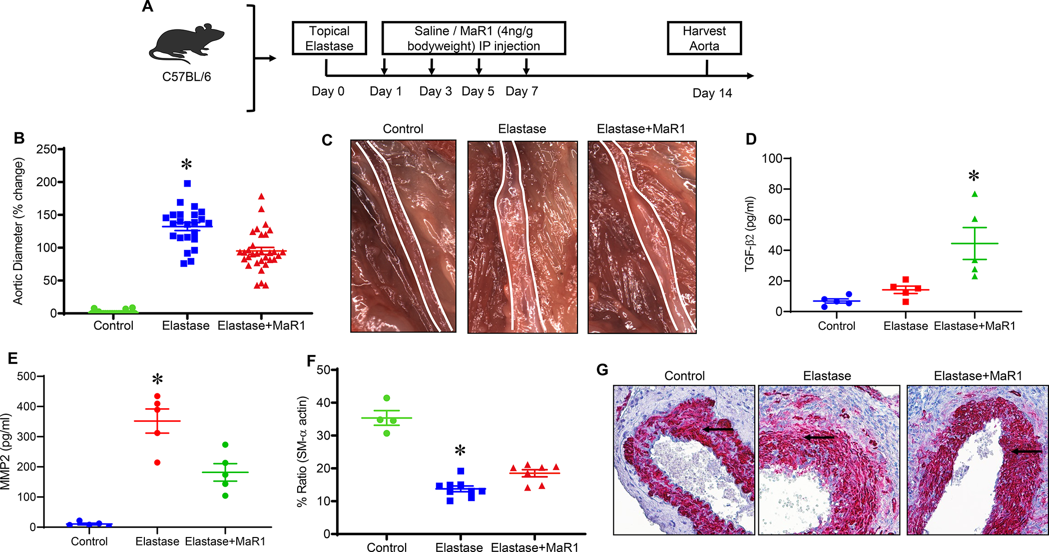
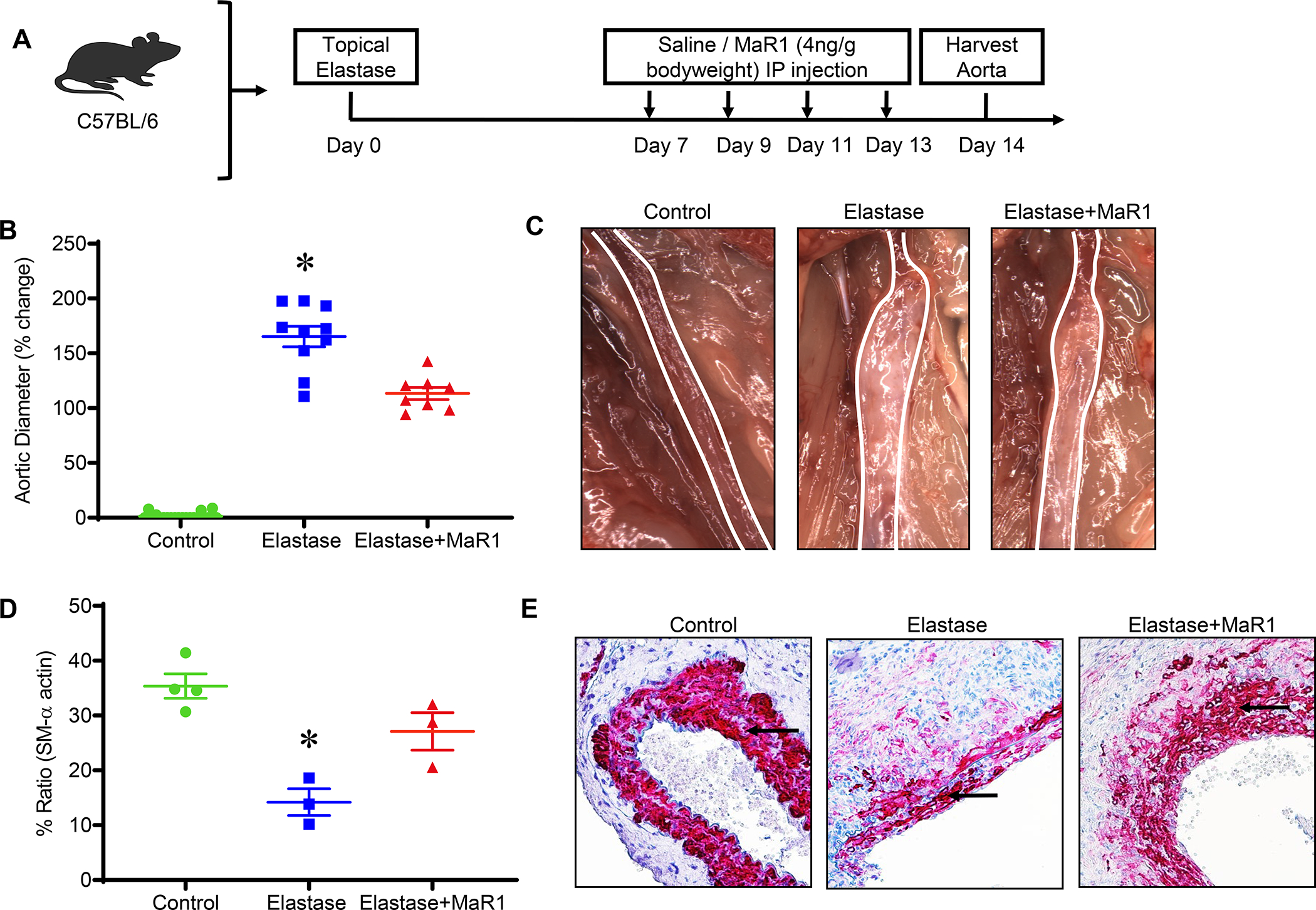
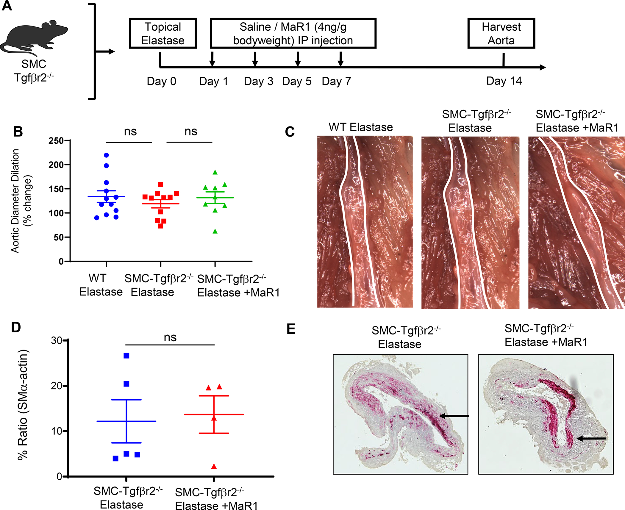
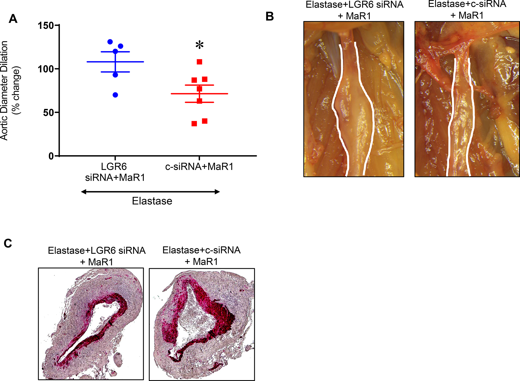
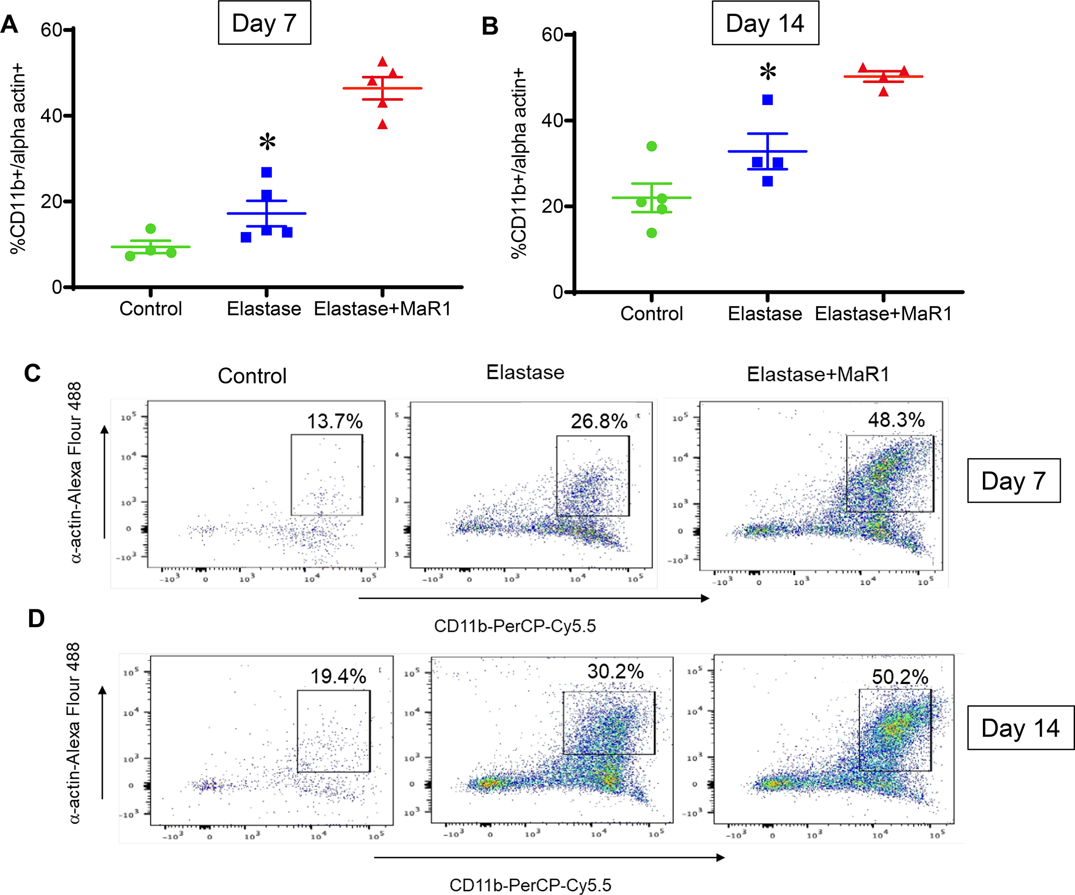
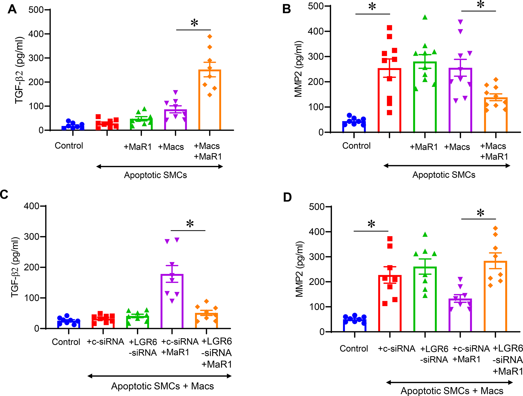
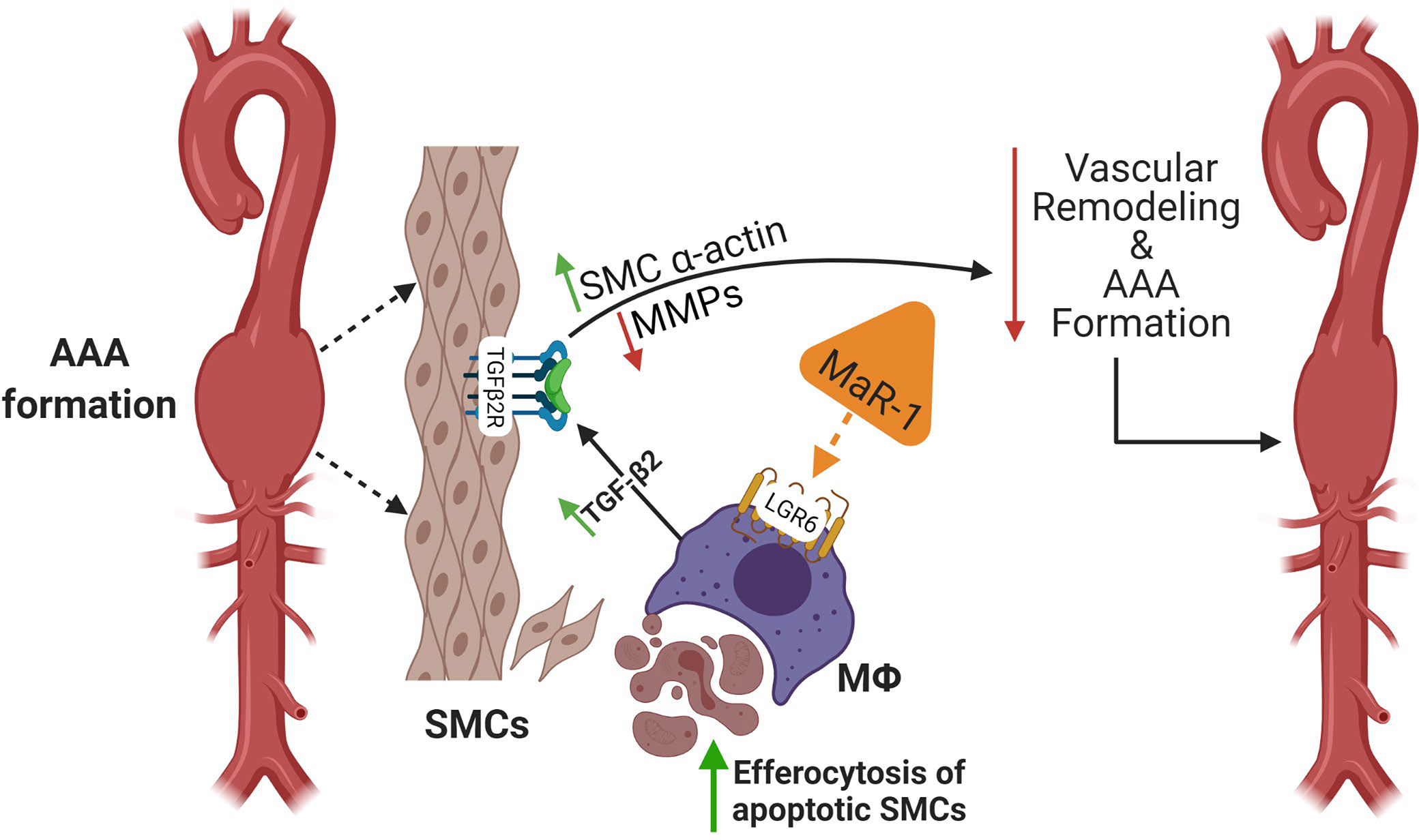
The specialized pro-resolving lipid mediator maresin 1 (MaR1) is involved in the resolution phase of tissue inflammation. It was hypothesized that exogenous administration of MaR1 would attenuate abdominal aortic aneurysm (AAA) growth in a cytokine-dependent manner via LGR6 receptor signaling and macrophage-dependent efferocytosis of smooth muscle cells (SMCs). AAAs were induced in C57BL/6 wild-type (WT) mice and smooth muscle cell specific TGF-β2 receptor knockout (SMC-TGFβr2-/- ) mice using a topical elastase AAA model. MaR1 treatment significantly attenuated AAA growth as well as increased aortic SMC α-actin and TGF-β2 expressions in WT mice, but not SMC-TGFβr2-/- mice, compared to vehicle-treated mice. In vivo inhibition of LGR6 receptors obliterated MaR1-dependent protection in AAA formation and SMC α-actin expression. Furthermore, MaR1 upregulated macrophage-dependent efferocytosis of apoptotic SMCs in murine aortic tissue during AAA formation. In vitro studies demonstrate that MaR1-LGR6 interaction upregulates TGF-β2 expression and decreases MMP2 activity during crosstalk of macrophage-apoptotic SMCs. In summary, these results demonstrate that MaR1 activates LGR6 receptors to upregulate macrophage-dependent efferocytosis, increases TGF-β expression, preserves aortic wall remodeling and attenuate AAA formation. Therefore, this study demonstrates the potential of MaR1-LGR6-mediated mitigation of vascular remodeling through increased efferocytosis of apoptotic SMCs via TGF-β2 to attenuate AAA formation.

Keywords: aneurysm; aorta; efferocytosis; macrophage; maresin; smooth muscle cells; transforming growth factor beta 2.


Figures

Figure 1.
Figure 1.
MaR1 administration attenuates AAA formation and preserves smooth muscle-α actin within the aortic wall. A, Schematic description of topical elastase treatment design. Mice were divided into three groups and treated with either heat inactivated elastase or elastase on day 0. Mice were then administered either vehicle or MaR1 on days 1, 3, 5, and 7. Aortic diameter was measured on day 14 and tissue harvested for additional analysis. B, MaR1 treated mice demonstrated a significant decrease in aortic diameter compared to vehicle treated mice; *p<0.001 vs. other groups; n=20–32 per group). C, Representative images of aortic phenotype in the respective groups. D, TGF-β2 expression in aortic tissue was significantly increased after MaR1 treatment in elastase-treated WT mice compared to untreated controls (*p<0.05 vs. other groups; n=5/group). E, Expression of MMP2 in aortic tissue was significantly attenuated in MaR1-treated mice compared to untreated controls (*p<0.01 vs. other groups; n=5/group). F, Expression of smooth muscle-α actin is significantly increased in mice treated with MaR1 compared to mice treated with vehicle alone (*p<0.02 vs. other groups; n=4–9 per group). G, Representative histological images of smooth muscle-α actin staining in the respective groups. Arrows indicate areas of immunostaining.
Figure 2.
Figure 2.
MaR1 attenuates the growth of pre-formed experimental murine AAA and preserves smooth muscle-α actin expression. A, Schematic description of treatment design. B, MaR1 treated mice demonstrated a significant decrease in aortic diameter compared to vehicle treated mice (*p<0.001 vs. other groups; n=8–10 per group). C, Representative images of aortic phenotype in the respective groups. D, Expression of smooth muscle-α actin is significantly increased in mice treated with MaR1 compared to mice treated with vehicle alone (*p<0.03 vs. Elastase+MaR1; n=3–4 per group). E, Representative histological images of smooth muscle-α actin staining in the respective groups is shown. Arrows indicate areas of immunostaining.
Figure 3.
Figure 3.
Maresin-1 mitigates AAA formation via smooth muscle cell-dependent TGF-β2 signaling. A, Schematic description of elastase AAA model in tamoxifen-treated SMC-Tgfβr2−/− mice. B, MaR1 treated SMC-Tgfβr2−/− mice showed no difference in aortic diameter compared to vehicle treated SMC-Tgfβr2−/− mice or elastase-treated WT mice (n=9–11 mice per group; ns, not significant). C, Representative images of aortic phenotype in respective groups. D, Expression of smooth muscle cell-α actin is similar in SMC-Tgfβr2−/− mice treated with MaR1 compared to mice treated with vehicle alone (n=4–5/group; ns, not significant). E, Representative histological images of smooth muscle-α actin staining in respective groups. Images were acquired using 20X magnification.
Figure 4.
Figure 4.
In vivo LGR6 knockdown reduces protective effect of MaR1. A, LGR6-siRNA treatment of WT mice demonstrated a significant increase in aortic diameter as compared to control (c)-siRNA treated mice after administration of MaR1 in respective groups (*p=0.04, n=5–7 per group). B, Representative images of aortic phenotype in respective groups. Images were acquired using 20X magnification. C, Expression of smooth muscle-α actin is decreased in mice administered with MaR1 and treated with LGR6-siRNAcompared to control-siRNA (n=5 per group).
Figure 5.
Figure 5.
MaR1 increases efferocytosis of SMCs in aortic tissue of murine AAA. Flow cytometry analysis of murine aortic tissue demonstrated increased levels of co-expression CD11b+SM-αA+ cell population in MaR1 treated mice compared to mice treated with vehicle alone at (A) post-operative day 7 (*p<0.001, n=4–5 per group) and (B) post-operative day 14 (*p=0.01, n=4–5 per group). Representative flow cytometry panels of day 7 (C) and day 14 (D) murine aortic tissue analysis.
Figure 6.
Figure 6.
Maresin-1 upregulates TGF-β2 secretion and decreases MMP2 expression. A, Co-culture of apoptotic SMCs with MaR1-treated macrophages upregulates TGF-β2 secretion compared to co-cultures of untreated macrophages and SMCs. B, Elastase-induced apoptotic SMCs demonstrate increased MMP2 expression which was inhibited by co-cultures with MaR1-treated macrophages. C, MaR1-treated macrophages upregulate TGF-β2 secretion via LGR6 receptors when co-cultured with apoptotic SMCs. MaR-1 dependent increase in TGF-β2 was abolished with LGR6-siRNA blockade of macrophages and co-culture with apoptotic SMCs, compared to pre-treatment with control siRNA. D, Inhibition of MMP2 expression by MaR1-treated macrophages is dependent on LGR6 receptors. Macrophages treated with LGR6-siRNA+MaR1 abolished the decrease in MMP2 expression observed in macrophages treated with control siRNA+MaR1. *p<0.05; n=8–10/group.
Figure 7.
Figure 7.
Schematic description of MaR1 mediated attenuation of vascular remodeling during AAA formation. The crosstalk between immune cells and aortic SMCs involves pro- and anti-inflammatory pathways that modulates vascular remodeling during AAA formation. Apoptosis and transformation of SMC architecture is a hallmark of aortic inflammation and remodeling that can be immunomodulated by SPMs. MaR1 acts on LGR6 receptors on macrophages to upregulate efferocytosis of apoptotic SMCs during AAA formation. This process upregulates TGF-β2 secretion that leads to SMC preservation, decrease in MMP2 activation and subsequent aortic remodeling as well as AAA formation. MaR1, maresin-1; LGR6, leucine-rich repeat-containing G-protein coupled receptor 6; SMCs; smooth muscle cells; MФ; macrophages; MMP; matrix metalloproteinases.

References

    1. Underlying Cause of Death 1999–2018 on CDC WONDER Online Database. United States, Centers for Disease Control and Prevention, http://wonder.cdc.gov/ucd-icd10.html. Accessed 10/14/2020.
    1. Harris LM, Faggioli GL, Fiedler R, Curl GR, and Ricotta JJ (1991) Ruptured abdominal aortic aneurysms: factors affecting mortality rates. Journal of vascular surgery 14, 812–818; discussion 819–820 - PubMed
    1. Thompson SG, Ashton HA, Gao L, Buxton MJ, and Scott RAP (2012) Final follow-up of the Multicentre Aneurysm Screening Study (MASS) randomized trial of abdominal aortic aneurysm screening. Br J Surg 99, 1649–1656 - PMC - PubMed
    1. Dale MA, Ruhlman MK, and Baxter BT (2015) Inflammatory cell phenotypes in AAAs; their role and potential as targets for therapy. Arterioscler Thromb Vasc Biol 35, 1746–1755 - PMC - PubMed
    1. Shimizu K, Mitchell RN, and Libby P (2006) Inflammation and cellular immune responses in abdominal aortic aneurysms. Arterioscler Thromb Vasc Biol 26, 987–994 - PubMed

Publication types

MeSH terms

Substances

LinkOut - more resources