. 2021 Jul 27;36(4):109421.
doi: 10.1016/j.celrep.2021.109421.

Methylation of dual-specificity phosphatase 4 controls cell differentiation

Affiliations

Methylation of dual-specificity phosphatase 4 controls cell differentiation

Hairui Su et al. Cell Rep. .

Abstract

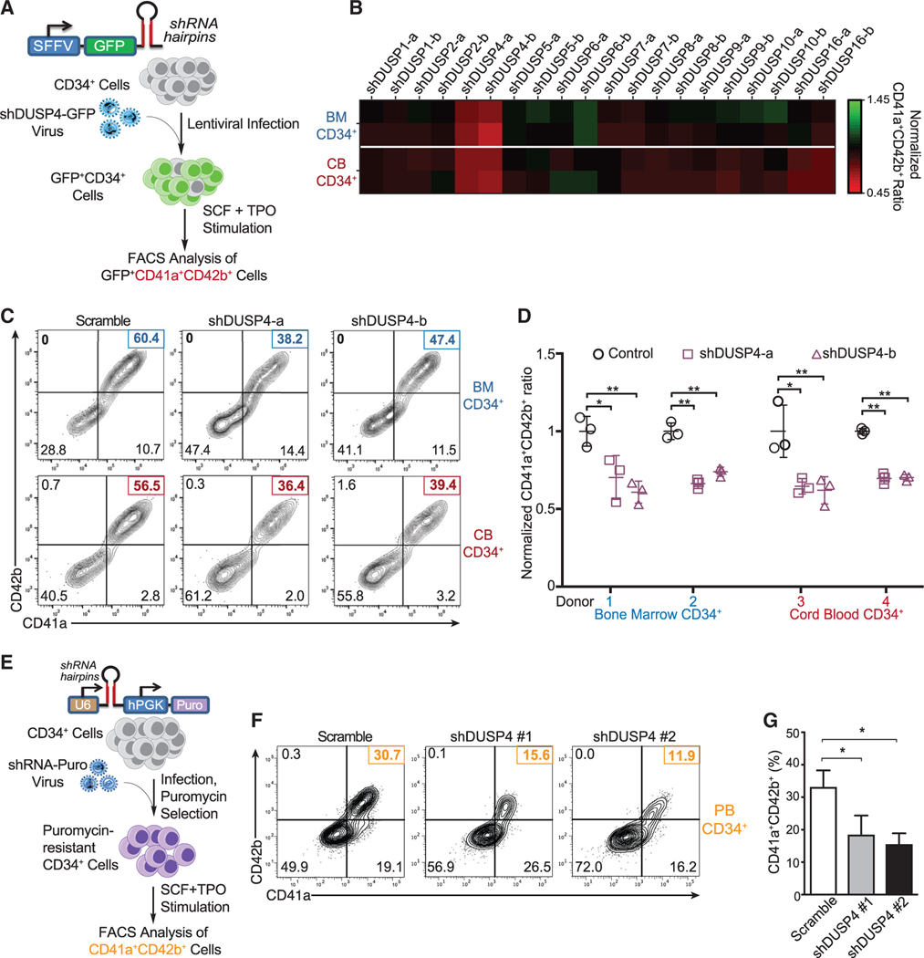
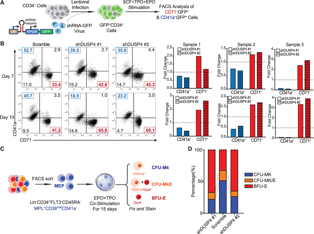
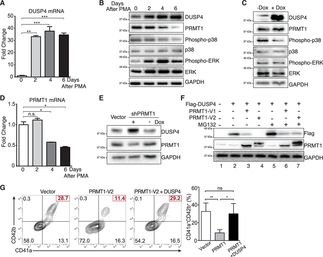
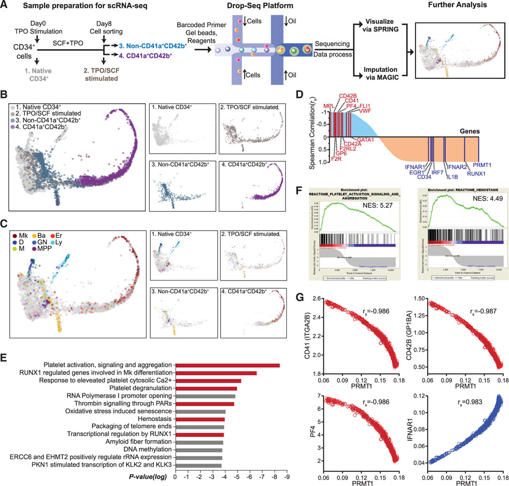
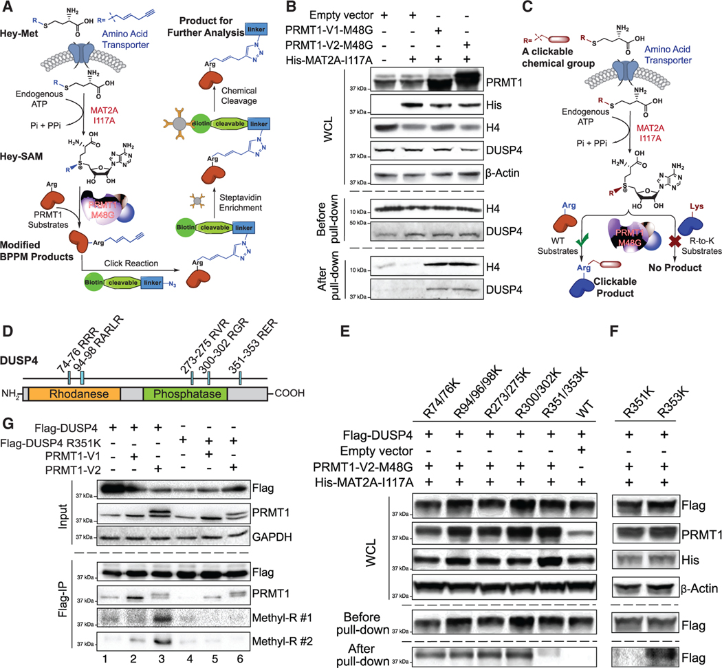
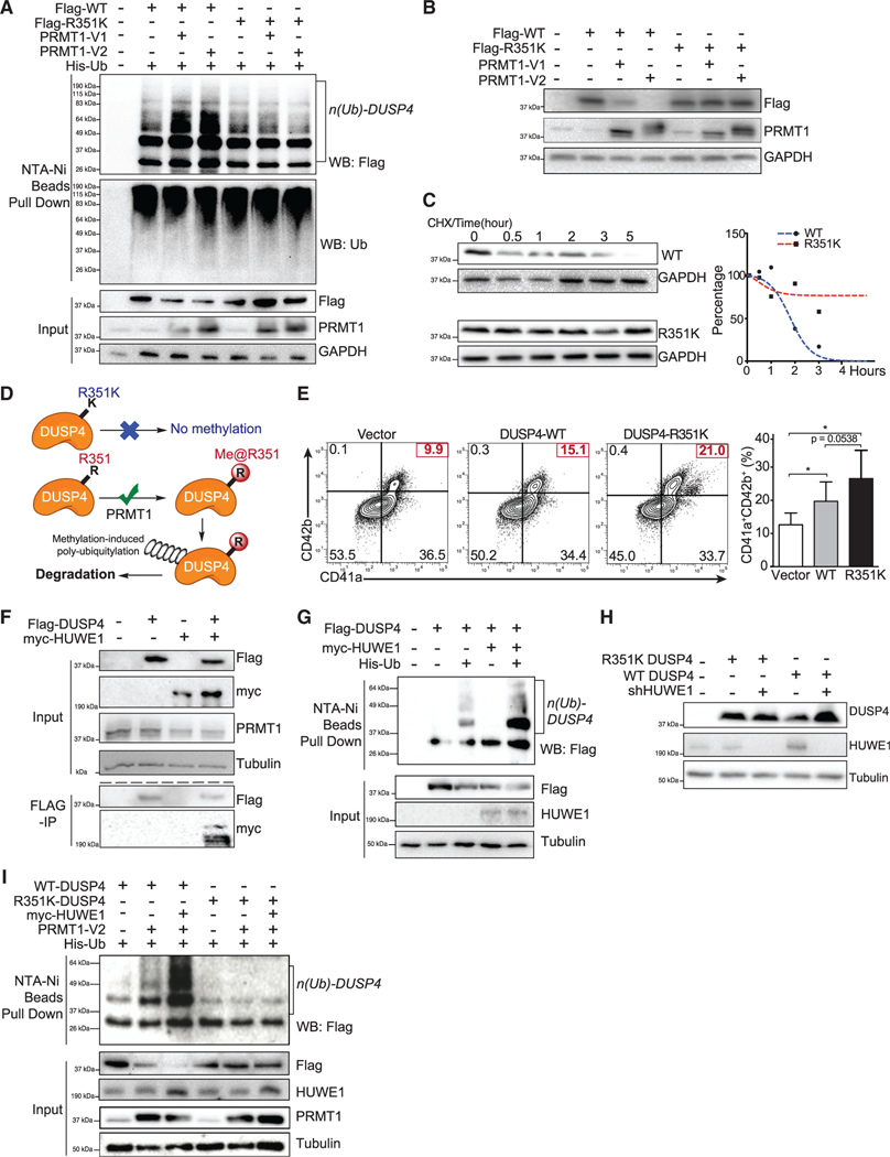
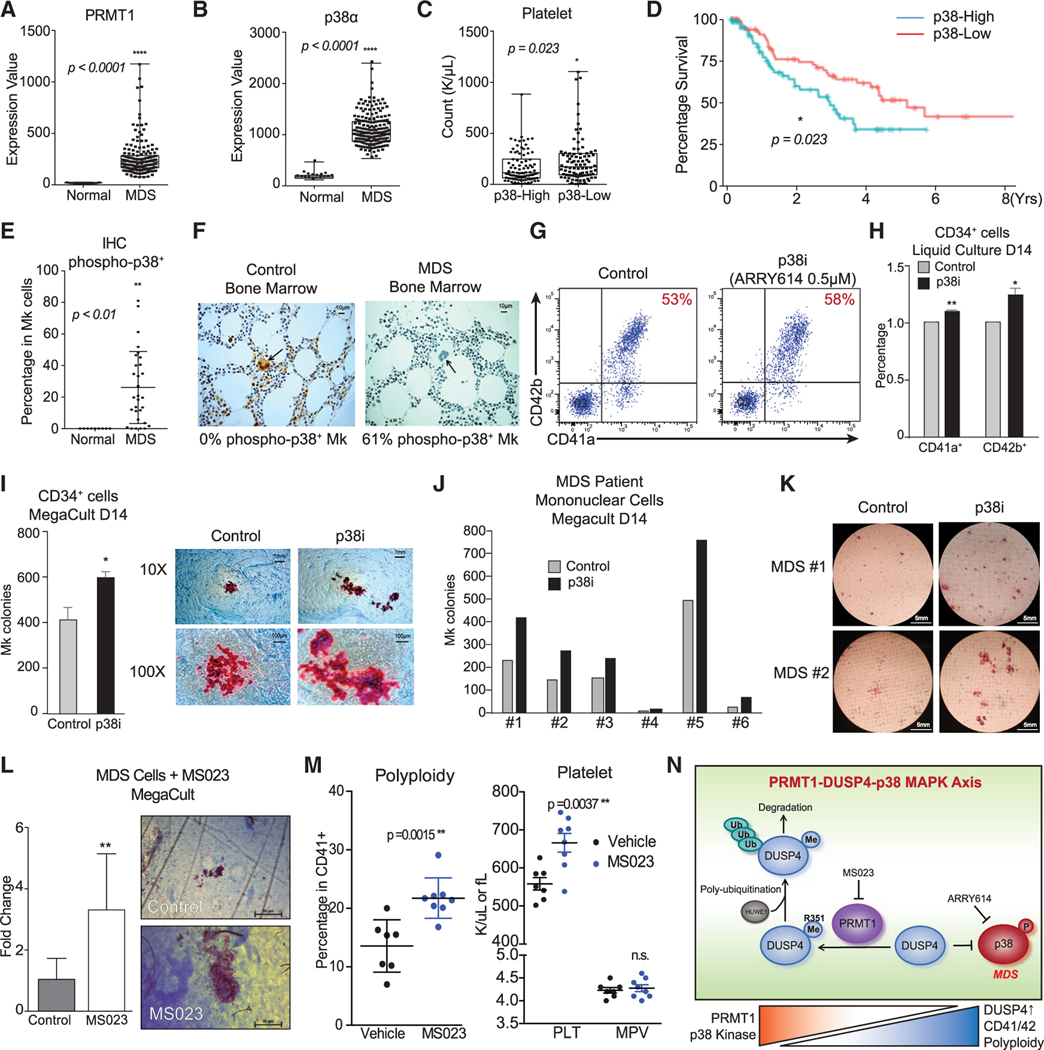
Mitogen-activated protein kinases (MAPKs) are inactivated by dual-specificity phosphatases (DUSPs), the activities of which are tightly regulated during cell differentiation. Using knockdown screening and single-cell transcriptional analysis, we demonstrate that DUSP4 is the phosphatase that specifically inactivates p38 kinase to promote megakaryocyte (Mk) differentiation. Mechanistically, PRMT1-mediated methylation of DUSP4 triggers its ubiquitinylation by an E3 ligase HUWE1. Interestingly, the mechanistic axis of the DUSP4 degradation and p38 activation is also associated with a transcriptional signature of immune activation in Mk cells. In the context of thrombocytopenia observed in myelodysplastic syndrome (MDS), we demonstrate that high levels of p38 MAPK and PRMT1 are associated with low platelet counts and adverse prognosis, while pharmacological inhibition of p38 MAPK or PRMT1 stimulates megakaryopoiesis. These findings provide mechanistic insights into the role of the PRMT1-DUSP4-p38 axis on Mk differentiation and present a strategy for treatment of thrombocytopenia associated with MDS.

Trial registration: ClinicalTrials.gov NCT01496495.

Keywords: DUSP4; HUWE1; MDS; PRMT1; leukemia; megakaryocyte; myelodysplasia syndrome; p38; platlet; trombocytopenia.


Conflict of interest statement

Declaration of interests M.L. has served on the Scientific Advisory Board for Epi One. A.V. has received research funding from GlaxoSmithKline, Incyte, MedPacto, Novartis, Curis, and Eli Lilly and Company; has received compensation as a scientific advisor to Novartis, Stelexis Therapeutics, Acceleron Pharma, and Celgene; and has equity ownership in Stelexis Therapeutics. The remaining authors declare no competing interests. Array BioPharma provided the p38 inhibitor pexmetinib (ARRY614) and participated in its phase I study (ClinicalTrials.gov: NCT01496495).

Figures

Figure 1.
Figure 1.. Identification of DUSP4 for optimal Mk differentiation
(A) Schematic of shRNA-based screening assay to identify essential DUSPs for Mk-induced Mk differentiation. Human CD34+ cells infected with lentiviruses expressing shRNAs against DUSPs were cultured for Mk differentiation. (B) Heatmap of the percentages of CD41a+CD42b+ cells upon DUSP knockdown on day 8. Fold changes were normalized to the percentage of double-positive cells with the group treated with control shRNA. (C) Representative flow chart of FACS analysis of Mk differentiation using BM cells (top panel) and CB cells (bottom panel) cultured in TPO-containing medium. (D) Summary of FACS analysis. Statistics are based on the data of three independent experiments (n = 3) with the bone marrow (BM) or cord blood (CB) cells from two donors. Data are shown as mean ± SD. Two-tailed paired t test, *p ≤ 0.05, **p ≤ 0.01. (E) Schematic description of Mk differentiation of human peripheral blood-derived CD34+ cells with DUSP4 knockdown. (F and G) Representative and complete FACS analysis of CD41a and CD42b markers for Mk differentiation with peripheral blood CD34+ cells upon DUSP4 knockdown on day 7. Representative plots (F) and statistics (G) are shown (n = 3, independent experiments). Data are shown as mean ± SD. Two-tailed paired t test, *p ≤ 0.05, **p ≤ 0.01.
Figure 2.
Figure 2.. DUSP4-regulated differentiation choices between Mk cells and Er cells
(A) Schematic of differentiation experiments using CB CD34+ cells. (B) FACS analysis of CD41a and CD71 on the cultured cells. Representative plots (top panel) and normalized statistics of three samples (bottom panel) are shown (n = 3, independent experiments). Fold changes were normalized with scramble controls. (C) Schematic description of colony-forming unit (CFU) assays using MEP. (D) Percentages of CFU-Mk, BFU-E, and CFU-MkE in each sorted population (n = 2, independent experiments).
Figure 3.
Figure 3.. Crosstalk between DUSP4 and PRMT1 for MAPK signaling in Mk differentiation
(A) DUSP4 mRNA level in PMA-treated MEG-01 cells. Cells were harvested at indicated time intervals, and the extracted mRNAs were quantified by real-time PCR. Representative statistics are shown as mean ± SD. Two-tailed unpaired t test, **p ≤ 0.01, ***p ≤ 0.001 (n = 3, independent experiments). (B) MAPK-related proteins and PRMT1 in MEG-01 cells after PMA stimulation. Protein extracts were collected at indicated time intervals for western blotting. (C) Regulation of MAPK signaling upon DUSP4 overexpression. MEG-01 cells were treated overnight with doxycycline to induce DUSP4 ectopically expressed from lentivirus. Cell extracts were collected for western blotting (n = 3, representative western blots). (D) PRMT1 mRNA level in MEG-01 cells during the course of PMA-stimulated Mk differentiation. Representative statistics were shown as mean ± SD, two-tailed unpaired t test, *p ≤ 0.05 (n = 3, independent experiments). (E and F) PRMT1-dependent regulation of DUSP4 protein. NB4 cells that conditionally express shRNA against PRMT1 were treated with doxycycline to induce PRMT1 knockdown (E). DUSP4- and PRMT1-encoding plasmids were transfected into HEK293T cells for their overexpression in the presence or absence of MG132 treatment (F). (G) Antagonistic roles of PRMT1 and DUSP4 on Mk differentiation of human CD34+ cells. Human CD34+ cells were infected with PRMT1 lentivirus (puromycin-R) and DUSP4 lentivirus (GFP), followed by puromycin selection and Mk differentiation. Representative plots and statistics are shown (n = 3, independent experiments). Data are shown as mean ± SD. Two-tailed paired t test, *p ≤ 0.05, **p ≤ 0.01.
Figure 4.
Figure 4.. Negative correlation between PRMT1 and Mk differentiation revealed by single-cell RNA-seq (scRNA-seq) analysis
(A) Experimental design for scRNA-seq analysis. Sample 1: Native CD34+ cells were cells isolated directly from BM. Sample 2: CD34+ cells were cells cultured in TPO and SCF for 8 days before sorting with CD41a and CD42b. Sample 3: The non-CD41a+CD42b+ cells were sorted from sample 2. Sample 4: The CD41a+CD42b+ cells were sorted from sample 2. (B and C) SPRING plots of single-cell transcriptomes. Individual cells are presented according to their origins (B) or transcriptome-associated cell types (C). Ba, basophilic or mast cell; D, dendritic; Er, erythroid; GN, granulocytic neutrophil; Ly, lymphocytic; M, monocytic; Mk, megakaryocytic; MPP, multipotential progenitor. (D) Pathway analysis of the top 400 genes with the strongest negative Spearman correlation to PRMT1 expression level in the TPO/SCF-stimulated CD41a+CD42b+ population. Red bars highlight Mk-relevant biological pathways. (E) Spearman correlation coefficients between PRMT1 and any gene revealed by scRNA-seq in TPO/SCF-stimulated CD41a+CD42b+ cells. Representative genes with significant correlation and functional relevance are annotated. (F) Gene set enrichment analysis (GSEA) of PRMT1-correlated genes in the TPO/SCF-stimulated CD41a+CD42b+ population. GSEA inputs are Spearman correlation coefficients of PRMT1 versus any gene with single-cell resolution. (G) Normalized expression of representative transcripts co-plotted against that of PRMT1 transcript in TPO/SCF-stimulated CD41a+CD42b+ cells with single-cell resolution.
Figure 5.
Figure 5.. R351 methylation of DUSP4 by PRMT1
(A) Schematic of the next-generation live-cell BPPM technology to uncover substrates of PRMT1. (B) Immunoblotting readouts of H4 and DUSP4 as PRMT1 targets enriched via the BPPM technology using HEK293T cells (n = 3, representative western blots). (C) Schematic description of the next-generation live-cell BPPM technology to reveal methylation sites of PRMT1. Candidates of methylated arginine (Arg) residues on a protein substrate are mutated to lysine (Lys). The resulting arginine-to-lysine mutation is expected to diminish or abolish the BPPM-associated labeling. (D) DUSP4 sequence with functional domains and RXR motifs highlighted. (E and F) Revealing PRMT1 methylation sites on DUSP4 with the next-generation live-cell BPPM technology. The DUSP4 variants contain dual arginine-to-lysine mutations at RXR motifs (E) and the point mutations at R351 and R353 (F), respectively (n = 3, representative western blots). (G) Validating PRMT1-invovled R351 methylation on DUSP4 with anti-methylarginine antibodies in HEK293T cells (n = 3, representative western blots). Two anti-methyl-arginine antibodies were used to determine the presence of arginine methylation in DUSP4.
Figure 6.
Figure 6.. Polyubiquitylation and instability of DUSP4 promoted by PRMT1-involved R351 methylation
For a Figure360 author presentation of this figure, see https://doi.org/10.1016/j.celrep.2021.109421. (A) Polyubiquitylation of DUSP4, but not its R351K variant, is stimulated by PRMT1. 293T cells were transfected with DUSP4 (wild-type and R351K mutant), 6× His-tagged ubiquitin, and PRMT1 (V1 and V2 isoforms). Cells were treated with MG132 for 6 h prior to harvest. Fractions of pull-downs (upper panel) and inputs (bottom panel) were applied for western blotting (n = 3, representative western blots). (B) PRMT1-dependent stability levels of wild-type and R351K DUSP4 were determined in 293T cells by western blotting (n = 3, representative western blots). (C) Half-life time of wild-type and R351K DUSP4. Normalized protein stability curves are plotted in the right panel (n = 3, representative western blots). (D) Mechanistic description of DUSP4 stability modulated by PRMT1-dependent R351 methylation. R351 methylation of DUSP4 by PRMT1 triggers its polyubiquitylation and thus its degradation; R351K mutation abolishes the methylation and thus suppresses polyubiquitylation and degradation (n = 3, representative western blots). (E) Mk differentiation in the presence of wild-type and R351K DUSP4. Human CD34+ cells infected with lentiviruses expressing DUSP4s (wild-type or R351K mutant) were induced for Mk differentiation. Percentage of CD41a+CD42b+ cells was determined by FACS after 7 days. Representative plots and statistics are shown (n = 3, independent experiments). Data are shown as mean ± SD. Two-tailed paired t test, *p ≤ 0.05. (F) Co-immunoprecipitation of HUWE1 and DUSP4. Flag-tagged DUSP4 was used to immunoprecipitate myc-tagged HUWE1 in co-transfected 293T cells (n = 3, representative western blots after immunoprecipitation). (G) The protein ubiquitylation of DUSP4 is measured in 293T cells transfected with plasmids as shown on top of the gels (n = 3, representative western blots). (H) Mutant and wild-type DUSP4 were expressed together with HUWE1 shRNA in 293T cells for western blotting with respective antibodies (n = 3, representative western blots). (I) Protein ubiquitylation assays with wild-type and mutant DUSP4. DUSP4 wild-type protein and mutant protein were expressed in 293T cells transfected with or without the plasmid combination of HUWE1 and PRMT1 as indicated on the top of the gel for affinity purification with Ni-NTA beads (n = 3, representative western blots).
Figure 7.
Figure 7.. Clinical implication and pharmacological targeting of the p38-DUSP4-PRMT1 axis in MDS
(A and B) Gene expression of PRMT1 and p38α in MDS patients and healthy donors with array-based analysis. The CD34+ HSPCs of MDS patients (N = 183) and age-matched healthy controls (N = 17) were analyzed for PRMT1 expression (A) and p38α expression (B). Data are shown as mean ± SD. Two-tailed unpaired t test, ****p ≤ 0.0001. (C) MDS cohorts as classified by low and high expression of p38α MAPK on the basis of median expression levels. The subjects with high p38α expression showed significantly lower platelet counts. Data are shown as mean ± SD. Two-tailed unpaired t test, *p ≤ 0.05. (D) Survival curves of MDS patients classified by low and high expression of p38α MAPK. The MDS patients with higher p38α expression in HSPCs showed significantly worse overall survival. *p ≤ 0.05. (E and F) Immunohistochemistry (IHC) analysis for phosphorylation-activated p38α MAPK of age-matched healthy controls and MDS BM samples from a clinical trial with the p38α inhibitor pexmetinib (ARRY614, labeled as p38i). MDS BMs showed significantly higher phospho-p38α staining in megakaryocytes (E). Representative stains are shown (F). Data are shown as mean ± SD. Two-tailed unpaired t test, **p ≤ 0.01. (G–I) Effects of the p38α inhibitor pexmetinib (labeled as p38i) on normal CD34+ cells. Normal CD34+ cells were grown in liquid culture conditions in the presence and absence of pexmetinib and analyzed for CD41 and CD42 expression by representative charts shown (G) and averaged FACS data (H) (n = 2, independent experiments). (I) Normal CD34+ cells were also grown for MegaCult assay for production of megakaryocyte colonies (n = 2, independent experiments). Data are shown as mean ± SD. Two-tailed paired t test, **p ≤ 0.05, **p ≤ 0.01. (J and K) Analysis of BM mononuclear cells (MNCs) of MDS patients with MegaCult assay for production of megakaryocyte colonies in the presence or absence of the p38α inhibitor pexmetinib (or p38i). Six MNC samples were examined (J) with the representative images shown (K). (L) Analysis of MNCs of MDS patients (n = 6) with MegaCult assay for production of megakaryocyte colonies in the presence or absence of a PRMT1 inhibitor MS023. Data are shown as mean ± SD. Two-tailed paired t test, **p ≤ 0.01. (M) Mk polyploidy and platelet count analysis in C57BL6/J mice treated with MS023. BM CD41+ cells were analyzed by FACS for polyploidy. The number of platelets in peripheral blood and MPV (mean platelet volume) were analyzed by a Hemavet machine. (N) Mechanistic description of Mk differentiation via the PRMT1-DUSP4-p38 axis. Mk progenitors undergo abnormal differentiation in MDS by upregulation of PRMT1, which leads to p38 kinase activation. The relative levels of phospho-p38 are regulated by DUSP4. DUSP4 R351 is subject to PRMT1-medidated methylation, which leads to polyubiquitylation by HUWE1 and then degradation. Collectively, the PRMT1-DUSP4-p38 axis determines generation of Mk progenitor cells and the maturation of Mk cells.

Similar articles

Cited by

References

    1. Al-Mutairi MS, Cadalbert LC, McGachy HA, Shweash M, Schroeder J, Kurnik M, Sloss CM, Bryant CE, Alexander J, and Plevin R (2010). MAP kinase phosphatase-2 plays a critical role in response to infection by Leishmania mexicana. PLoS Pathog. 6, e1001192. - PMC - PubMed
    1. Auger-Messier M, Accornero F, Goonasekera SA, Bueno OF, Lorenz JN, van Berlo JH, Willette RN, and Molkentin JD (2013). Unrestrained p38 MAPK activation in Dusp1/4 double-null mice induces cardiomyopathy. Circ. Res. 112, 48–56. - PMC - PubMed
    1. Bachegowda L, Morrone K, Winski SL, Mantzaris I, Bartenstein M, Ramachandra N, Giricz O, Sukrithan V, Nwankwo G, Shahnaz S, et al. (2016). Pexmetinib: A novel dual inhibitor of Tie2 and p38 MAPK with efficacy in preclinical models of myelodysplastic syndromes and acute myeloid leuke mia. Cancer Res. 76, 4841–4849. - PMC - PubMed
    1. Baldwin RM, Morettin A, Paris G, Goulet I, and Côté J (2012). Alternatively spliced protein arginine methyltransferase 1 isoform PRMT1v2 promotes the survival and invasiveness of breast cancer cells. Cell Cycle 11, 4597–4612. - PMC - PubMed
    1. Balko JM, Cook RS, Vaught DB, Kuba MG, Miller TW, Bhola NE, Sanders ME, Granja-Ingram NM, Smith JJ, Meszoely IM, et al. (2012). Profiling of residual breast cancers after neoadjuvant chemotherapy identifies DUSP4 deficiency as a mechanism of drug resistance. Nat. Med. 18, 1052–1059. - PMC - PubMed

Publication types

MeSH terms

Associated data