. 2021 Aug 3;36(5):109493.
doi: 10.1016/j.celrep.2021.109493. Epub 2021 Jul 20.

Development of safe and highly protective live-attenuated SARS-CoV-2 vaccine candidates by genome recoding

Affiliations

Development of safe and highly protective live-attenuated SARS-CoV-2 vaccine candidates by genome recoding

Jakob Trimpert et al. Cell Rep. .

Abstract

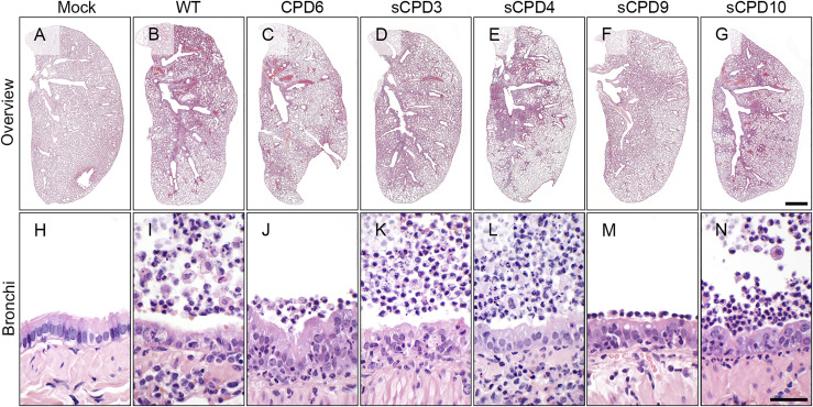
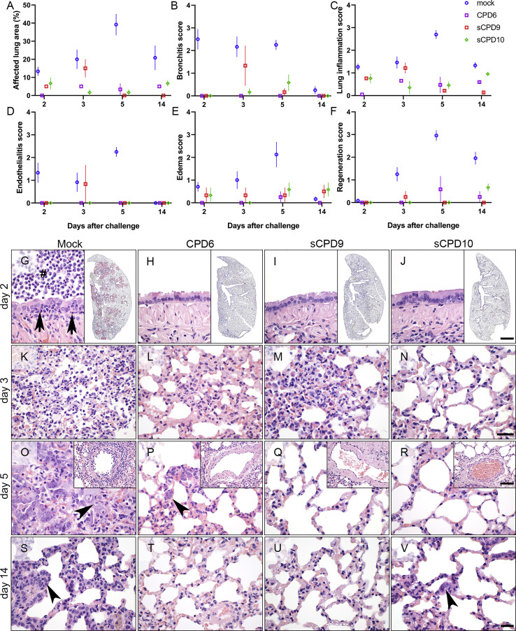
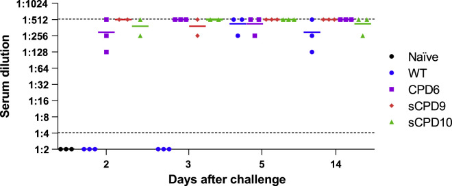
Safe and effective vaccines are urgently needed to stop the pandemic caused by severe acute respiratory syndrome coronavirus 2 (SARS-CoV-2). We construct a series of live attenuated vaccine candidates by large-scale recoding of the SARS-CoV-2 genome and assess their safety and efficacy in Syrian hamsters. Animals were vaccinated with a single dose of the respective recoded virus and challenged 21 days later. Two of the tested viruses do not cause clinical symptoms but are highly immunogenic and induce strong protective immunity. Attenuated viruses replicate efficiently in the upper but not in the lower airways, causing only mild pulmonary histopathology. After challenge, hamsters develop no signs of disease and rapidly clear challenge virus: at no time could infectious virus be recovered from the lungs of infected animals. The ease with which attenuated virus candidates can be produced and administered favors their further development as vaccines to combat the ongoing pandemic.

Keywords: COVID-19; Roborovski dwarf hamster; SARS-CoV-2; Syrian hamster; codon pair deoptimization; coronavirus; genome recoding; live attenuated vaccine; synthetic attenuated virus engineering.


Conflict of interest statement

Declaration of interests Freie Universität Berlin and the University of Bern received funding from a commercial partner for research similar to that described in this manuscript.

Figures

None
Graphical abstract
Figure 1
Figure 1
Structure and recoding of the SARS-CoV-2 genome (A) The SARS-CoV-2 genome is a single-stranded, positive-sense RNA molecule of about 30,000 nt, which encodes 11 canonical ORFs. (B) After infection, ORF 1a/1ab is directly translated and cleaved into 15 proteins of the replication-transcription complex. (C) Recoded SARS-CoV-2 mutants were constructed using a recently established reverse genetics system of SARS-CoV-2, which consists of 12 subgenomic fragments. Fragments 1, 11, and 12 were not recoded. The purple boxes indicate recoded sequences, and the green boxes indicate parental, non-recoded sequences in fragments CPD2-10. The frameshifting element contained in fragment 6 and the transcription regulatory sequence (TRS) of the spike gene in fragment 9 were excluded from the recoding process (green boxes present between two purple boxes in CPD6 and CPD9). (D) The pink boxes indicate recoded sequences in fragments sCPD3-5 and sCPD8-10. 3CL-Pro, 3C-like proteinase; RdRp, RNA-dependent RNA polymerase; ExoN, 3′-to-5′ exoribonuclease; EndoRNase, endoribonuclease; 2′-O-MT, 2′O-ribose methyltransferase.
Figure 2
Figure 2
Areas of infected cell foci and multi-step growth kinetics of the parental and recoded viruses in Vero E6 cells (A) Relative areas of infected cell foci at 48 hpi. Focus areas were normalized against the average focus size of the parental virus. Bars show geometric mean and SD. p values were calculated using Kruskal-Wallis test with Dunn’s post hoc test, p = 0.0135 and ∗∗∗∗p < 0.0001. See also Figure S1. (B) Multi-step growth kinetics of the parental and recoded viruses. Vero E6 cells were infected with the parental or recoded viruses at a multiplicity of infection of 0.01. Cell-culture medium was collected 6, 12, 24, 48, and 72 hpi, and virus titers were determined by focus-forming assay. Data are represented as means of three independent biological replicates ± SD. Comparison of growth curves by Friedman test with Dunn’s post hoc test showed that sCPD9 and sCPD10 viruses replicated significantly worse than the parental virus, p = 0.0264 and ∗∗p = 0.0049, respectively.
Figure 3
Figure 3
Attenuation of recoded SARS-CoV-2 mutants in Syrian hamsters (A–P) The attenuation level of recoded virus vaccine candidates was evaluated in two sequential trials. In the first trial, Syrian hamsters were either mock-vaccinated or vaccinated with CPD6, sCPD3, or sCPD4 viruses (A)–(E) and (K)–(M). In the second trial, Syrian hamsters were either mock-vaccinated, or vaccinated with sCPD9 or sCPD10 viruses (F–J and N–P). Twenty-one days after vaccination, all animals were challenged by infection with WT SARS-CoV-2. (A, B, F, and G) Change in body weight of Syrian hamsters after vaccination (n = 15); (A) and (F) and challenge (n = 12); (B) and (G). Data are represented as mean ± SD. (C, D, H, and I) Viral load in the upper (C) and (H) and lower (D) and (H) respiratory tract on day 3 after vaccination. (E and J) The number of infectious virus particles detected in 50 mg of lung tissue on day 3 after vaccination. (K, L, N, and O) Viral load in the upper (K) and (N) and lower (L) and (O) respiratory tract of animals on days 2, 3, 5, and 14 after challenge. (M and P) The number of infectious virus particles detected in 50 mg of lung tissue on days 2, 3, and 5 after challenge. The Kruskal-Wallis test was used to determine whether the differences in viral load among the different groups were significant (N–P), p < 0.05.
Figure 4
Figure 4
Lung histopathology of infected Syrian hamsters 3 days after vaccination Representative whole cross-sectional scans of left lung lobes (upper row, A–G) and micrographs of bronchial epithelium (bottom row, H–N) of formalin-fixed, paraffin embedded, hematoxylin and eosin-stained tissues. Hamsters were either mock-vaccinated (Mock), infected with SARS-CoV-2 (WT) or vaccinated with viruses CPD6, sCPD3, sCPD4, sCPD9, or sCPD10. Bars: 1 mm (A–G) or 100 μm (H–N).
Figure 5
Figure 5
Lung histopathology of vaccinated Syrian hamsters on days 2–14 after challenge (A–F) Histopathological evaluation and scoring of lung pathology. Parameters assessed: estimated percentages of affected lung areas (A), degree of bronchitis (B), lung inflammation (C), endothelialitis (D), edema (E), and epithelial hyperplasia (F). Scores and parameters in (B)–(F) were classified as absent (0), minimal (1), mild (2), moderate (3), or severe (4). n = 3 for each treatment at each time point. (G–V) Representative photomicrographs of formalin-fixed, paraffin-embedded, hematoxylin and eosin-stained lung tissues: bronchial epithelium (G)–(J), air spaces (K)–(V). Insets in (G)–(J) show the distribution of SARS-CoV-2 RNA, visualized by in situ hybridization in lungs of Syrian hamsters on day 2 after challenge. Red signal: viral RNA; blue: hemalaun counterstain. Insets in (O)–(R) show blood vessels affected by endothelialitis on day 5 after challenge. Pathological changes are shown from representative animals that had been either mock-vaccinated (Mock) or vaccinated with CPD6, sCPD9, or sCPD10. Bars: 50 μm (G–V), 1 mm (insets G–J), 100 μm (insets O–R).
Figure 6
Figure 6
Titers of SARS-CoV-2 neutralizing antibodies in vaccinated Syrian hamsters SARS-CoV-2 neutralizing antibodies were quantified in sera of mock-vaccinated hamsters and in hamsters vaccinated with CPD6, sCPD9, and sCPD10 after challenge infection with the WT virus. Sera of naive hamsters were used as a negative control. The dashed lines represents the detection limits of the assay.
Figure 7
Figure 7
Recoded SARS-CoV-2 mutant sCPD9s is strongly attenuated in Roborovski dwarf hamsters (A) Weights change of mock- (n = 12) and sCPD9-infected hamsters (n = 30). Data show mean ± SD. (B) Daily body temperature of mock- (n = 12) and sCPD9-infected hamsters (n = 30). Data show mean ± SD. (C) Viral load in the upper (oropharyngeal swab) and lower (lung) airways and infectious virus particles detected in 50 mg of lung tissue on day 3 after infection (lung titers). (D–O) Histopathological evaluation of the infection with sCPD9 (D–H) or SARS-CoV-2 variant (WT; I–N) on lung tissues at day 3 after infection. Left lung lobe with mild inflammatory lesions (D). Bronchioli had virtually normal columnar epithelium (E) and only occasional mild necrosuppurative bronchiolitis (F); neutrophil (black arrow). The alveolar septa showed mildly increased numbers of macrophages, and few neutrophils (G) with smaller areas of apparent pneumonia with macrophages (white arrows), neutrophils and necroses of alveolar epithelial cells (H). Normal pulmonary blood vessel (I). Typical lung of hamsters infected with WT virus (J), necrosuppurative bronchiolitis with early hyperplasia of bronchiolar epithelial cells (K), intraluminal cellular debris (hash), infiltrating neutrophils (black arrow). Bronchointerstitial pneumonia (L) with necrosis of alveolar epithelial cells, infiltration by macrophages and neutrophils (M), black arrow) and alveolar edema (M), asterisk). Endothelialitis (arrowhead) with mild to moderate perivascular edema (N, O, asterisk;). Scale bars: 1 mm (D) and (J), 50 μm (E, G, K, and L), 100 μm (I) and (N), 20 μm (insets F, H, M, and O).

Similar articles

Cited by

References

    1. Abu-Raddad L.J., Chemaitelly H., Butt A.A., National Study Group for COVID-19 Vaccination Effectiveness of the BNT162b2 Covid-19 Vaccine against the B.1.1.7 and B.1.351 Variants. N. Engl. J. Med. 2021;385:187–189. - PMC - PubMed
    1. Almazán F., González J.M., Pénzes Z., Izeta A., Calvo E., Plana-Durán J., Enjuanes L. Engineering the largest RNA virus genome as an infectious bacterial artificial chromosome. Proc. Natl. Acad. Sci. USA. 2000;97:5516–5521. - PMC - PubMed
    1. Anderson E.J., Rouphael N.G., Widge A.T., Jackson L.A., Roberts P.C., Makhene M., Chappell J.D., Denison M.R., Stevens L.J., Pruijssers A.J., mRNA-1273 Study Group Safety and Immunogenicity of SARS-CoV-2 mRNA-1273 Vaccine in Older Adults. N. Engl. J. Med. 2020;383:2427–2438. - PMC - PubMed
    1. Baden L.R., El Sahly H.M., Essink B., Kotloff K., Frey S., Novak R., Diemert D., Spector S.A., Rouphael N., Creech C.B., COVE Study Group Efficacy and Safety of the mRNA-1273 SARS-CoV-2 Vaccine. N. Engl. J. Med. 2021;384:403–416. - PMC - PubMed
    1. Bazin H. A brief history of the prevention of infectious diseases by immunisations. Comp. Immunol. Microbiol. Infect. Dis. 2003;26:293–308. - PubMed

Publication types