. 2021 Oct:106:241-256.
doi: 10.1016/j.neurobiolaging.2021.06.012. Epub 2021 Jun 20.

Age-related neuroinflammation and pathology in the locus coeruleus and hippocampus: beta-adrenergic antagonists exacerbate impairment of learning and memory in aged mice

Affiliations

Age-related neuroinflammation and pathology in the locus coeruleus and hippocampus: beta-adrenergic antagonists exacerbate impairment of learning and memory in aged mice

Andrew K Evans et al. Neurobiol Aging. 2021 Oct.

Abstract

The locus coeruleus (LC) provides the primary noradrenergic input to the forebrain and hippocampus, and may be vulnerable to degeneration and contribute to age-related cognitive decline and neuroinflammation. Additionally, inhibition of noradrenergic transmission by brain-permeable beta-blockers could exacerbate cognitive impairment. This study examined effects of age and acute beta-blocker administration on LC and hippocampus pathology, neuroinflammation and learning and memory behavior in mice. Male mice, 3 and 18 months old, were administered propranolol (beta-blocker) or mabuterol (beta-adrenergic agonist) acutely around behavioral assessment. Terminal inflammatory markers in plasma, hippocampus and LC were assessed alongside histopathology. An increase in hippocampal and LC microgliosis and inflammatory proteins in the hippocampus was detected in aged mice. We report pathological hyperphosphorylation of the postsynaptic NMDA receptor subunit 2B (NR2B) in the hippocampus, suggesting neuronal hyperexcitability. Furthermore, the aged proteome revealed an induction in proteins related to energy metabolism, and mitochondria dysfunction in the LC and hippocampus. In a series of hippocampal dependent behavioral assessment tasks acute beta-adrenergic agonist or beta blocker administration altered learning and memory behavior in both aged and young mice. In Y-maze, propranolol and mabuterol differentially altered time spent in novel versus familiar arms in young and aged mice. Propranolol impaired Novel Object Recognition in both young and aged mice. Mabuterol enhanced trace learning in fear conditioning. Aged mice froze more to context and less to cue. Propranolol impaired contextual recall in aged mice. Concluding, aged mice show LC and hippocampus pathology and heightened effects of beta-adrenergic pharmacology on learning and memory.

Keywords: Aging; Behavior; Beta-blocker; Inflammation; Locus coeruleus; proteomics.


Conflict of interest statement

Declaration of Interest

None declared

Figures

Figure 1.
Figure 1.
Experimental Design depicts timing of dosing and behavioral testing for young (3–4 months) and aged (18–19 months) mice. Mice were dosed with vehicle, propranolol or mabuterol around each behavioral task as well as daily for 9 days prior to termination. Timing of dosing around each specific behavioral task is indicated in the respective figures. Sample size is 6–7 per group; 2 × 3 factor, age x treatment design.
Figure 2.
Figure 2.
Aged (19 months) and young (4 months) mice have equivalent tyrosine hydroxylase (TH) immunoreactive (-ir) cell counts across the rostrocaudal extent of the locus coeruleus. Aged mice have increased iba1-ir in the locus coeruleus. Photomicrographs depict representative TH-ir in the locus coeruleus at −5.62 mm bregma in A) young versus B) aged mice. White scale bars equal 100 micrometers. C) Line graph shows quantification of TH-ir cell counts. D-F) Photomicrographs show representative iba1-ir staining in D) young versus E) aged mice. Insets in C and D show higher magnification iba-ir staining. F) No primary antibody control for the iba1 channel in aged mice show age-related autofluorescence that is distinct from iba1-ir. Iba1-ir can be thresholded out above age-related background staining. G) Scatter bar graph depicts quantification of thresholded iba-ir % area in locus coeruleus averaged across 3 rostrocaudal levels.
Figure 3.
Figure 3.
Volcano plots depict age-related fold-change and significance for proteins detected in the A) locus coeruleus and C) hippocampus with proteomics. A,B) In locus coeruleus (1043 proteins detected), 186 proteins were identified to be 2-fold up- (red) or down-regulated (blue) with age (fold change as old/young; p-value cutoff p<.05; -Log10(p-value)>1.3). B) Clustergram analysis for locus coeruleus identifies age-modulated proteins in specific pathways. Locus coeruleus pathways include metabolic pathways, oxidative phosphorylation and neurodegeneration as indicated by KEGG analysis. C,D) In hippocampus (2603 proteins detected), only 24 proteins met the 2-fold change criteria, so for hippocampal pathway analysis criteria was expanded to include all proteins that were significantly up- or down-regulated (p<.05; -Log10(p-value)>1.3; 118 proteins) independent of fold-change. D) Clustergram analysis for hippocampus identifies age-modulated proteins in specific pathways. Hippocampal pathways modulated with aging include retrograde endocannabinoid signaling, synaptic plasticity, oxidative phosphorylation, and phagosomes indicated by KEGG analysis. Blue indicates downregulation with age and red indicates upregulation with age. Numbers in clustergram indicate Log2 fold-change (aged/young) for each protein.
Figure 4.
Figure 4.
Aged mice have increased iba1-immunoreactivity (-ir) in the hippocampus. A) Scatter bar graph depicts quantification of iba1-ir in CA1. Photomicrographs depict representative immunoreactivity in the iba1 channel for B) young versus C) aged mice in the CA1 region with representative no antibody controls for young D) versus E) old mice. F-J) shows the quantification and representative images for CA3 and K-O) shows the quantification and representative images for dentate gyrus (DG). Iba1-immunoreactivity is morphologically distinct from age-related autofluorescence and can be thresholded out above age-related autofluorescence. White scale bars equal 100 micrometers.
Figure 5.
Figure 5.
Aged mice have elevated markers of inflammation in hippocampus as compared to young mice. Log2-fold change bar graphs (mean ± SEM, n=6) indicate changes in immune related proteins in plasma and hippocampus. A) Plasma: No effects of age or drug were observed on plasma markers of inflammation. A general trend is observed for 18 month (18M) mice to have lower concentrations of most markers of inflammation measured (40 out of 48), although no individual markers reach significance. B) Hippocampus: 18 month (18M) mice have elevated markers of inflammation in hippocampus as compared to 3M mice. There was no further effect of beta-adrenergic pharmacology on these age-related effect (see related supplementary Figure S1 and Figure S2). * indicates p < .05, **p < .01, ***p < .001; Sidak’s posthoc following one-way ANOVA within each protein.
Figure 6.
Figure 6.
Western blot analysis of young and aged hippocampus shows no change in the expression level of the NMDA receptor 2B (NR2B) but an increase in the phosphorylation state of the NR2B receptor protein at the Ser1303 site in aged mice. A,B) Scatter bar graphs depict quantification of western blot for NR2B relative to tubulin and pNR2B relative to NR2B. C,D,E) Representative western blot images for NR2B, pNR2B, and tubulin. ** indicates p < .01, t-test.
Figure 7.
Figure 7.
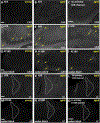
Aged mice have evidence for AT8 and AT180-immunoreactivity in the hippocampus and locus coeruleus in addition to a distinctive age-related autofluorescence. Photomicrographs of the CA3 region of the hippocampus show representative AT8-immunoreactivity in A,B) young and aged mice as compared to C) no primary control in aged mice. D-F) Distinctive AT8-immunoreactivity is depicted in CA3 and CA1 of two independent aged mice (2 out of 10 mice had this type of staining). G-I) AT180 immunoreactivity is depicted in young and aged tissue treated with Sudan Black in order to reduce non-specific background staining. Panels H and I show a similar immunoreactivity pattern with AT180 as observed with the AT8 antibody in the same subjects as panels D-F. Photomicrographs of the locus coeruleus at −5.62 mm Bregma show representative J,K) AT8-immunoreactivity in young versus aged mice alongside L) a no primary control in aged mice. M,N) AT180 immunoreactivity is depicted in young and aged tissue treated with Sudan Black. O) No antibody control for the AT180 immunostaining conditions. White scale bars equal 100 micrometers.
Figure 8.
Figure 8.
Mabuterol decreased distance moved in Y-maze and increased time spent in novel versus familiar arms in young versus aged mice respectively. A) Y-Maze Forced Alternation experimental design. B) 18 month (18M) mice tended to move less than 3M mice during Y-Maze forced alternation testing. C,D) Mabuterol decreased distance moved in both 3M and 18M mice and D) propranolol tended to increase distance moved in 18M mice, but increase did not reach significance. E) 3M mice explore both novel and familiar arms, while 18M mice show a preference for the familiar arm. F) Mabuterol increases time spent in the novel arm in 3M mice. G) Propranolol decreases preference for familiar arm in 18M mice. H) 18M mice but not 3M mice had more entries into the familiar versus novel arms and J) propranolol blocked the increased number of entries into the familiar arm in 18M mice. * indicates p < .05, **p < .01, ***p < .001; Dunnett’s posthoc after one-way ANOVA (C, D), or Sidak’s posthoc after two-way ANOVA (E-J).
Figure 9.
Figure 9.
In Novel Place Recognition (NPR), propranolol tends to increase distance moved in old mice, but all groups successfully show novelty discrimination. A) NPR experimental design. B-D) 18 month (18M) mice tend to move less during NPR testing, but E-G) both 3 and 18 month old mice show preference for novel place in terms of % time interaction during testing. ** indicates p < .01, ***p < .001; Dunnett’s posthoc after one-way ANOVA (C, D), or Sidak’s posthoc after two-way ANOVA (E-G).
Figure 10.
Figure 10.
In Novel Object Recognition (NOR), young and old mice show novelty discrimination, but propranolol blocks discrimination in young mice and impairs discrimination in old mice. A) NOR experimental design. B-D) 3 month (3M) but not 18M mice move greater distance during the testing phase of NOR relative to habituation and training. E-G) Both 3M and 18M mice show preference for novel object in terms of % time interaction. F,G) Propranolol impairs novel object preference in 3M and 18M mice, and G) mabuterol impairs novel object preference in 18M mice. * indicates p < .05, ** p < .01, *** p < .001; Sidak’s posthoc after two-way ANOVA.
Figure 11.
Figure 11.
Young and old mice both learn to associate a tone with freezing behavior following tone-shock pairings, but old mice show greater freezing in response to context and less freezing response to cue when tested for recall. A) Fear conditioning experimental design. B-D) Training. B) 3 month (3M) and 18M mice learn to freeze in response to a tone paired with shock. C,D) Mabuterol enhances this freezing during training in both 3M and 18M mice and D) propranol impairs this freezing during training but only in 18M mice. E,F,G) Context. E) 18 M mice freeze more in response to re-exposure to context. F) Propranol and metoprolol increase freezing to context in 3M mice. G) Propranolol reduces freezing to context in 18 M mice. H,I,J) Cue. H) 3M mice freeze more in response to cue, I,J) with no effect of drug in either 3M or 18M. * indicates p < .05, ** p < .01; Dunnett’s after two-way ANOVA (B-D), t-test (E), Dunnett’s after one-way ANOVA (F-G) or Sidak’s posthoc after two-way ANOVA (H-J).
Figure 12.
Figure 12.
Summary schematic illustrates hypothetical model in which oxidative stress and metabolic demand from neurons in the LC and hippocampus accompany an increase in inflammatory signaling from astrocytes and microglia. Excessive glutamate release in aging brain leads to activation of extrasynaptic NMDA receptors predominantly containing NR2B subunits leading to impairment of neuronal function and behavioral impairment.

Similar articles

Cited by

References

    1. Amrhein V, et al., 2019. Scientists rise up against statistical significance. Nature. 567, 305–307. - PubMed
    1. Betts MJ, et al., 2019. Locus coeruleus imaging as a biomarker for noradrenergic dysfunction in neurodegenerative diseases. Brain. 142, 2558–2571. - PMC - PubMed
    1. Bialik S, Kimchi A, 2006. The death-associated protein kinases: structure, function, and beyond. Annu Rev Biochem. 75, 189–210. - PubMed
    1. Braak H, Del TK, 2011. The pathological process underlying Alzheimer’s disease in individuals under thirty. Acta Neuropathol. 121, 171–181. - PubMed
    1. Braak H, et al., 2011. Stages of the pathologic process in Alzheimer disease: age categories from 1 to 100 years. J. Neuropathol. Exp. Neurol. 70, 960–969. - PubMed

Publication types

MeSH terms