. 2021 Sep 1;126(3):827-839.
doi: 10.1152/jn.00184.2021. Epub 2021 Jul 28.

A novel gain-of-function sodium channel β2 subunit mutation in idiopathic small fiber neuropathy

Affiliations

A novel gain-of-function sodium channel β2 subunit mutation in idiopathic small fiber neuropathy

Matthew Alsaloum et al. J Neurophysiol. .

Abstract

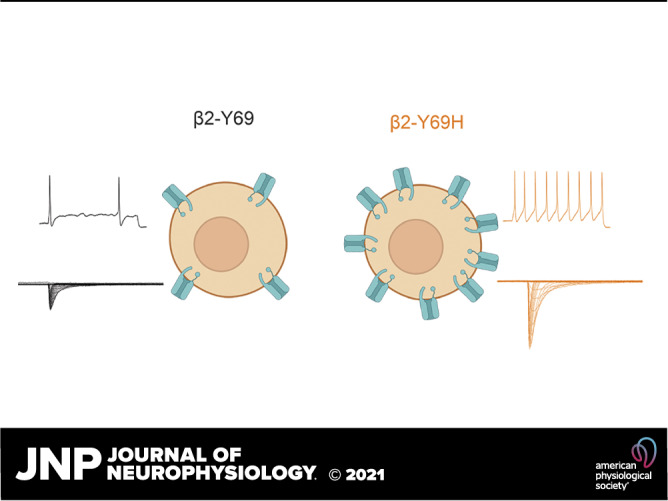
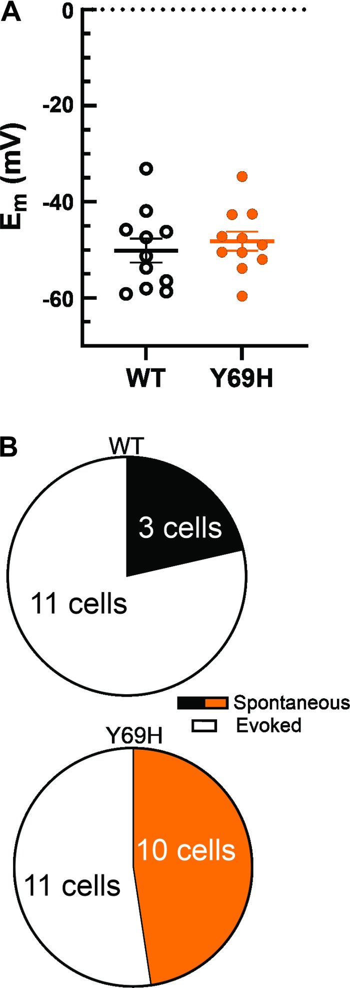
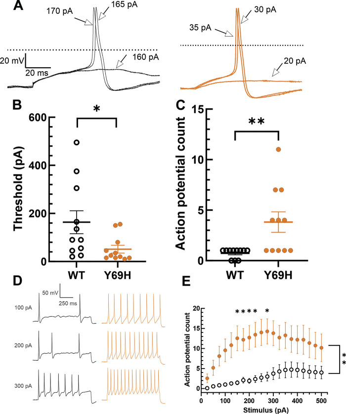
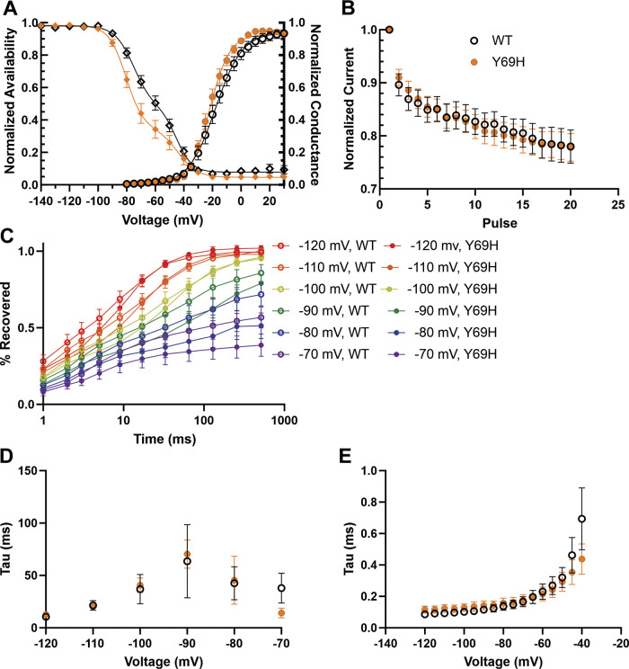
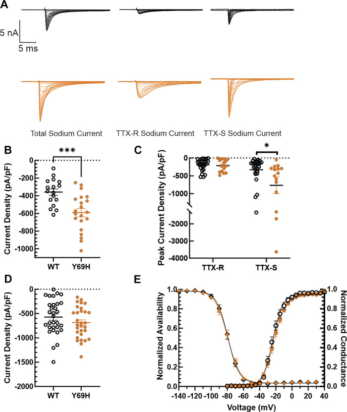
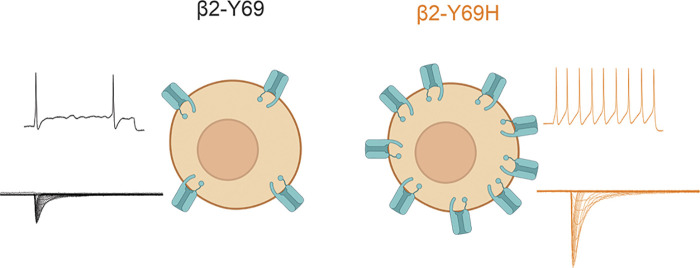
Small fiber neuropathy (SFN) is a common condition affecting thinly myelinated Aδ and unmyelinated C fibers, often resulting in excruciating pain and dysautonomia. SFN has been associated with several conditions, but a significant number of cases have no discernible cause. Recent genetic studies have identified potentially pathogenic gain-of-function mutations in several pore-forming voltage-gated sodium channel α subunits (NaV) in a subset of patients with SFN, but the auxiliary sodium channel β subunits have been less implicated in the development of the disease. β subunits modulate NaV trafficking and gating, and several mutations have been linked to epilepsy and cardiac dysfunction. Recently, we provided the first evidence for the contribution of a mutation in the β2 subunit to pain in human painful diabetic neuropathy. Here, we provide the first evidence for the involvement of a sodium channel β subunit mutation in the pathogenesis of SFN with no other known causes. We show, through current-clamp analysis, that the newly identified Y69H variant of the β2 subunit induces neuronal hyperexcitability in dorsal root ganglion neurons, lowering the threshold for action potential firing and allowing for increased repetitive action potential spiking. Underlying the hyperexcitability induced by the β2-Y69H variant, we demonstrate an upregulation in tetrodotoxin-sensitive, but not tetrodotoxin-resistant sodium currents. This provides the first evidence for the involvement of β2 subunits in SFN and strengthens the link between sodium channel β subunits and the development of neuropathic pain in humans.NEW & NOTEWORTHY Small fiber neuropathy (SFN) often has no discernible cause, although mutations in the voltage-gated sodium channel α subunits have been implicated in some cases. We identify a patient suffering from SFN with a mutation in the auxiliary β2 subunit and no other discernible causes for SFN. Functional assessment confirms this mutation renders dorsal root ganglion neurons hyperexcitable and upregulates tetrodotoxin-sensitive sodium currents. This study strengthens a newly emerging link between sodium channel β2 subunit mutations and human pain disorders.

Keywords: small fiber neuropathy (SFN); sodium channel β subunits; tetrodotoxin-sensitive voltage-gated sodium channels; voltage-gated sodium (Nav) channels; β2 subunit.


Conflict of interest statement

No conflicts of interest, financial or otherwise, are declared by the authors.

Figures

None
Graphical abstract
Figure 1.
Figure 1.
Molecular modeling of the β2-Y69H variant. The immunoglobulin domain of the human β2 subunit (cyan) is linked to the NaV1.7 channel α subunit (green) via a disulfide bond with the cystine at position 55 (C55, yellow). The sidechain of the tyrosine at position 69 (Y69, magenta) is shown, facing away from the α subunit. The star in the cartoon diagram of the β2 subunit (top right) indicates the relative position of the mutation. The sidechain of the aspartic acid at position 109 (D109, orange) is also shown. The α subunit is embedded in a lipid bilayer membrane (red, white, and green spheres). A small portion of the β1 subunit (red, top left) may be seen, as well.
Figure 2.
Figure 2.
The Y69H variant does not alter resting membrane potential of dorsal root ganglion (DRG) neurons. A: comparison of resting membrane potentials for DRG neurons expressing either the wild-type human β2 subunit (monochrome) or the Y69H variant (orange) illustrates no change in neuronal resting membrane potentials (Em, RMP). B: a numerically larger, but not statistically significant, proportion of DRG neurons expressing the Y69H variant fire spontaneous action potentials at rest than DRG neurons expressing the wild-type human β2 subunit. WT, wild type.
Figure 3.
Figure 3.
The Y69H variant reduces the threshold of action potential firing and enhances repetitive spiking in dorsal root ganglion (DRG) neurons. A: representative traces depicting the action potential response of DRG neurons expressing the wild-type (black) or Y69H variant (orange) human β2 subunit during stimulation with current injections of 200 ms duration. The 0 mV membrane potential is indicated by the dashed line. B: DRG neurons expressing the Y69H variant displayed a significantly lower threshold to action potential firing than did neurons expressing the wild-type β2 subunit. C: when neurons were stimulated with a 1,000-ms pulse at the threshold amplitude as determined by a 200-ms injection in B, DRG neurons expressing the Y69H variant fired significantly more action potentials than those expressing the wild-type β2 subunit. D: representative traces depicting repetitive action potential firing of DRG neurons expressing the Y69H variant (orange) compared with those expressing the wild-type β2 subunit (black). E: neurons expressing the Y69H variant (orange) fired significantly more action potentials during a 1,000-ms stimulus of graded amplitude than did neurons expressing the wild-type β2 subunit (monochrome). WT, wild type. *P ≤ 0.05, **P ≤ 0.01.
Figure 4.
Figure 4.
The Y69H variant does not alter total NaV channel gating properties. A: the voltage-dependence of channel inactivation (left; diamonds) are not statistically different between dorsal root ganglion (DRG) neurons expressing the wild-type and Y69H variant subunits. In addition, the voltage-dependence of activation (right, circles) is also not statistically different. B: there is no difference in the use-dependent inhibition of sodium channels in DRG neurons expressing either the wild-type or the Y69H β2 subunit. C: the extent of sodium channel recovery from inactivation at voltages between −70 and −120 mV is comparable for DRG neurons expressing either the wild-type or the variant β2 subunits. D: the rate of recovery from channel inactivation is also comparable in both groups. E: there is no difference in the kinetics of entry into deactivation between DRG neurons expressing the wild-type or Y69H variant. WT, wild type.
Figure 5.
Figure 5.
Tetrodotoxin-sensitive (TTX-S) current is upregulated in dorsal root ganglion (DRG) neurons expressing Y69H β2 subunits. A: representative traces illustrating reference series subtraction to determine TTX-S current (right) from the difference between total sodium current (left) and tetrodotoxin-resistant (TTX-R) current (middle) recorded in 1 μM TTX. Example traces are shown for sodium currents in both neurons expressing the wild-type β2 variant (top) and the Y69H variant (bottom, orange). B: there is a statistically significant increase in total sodium current passed by DRG neurons expressing the Y69H variant compared with DRG neurons expressing the wild-type β2 subunit. C: TTX-S sodium currents in DRG neurons are upregulated in the presence of the Y69H variant (orange), relative to the wild-type (monochrome) β2 subunit. However, there is no change in TTX-R sodium current. D: the voltage dependence of NaV1.7 channel inactivation (left; diamonds) is not statistically different between HEK293 cells expressing the wild-type β2 (monochromatic) and the Y69H variant (orange) subunits. In addition, the voltage-dependence of activation (right, circles) is also not statistically different. E: the Y69H variant does not significantly upregulate.7 current density in HEK293 cells stably expressing NaV1.7 channels when compared with the wild-type β2 subunit. WT, wild type. *P ≤ 0.05, ***P ≤ 0.001.
Figure 6.
Figure 6.
Schematic illustrating the proposed mechanism of action of the β2-Y69H (right) on small-diameter dorsal root ganglion neurons. Compared to the wild-type β2 subunit (left), neurons expressing the Y69H variant are hyperexcitable (top traces) and display a larger tetrodotoxin-sensitive sodium current density (bottom traces), suggesting increased trafficking of sodium channels to the neuronal membrane.

References

    1. Faber CG, Hoeijmakers JG, Ahn HS, Cheng X, Han C, Choi JS, Estacion M, Lauria G, Vanhoutte EK, Gerrits MM, Dib-Hajj S, Drenth JP, Waxman SG, Merkies IS. Gain of function Naν1.7 mutations in idiopathic small fiber neuropathy. Ann Neurol 71: 26–39, 2012. doi:10.1002/ana.22485. - DOI - PubMed
    1. Sopacua M, Hoeijmakers JGJ, Merkies ISJ, Lauria G, Waxman SG, Faber CG. Small-fiber neuropathy: expanding the clinical pain universe. J Peripher Nerv Syst 24: 19–33, 2019. doi:10.1111/jns.12298. - DOI - PubMed
    1. Tesfaye S, Boulton AJ, Dyck PJ, Freeman R, Horowitz M, Kempler P, Lauria G, Malik RA, Spallone V, Vinik A, Bernardi L, Valensi P; Toronto Diabetic Neuropathy Expert Group. Diabetic neuropathies: update on definitions, diagnostic criteria, estimation of severity, and treatments. Diabetes Care 33: 2285–2293, 2010[Erratum inDiabetes Care33: 2725, 2010]. doi:10.2337/dc10-1303. - DOI - PMC - PubMed
    1. Han Y, Smith MT. Pathobiology of cancer chemotherapy-induced peripheral neuropathy (CIPN). Front Pharmacol 4: 156, 2013. doi:10.3389/fphar.2013.00156. - DOI - PMC - PubMed
    1. Devigili G, Rinaldo S, Lombardi R, Cazzato D, Marchi M, Salvi E, Eleopra R, Lauria G. Diagnostic criteria for small fibre neuropathy in clinical practice and research. Brain 142: 3728–3736, 2019. doi:10.1093/brain/awz333. - DOI - PMC - PubMed

Substances