

Basics for performing a high-quality color Doppler sonography of the vascular access
- PMID: 34320855
- PMCID: PMC8607315
- DOI: 10.1177/11297298211018060
Basics for performing a high-quality color Doppler sonography of the vascular access
Abstract
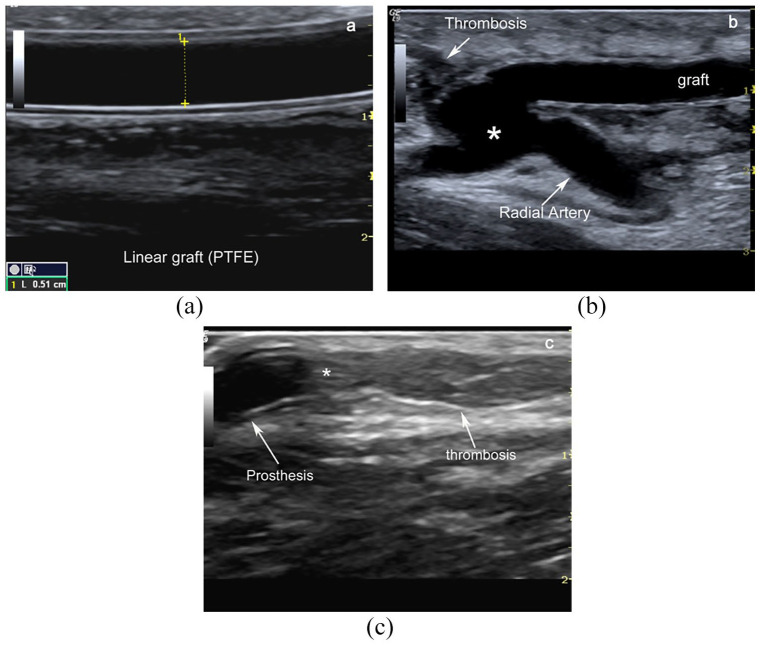
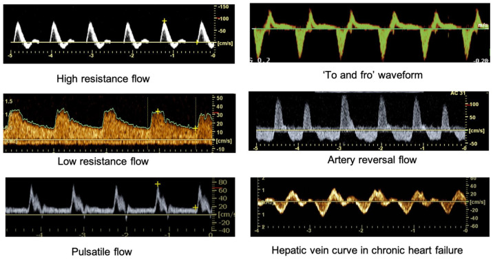
In the last years, the systematic use of ultrasound mapping of the upper limb vascular network before the arteriovenous fistula (AVF) implantation, access maturation, and clinical management of late complications is widespread and expanding. Therefore, a good knowledge of theoretical outlines, instrumentation, and operative settings is undoubtedly required for a thorough examination. In this review, the essential Doppler parameters, B-Mode setting, and Doppler applications are considered. Basic concepts on the Doppler shift equation, angle correction, settings on pulse repetition frequency, operative Doppler frequency, gain are reported to ensure adequate and correct sampling of blood flow velocity. A brief analysis of the Doppler inherent artefacts (as random noise, blooming, aliasing, and motion artefacts) and the adjustment setting to minimize or eliminate the confounding artefacts are also considered. Doppler aliasing occurs when the pulse repetition frequency is set too low. This artefact is particularly frequent in vascular access sampling due to the high velocities range registered in the fistula's different segments. Aliasing should be recognized because its correction is crucial to analyse the Doppler signals correctly. Recent advances in instrumentation are also considered about a potential purchase of a portable ultrasound machine or a top-of-line, high-end, or mid-range ultrasound system. Last, the pulse wave Doppler setting for vascular access B-Mode and Doppler assessment is summarized.
Keywords: B-mode ultrasound; Doppler parameters; Doppler/B-mode setting; arterio-venous fistula; pulse wave Doppler.
Conflict of interest statement
Figures


Similar articles
-
Settings and artefacts relevant for Doppler ultrasound in large vessel vasculitis.Arthritis Res Ther. 2017 Jul 20;19(1):167. doi: 10.1186/s13075-017-1374-1. Arthritis Res Ther. 2017. PMID: 28728567 Free PMC article. Review.
-
Farbkodierte Duplexsonografie der Gefässe in der Praxis: Pitfalls.Praxis (Bern 1994). 2019 Sep;108(12):807-813. doi: 10.1024/1661-8157/a003295. Praxis (Bern 1994). 2019. PMID: 31530132 German.
-
[Color Doppler Sonography: Device Settings Determine the Quality of Diagnosis and the Occurrence of Artifacts].Praxis (Bern 1994). 2020;109(8):566-571. doi: 10.1024/1661-8157/a003499. Praxis (Bern 1994). 2020. PMID: 32517601 German.
-
Advances in vascular anatomy and pathophysiology using high resolution and multiparametric sonography.J Vasc Access. 2021 Nov;22(1_suppl):1-8. doi: 10.1177/11297298211020150. Epub 2021 Aug 2. J Vasc Access. 2021. PMID: 34338066 Free PMC article. Review.
-
[Sonographic findings in the diagnostic course of vascular access for hemodialysis].Med Klin (Munich). 2005 Jan 15;100(1):1-5. doi: 10.1007/s00063-005-1112-3. Med Klin (Munich). 2005. PMID: 15654536 German.
Cited by
-
Non-invasive real-time pulsed Doppler assessment of blood flow in mouse ophthalmic artery.Cell Rep Methods. 2025 Feb 24;5(2):100983. doi: 10.1016/j.crmeth.2025.100983. Epub 2025 Feb 14. Cell Rep Methods. 2025. PMID: 39954674 Free PMC article.
-
Protocol for non-invasive pulsed-Doppler evaluation of blood flow in the mouse ophthalmic artery.STAR Protoc. 2025 Jun 16;6(3):103893. doi: 10.1016/j.xpro.2025.103893. Online ahead of print. STAR Protoc. 2025. PMID: 40526471 Free PMC article.
-
Current State of Knowledge in Ocular Blood Flow in Glaucoma: A Narrative Review.Clin Ophthalmol. 2023 Aug 31;17:2599-2607. doi: 10.2147/OPTH.S426709. eCollection 2023. Clin Ophthalmol. 2023. PMID: 37671333 Free PMC article. Review.
-
Superb microvascular ultrasound is a promising non-invasive diagnostic tool to assess a ventriculoperitoneal shunt system function: a feasibility study.Neurosurg Rev. 2024 Sep 2;47(1):521. doi: 10.1007/s10143-024-02665-w. Neurosurg Rev. 2024. PMID: 39222164 Free PMC article.
-
Quantification of Portal Vein Vascularization Using an Automated Post-Processing Video Analysis Tool.Ultrasound Int Open. 2023 Mar 20;9(1):E2-E10. doi: 10.1055/a-1999-7818. eCollection 2023 Jan. Ultrasound Int Open. 2023. PMID: 36950090 Free PMC article.
References
-
- Mannaerts CK, Wildeboer RR, Remmers S, et al. Multiparametric ultrasound for prostate cancer detection and localization: correlation of B-mode, shear wave elastography and contrast enhanced ultrasound with radical prostatectomy specimens. J Urol 2019; 202: 1166–1173. - PubMed
-
- Harvey CJ, Pilcher JM, Eckersley RJ, et al. Advances in ultrasound. Clin Radiol 2002; 57: 157–177. - PubMed
-
- Claudon M, Tranquart F, Evans DH, et al. Advances in ultrasound. Eur Radiol 2002; 12: 7–18. - PubMed
-
- Lawrence JP. Physics and instrumentation of ultrasound. Crit Care Med 2007; 35: S314–S322. - PubMed
-
- Meola M, PetruccI I. Ultrasound and color Doppler in nephrology. Physical and technical principles. G Ital Nefrol. 2012; 29: 81–91. - PubMed
Publication types
MeSH terms
LinkOut - more resources
Full Text Sources
Other Literature Sources
