. 2021 Jul 28;19(1):223.
doi: 10.1186/s12951-021-00963-9.

Intratumoral administration of astatine-211-labeled gold nanoparticle for alpha therapy

Affiliations

Intratumoral administration of astatine-211-labeled gold nanoparticle for alpha therapy

Hiroki Kato et al. J Nanobiotechnology. .

Abstract

Background: 211At is a high-energy α-ray emitter with a relatively short half-life and a high cytotoxicity for cancer cells. Its dispersion can be imaged using clinical scanners, and it can be produced in cyclotrons without the use of nuclear fuel material. This study investigated the biodistribution and the antitumor effect of 211At-labeled gold nanoparticles (211At-AuNP) administered intratumorally.

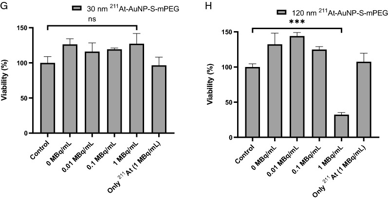
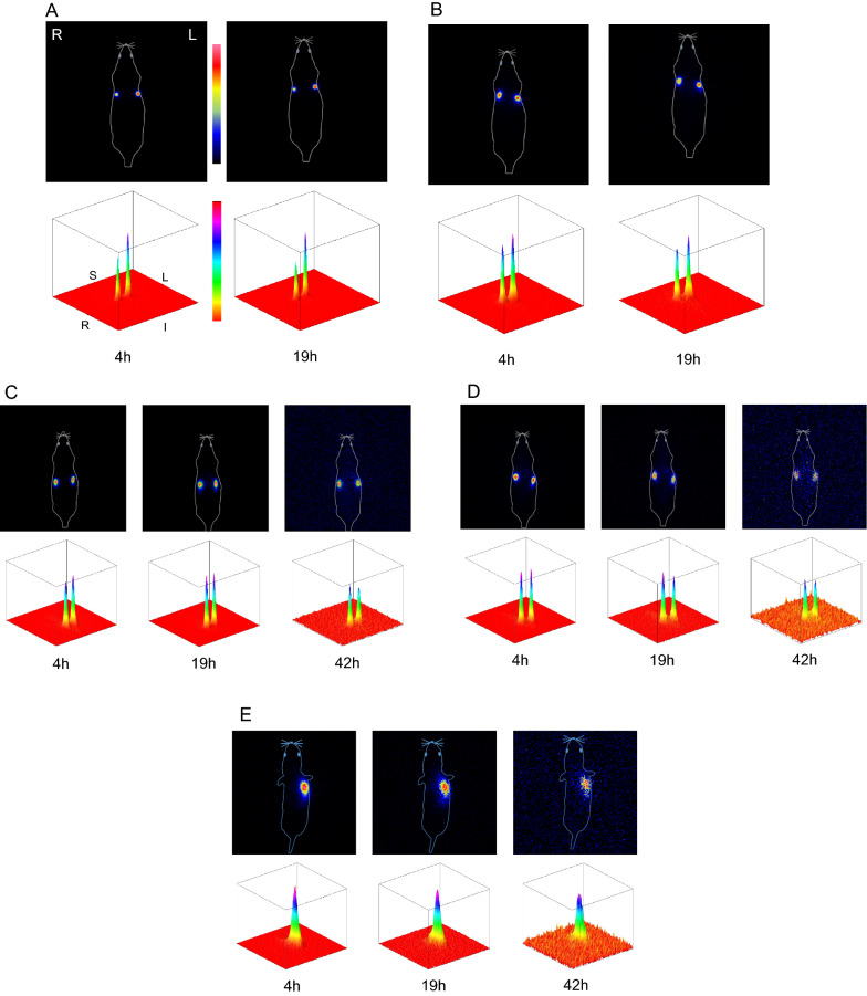
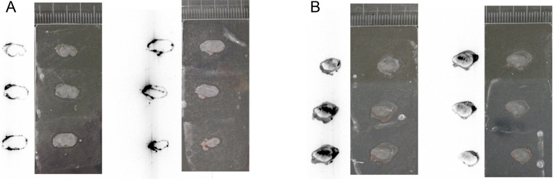
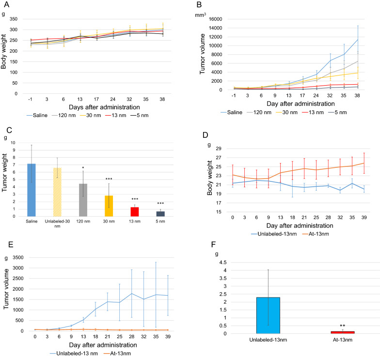
Results: AuNP with a diameter of 5, 13, 30, or 120 nm that had been modified with poly (ethylene glycol) methyl ether (mPEG) thiol and labeled with 211At (211At-AuNP-S-mPEG) were incubated with tumor cells, or intratumorally administered to C6 glioma or PANC-1 pancreatic cancers subcutaneously transplanted into rodent models. Systemic and intratumoral distributions of the particles in the rodents were then evaluated using scintigraphy and autoradiography, and the changes in tumor volumes were followed for about 40 days. 211At-AuNP-S-mPEG was cytotoxic when it was internalized by the tumor cells. After intratumoral administration, 211At-AuNP-S-mPEG became localized in the tumor and did not spread to systemic organs during a time period equivalent to 6 half-lives of 211At. Tumor growth was strongly suppressed for both C6 and PANC-1 by 211At-AuNP-S-mPEG. In the C6 glioma model, the strongest antitumor effect was observed in the group treated with 211At-AuNP-S-mPEG with a diameter of 5 nm.

Conclusions: The intratumoral single administration of a simple nanoparticle, 211At-AuNP-S-mPEG, was shown to suppress the growth of tumor tissue strongly in a particle size-dependent manner without radiation exposure to other organs caused by systemic spread of the radionuclide.

Keywords: Alpha emitters; Astatine-211; Cancer therapy; Gold nanoparticles; Radiolabeling.


Conflict of interest statement

The author(s) have no potential competing of interest to declare with respect to the conduct of the research, the authorship, and/or the publication of this article.

Figures

Fig. 1
Fig. 1
C6 glioma cells (A, B, C, D) and PANC-1 cells (E, F, G, H) were cultured for 24 h with saline as a control, 1 MBq/mL of 211At, or 211At-AuNP-S-mPEG labeled with 0 to 1 MBq/mL of radioactivity and a diameter of 5 nm (A, E), 13 nm (B, F), 30 nm (C, G) or 120 nm (D, H). For the evaluation of cell viability, CCK8 Kit was used. WTS-8 solution was added and the cells were cultured for 2 h. Viability was calculated by measuring the absorbance as shown in the formula below: Viability%=T-T0C-C0, where T is absorbance of the test cells, T0 is background of the test cells, C is the absorbance of the control cells, and C0 is the background of the control cells. The mass concentrations of AuNP-S-mPEG labeled with 1 MBq/mL 211At in (A, B, C, D) were almost the same level as those in (A, C, E, G) in Fig. 2, respectively (see Additional file 1: Table S3).
Fig. 1
Fig. 1
C6 glioma cells (A, B, C, D) and PANC-1 cells (E, F, G, H) were cultured for 24 h with saline as a control, 1 MBq/mL of 211At, or 211At-AuNP-S-mPEG labeled with 0 to 1 MBq/mL of radioactivity and a diameter of 5 nm (A, E), 13 nm (B, F), 30 nm (C, G) or 120 nm (D, H). For the evaluation of cell viability, CCK8 Kit was used. WTS-8 solution was added and the cells were cultured for 2 h. Viability was calculated by measuring the absorbance as shown in the formula below: Viability%=T-T0C-C0, where T is absorbance of the test cells, T0 is background of the test cells, C is the absorbance of the control cells, and C0 is the background of the control cells. The mass concentrations of AuNP-S-mPEG labeled with 1 MBq/mL 211At in (A, B, C, D) were almost the same level as those in (A, C, E, G) in Fig. 2, respectively (see Additional file 1: Table S3).
Fig. 2
Fig. 2
C6 glioma cells were seeded onto 35-mm glass bottom dishes and the cells were incubated with 5 nm, 13 nm, 30 nm or 120 nm AuNP-S-mPEG for 24 h. The intracellular uptake of these nanoparticles was examined using reflectance imaging. In each image, the leftmost section shows the bright field image of the cells, the second section shows the reflectance image of the particles taken up by the cells, the third section shows the fluorescent Hoechst staining for DNA, and the rightmost section shows the fused image. The C6 glioma cells were incubated with AuNP-S-mPEG with the following sizes and mass concentrations: (A) 5 nm and 12.7 mg/L, (B) 5 nm and 138.4 mg/L, (C) 13 nm and 6.9 mg/L, (D) 13 nm and 68.6 mg/L, (E) 30 nm and 8.7 mg/L, (F) 30 nm and 51.6 mg/L, and (G) 120 nm and 33.6 mg/L. The mass concentrations in (A), (C), (E), and (G) were almost the same as those of 1-MBq 211At-AuNP-S-mPEG in the cytotoxicity studies (A), (B), (C), and (D) illustrated in Fig. 1, respectively (see Additional file 1: Table S3)
Fig. 3
Fig. 3
Scintigraphy of C6 glioma-bearing rats was performed at 4 and 19 h after the administration of 120 nm (A), 30 nm (B), 13 nm (C), or 5 nm (D) 211At-AuNP-S-mPEG. For the 13 nm (C) and 5 nm (D) 211At-AuNP-S-mPEG, imaging was also performed at 42 h after administration. Scintigraphy of PANC-1 bearing mice was also performed in a same way as rats at 4, 19, and 42 h after the administration of 13 nm 211At-AuNP-S-mPEG (E). The radioactivity distributions (upper rows) are also shown using surface plots (lower rows) that display a square field of view of the dorsal detector with side length 30 cm for rats (AD), or 15 cm for mice (E), which covers the whole body of an animal. No systemic accumulation of radioactivity was observed in any of the organs. S: Superior, I: inferior, R: right, L: left
Fig. 4
Fig. 4
Twenty-four hours after the administration of 120 nm (A) and 30 nm (B) 211At-AuNP-S-mPEG, the tumors were excised, sliced and attached to a slide glass (right). Autoradiography (left) was performed after placing both tumor sections in contact with the same imaging plate for an hour
Fig. 5
Fig. 5
Changes in the body weights of the rats (A) and mice (D) and changes in the tumor volumes of the C6 glial cells (B) and PANC-1 cells (E) after intratumoral administration (Tumor size at the time of administration and the 211At dose were 334 ± 152 mm3 and 1.4 ± 0.4 MBq/tumor for rats and 72 ± 11 mm3 and 1.2 ± 0.1 MBq/tumor for mice, respectively). The C6 glial (C) and PANC-1 (F) tumors were excised 40 days after administration and their masses were measured. The error bars indicate the standard deviation. The tumor mass after the administration of 211At was significantly smaller than that seen in the controls. In PANC-1 xenografts, necrosis inside the tumor increased the size variability, leading to an increased standard deviation. The body weights of the mice in the control group were significantly lower than those in the group treated with 211At (*0.01 ≤ p < 0.05, **0.001 ≤ p < 0.01, ***p < 0.001)

Similar articles

Cited by

References

    1. Smith GD, Pickles T, Crook J, Martin AG, Vigneault E, Cury FL, Morris J, Catton C, Lukka H, Warner A, et al. Brachytherapy improves biochemical failure-free survival in low- and intermediate-risk prostate cancer compared with conventionally fractionated external beam radiation therapy: a propensity score matched analysis. Int J Radiat Oncol Biol Phys. 2015;91:505–516. doi: 10.1016/j.ijrobp.2014.11.018. - DOI - PubMed
    1. Skowronek J, Chichel A. Brachytherapy in breast cancer: an effective alternative. Prz Menopauzalny. 2014;13:48–55. - PMC - PubMed
    1. Roddiger SJ, Kolotas C, Filipowicz I, Kurek R, Kuner RP, Martin T, Baltas D, Rogge B, Kontova M, Hoffmann G, et al. Neoadjuvant interstitial high-dose-rate (HDR) brachytherapy combined with systemic chemotherapy in patients with breast cancer. Strahlenther Onkol. 2006;182:22–29. doi: 10.1007/s00066-006-1454-7. - DOI - PubMed
    1. Lin AJ, Hassanzadeh C, Markovina S, Schwarz J, Grigsby P. Brachytherapy and survival in small cell cancer of the cervix and uterus. Brachytherapy. 2019;18:163–170. doi: 10.1016/j.brachy.2018.11.006. - DOI - PMC - PubMed
    1. Robin TP, Amini A, Schefter TE, Behbakht K, Fisher CM. Brachytherapy should not be omitted when treating locally advanced neuroendocrine cervical cancer with definitive chemoradiation therapy. Brachytherapy. 2016;15:845–850. doi: 10.1016/j.brachy.2016.08.007. - DOI - PubMed

LinkOut - more resources