. 2021 Jul 28;23(1):202.
doi: 10.1186/s13075-021-02580-1.

Urate-induced epigenetic modifications in myeloid cells

Affiliations

Urate-induced epigenetic modifications in myeloid cells

M Badii et al. Arthritis Res Ther. .

Abstract

Objectives: Hyperuricemia is a metabolic condition central to gout pathogenesis. Urate exposure primes human monocytes towards a higher capacity to produce and release IL-1β. In this study, we assessed the epigenetic processes associated to urate-mediated hyper-responsiveness.

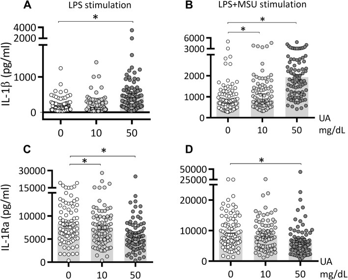
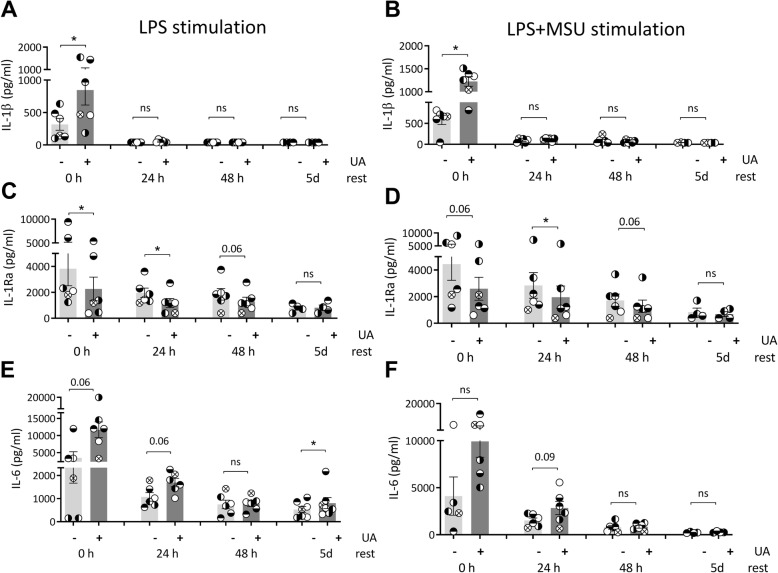
Methods: Freshly isolated human peripheral blood mononuclear cells or enriched monocytes were pre-treated with solubilized urate and stimulated with LPS with or without monosodium urate (MSU) crystals. Cytokine production was determined by ELISA. Histone epigenetic marks were assessed by sequencing immunoprecipitated chromatin. Mice were injected intraarticularly with MSU crystals and palmitate after inhibition of uricase and urate administration in the presence or absence of methylthioadenosine. DNA methylation was assessed by methylation array in whole blood of 76 participants with normouricemia or hyperuricemia.

Results: High concentrations of urate enhanced the inflammatory response in vitro in human cells and in vivo in mice, and broad-spectrum methylation inhibitors reversed this effect. Assessment of histone 3 lysine 4 trimethylation (H3K4me3) and histone 3 lysine 27 acetylation (H3K27ac) revealed differences in urate-primed monocytes compared to controls. Differentially methylated regions (e.g. HLA-G, IFITM3, PRKAB2) were found in people with hyperuricemia compared to normouricemia in genes relevant for inflammatory cytokine signaling.

Conclusion: Urate alters the epigenetic landscape in selected human monocytes or whole blood of people with hyperuricemia compared to normouricemia. Both histone modifications and DNA methylation show differences depending on urate exposure. Subject to replication and validation, epigenetic changes in myeloid cells may be a therapeutic target in gout.

Keywords: Cytokines; DNA methylation; Epigenetics; Gout; Hyperuricemia.


Conflict of interest statement

The authors declare that they have no competing interests.

Figures

Fig. 1
Fig. 1
IL-1β and IL-1Ra production after urate priming of PBMCs in vitro. Freshly isolated PBMCs from 85 healthy volunteers were exposed to culture medium (RPMI 1640 supplemented with 10% human pooled serum) in the presence or absence of urate (UA) 10 or 50 mg/dL. After 24 h, urate was removed, and cells were stimulated with LPS 10 ng/mL in the presence or absence of MSU crystals (300 μg/mL). IL-1β (A-B) and IL-1Ra (C-D) were measured in the supernatants of cells, data are representative of 3 independent experiments using a total of 85 different healthy volunteers of the 200FG cohort, graphs depict means+/−SEM. UA, uric acid/urate. *, Friedman test and post-hoc analysis p < 0.05
Fig. 2
Fig. 2
Persistence of urate priming effects in vitro. Freshly isolated PBMCs from 6 healthy volunteers were exposed to culture medium (RPMI 1640 supplemented with 10% human pooled serum) in the presence or absence of urate (UA) 50 mg/dL. After 24 h, urate was removed, and cells were stimulated with LPS 10 ng/mL in the presence or absence of MSU crystals (300 μg/mL). The second stimulation was performed at different times after urate washout: immediately (0 h resting time), or after increasing the number of days of resting in 10% serum RPMI (24 h, 48 h, 5 days). IL-1β (A-B), IL-1Ra (C-D), and IL-6 (E-F) were measured in the supernatants of cells, data are representative for 3 independent experiments and 6 different volunteers, graphs depict individual values with paired samples shown in identical symbols, bars and error bars represent means+/−SEM. UA, uric acid/urate 50 mg/dL. *, Wilcoxon p < 0.05
Fig. 3
Fig. 3
Methyltransferase inhibition limits gout inflammation in mice. Macroscopic (A) scores of the knees in mice treated with vehicle control or oxonic acid + urate in the presence or absence of methyl transferase inhibitor MTA (methyl-thio-adenosine) followed by intraarticular injection of MSU+C16:0. Inflammation was scored at 24 h. Histology (H&E staining) of joints treated with MSU+C16:0 in oxonic acid + urate mice (B) and in the presence of MTA (C)
Fig. 4
Fig. 4
ChIP-sequencing in urate-stimulated monocytes reveals no urate-dependent clustering based on phenotype for H3K4 trimethylation and H3K27 acetylation. Cluster and principal component analysis of datasets obtained on ChIP-sequencing for H3K4me3 (A, B) or H3K27ac (C, D) in 4 different donors (labeled D1-4) and 4 different conditions (medium, urate, medium+LPS, urate+LPS). Venn diagram of regions showing differential enrichment of either H3K27ac or H3K4me3 in urate primed cells (E) and list of overlapping genes based on histone marks at promoter regions, including log 2 fold change values for each of the two histone marks (F)
Fig. 5
Fig. 5
Differential DNA methylation in hyperuricemic versus normouricemic people. Principal component analysis of whole blood DNA methylation data, obtained using Illumina InfiniumMethylationEPIC BeadChips in whole blood of 26 people with hyperuricemia compared to 50 normouricemic individuals (A). Differentially methylated regions (DMRs) at the HLA-G locus (B) and average DNA methylation levels (beta values) for all CpG probes found within HLA-G (ticks on the x-axis represent individual probes and DNA chromosome position is indicated) (C). Transcription factors are known to bind at the highlighted DMR1 and DMR3 regions according to the transcription factor ChIP-seq clusters from ENCODE with factorbook motifs (D)

References

    1. Shi Y, Evans JE, Rock KL. Molecular identification of a danger signal that alerts the immune system to dying cells. Nature. 2003;425:516–521. doi: 10.1038/nature01991. - DOI - PubMed
    1. Kono H, Chen CJ, Ontiveros F, Rock KL. Uric acid promotes an acute inflammatory response to sterile cell death in mice. J Clin Invest. 2010. 10.1172/JCI40124. - PMC - PubMed
    1. Mandel NS, Mandel GS. Monosodium Urate Monohydrate, the Gout Culprit. J Am Chem Soc. 1976. 10.1021/ja00424a054. - PubMed
    1. Martinon F, Pétrilli V, Mayor A, Tardivel A, Tschopp J. Gout-associated uric acid crystals activate the NALP3 inflammasome. Nature. 2006;440:237–241. doi: 10.1038/nature04516. - DOI - PubMed
    1. Misawa T, Takahama M, Kozaki T, Lee H, Zou J, Saitoh T, et al. Microtubule-driven spatial arrangement of mitochondria promotes activation of the NLRP3 inflammasome. Nat Immunol. 2013. 10.1038/ni.2550. - PubMed

Publication types