Review
. 2021 Jul 28;25(1):24.
doi: 10.1186/s40824-021-00226-6.

Recent advances in transdermal drug delivery systems: a review

Affiliations
Review

Recent advances in transdermal drug delivery systems: a review

Woo Yeup Jeong et al. Biomater Res. .

Abstract

Various non-invasive administrations have recently emerged as an alternative to conventional needle injections. A transdermal drug delivery system (TDDS) represents the most attractive method among these because of its low rejection rate, excellent ease of administration, and superb convenience and persistence among patients. TDDS could be applicable in not only pharmaceuticals but also in the skin care industry, including cosmetics. Because this method mainly involves local administration, it can prevent local buildup in drug concentration and nonspecific delivery to tissues not targeted by the drug. However, the physicochemical properties of the skin translate to multiple obstacles and restrictions in transdermal delivery, with numerous investigations conducted to overcome these bottlenecks. In this review, we describe the different types of available TDDS methods, along with a critical discussion of the specific advantages and disadvantages, characterization methods, and potential of each method. Progress in research on these alternative methods has established the high efficiency inherent to TDDS, which is expected to find applications in a wide range of fields.

Keywords: Active/passive method; Characterization; Skin; Transdermal drug delivery.


Conflict of interest statement

The authors declare no conflict of interest.

Figures

Fig. 1
Fig. 1
The structure of skin
Fig. 2
Fig. 2
A Experimental set up for skin permeation test using iontophoresis. B In vitro drug release profiles of drug-loaded AuNP oleogels (d-AuNP) on skin. C Fluorescence spectroscopy images obtained from skin permeation experiment after 1 h of application. Arrows mark the top surface of the skin segment treated with d-AuNP. D A schematic illustration of sonophoresis-assisted transdermal drug delivery. E Penetration pathways of LaNO3 after treatment with low frequency sonophoresis, and TEM images of SC after treatment with low frequency sonophoresis using RuO4 fixation in the absence of low frequency sonophoresis (left) and after 5 min (middle) and 10 min (right) of treatment with low frequency sonophoresis. F Schematic Illustration Showing the Fabrication Process of the MTX-Loaded HA-Based Dissolving MN Patch. G Quantitative analysis of epidermal thickness. H Therapeutic effects of MTX-loaded MNs and oral administration of the same dose and a double dose of MTX on IMQ-induced psoriasis-like skin inflammation. Representative photographs of left ear lesions and skin sections stained with H&E and Ki67 on day 7. A, B, C Reproduced from [29], copyright permission by American Chemical Society 2020. D Reproduced from [74], copyright permission by Springer Nature 2021. E Reproduced from [40], copyright permission by Elsevier 2010. F, G, H Reproduced from [54], copyright permission by American Chemical Society 2019
Fig. 3
Fig. 3
A SEM images of the prepared ALA-ES gels. B TEM images of ALA-ES in human HS tissue dermis. ALA-ES is indicated using black arrows. C TEM image of OA-UCNP. The nanoparticles show near-spherical shape with an average diameter around 25 nm. D Microscopy images of a section of a sample pig ear skin under 980 nm excitation laser. E Schematic illustration of W/O/W emulsification of HA-PLGA. F Fluorescence microscopic images of histological sections of rat skin at 4 and 12 h after topical application of Rho B-encapsulated HA-PLGA NPs. Scale bar, 100 μm. A, B Reproduced from [63], copyright permission by American Chemical Society 2018. C, D Reproduced from [67], copyright permission by IOP Publishing Ltd. 2020. E, F, G Reproduced from [68], copyright permission by BioMed Central Ltd. 2019
Fig. 4
Fig. 4
A Schematic illustrations of the static Franz diffusion cell. B Permeation profiles of ketoprofen (KTP) for 24 h in different conditions of matrix, medium, pH, and type of membrane. A, B Reproduced from [83], copyright permission by MDPI 2018
Fig. 5
Fig. 5
A Diagram illustrating the process of skin tape stripping. B Imaging of a 4-mm volar skin surface area of a healthy forearm with optical coherence tomography (OCT). A Reproduced from [66], copyright permission by Springer Nature 2021. B Reproduced from [74], copyright permission by Frontiers Media 2019
Fig. 6
Fig. 6
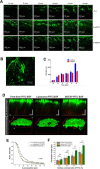
A CLSM images (100× magnification) of skin samples treated with free C-6, C-6/NLC, and C-6/SLN. B Enlarged CLSM Fig. (200× magnification). C Fluorescence intensity in receptor fluid at various times. D Reconstructed two-photon images in XZ orthogonal and 3D views. E Averaged normalized FITC-EGF signal intensity along the z-axis from the surface to the dermal layer of human skin samples. F Penetration depth of FITC-EGF with different thresholds of fluorescence intensity (50, 20, 10, and 5%) measured at the skin surface. A, B, C Reproduced from [88], copyright permission by Springer Nature 2018. D, E, F Reproduced from [89], copyright permission by OSA Publishing 2018

Similar articles

Cited by

References

    1. Vega-Vásquez P, Mosier NS, Irudayaraj J. Nanoscale drug delivery systems: from medicine to agriculture. Front Bioeng Biotechnol. 2020;8:79. doi: 10.3389/fbioe.2020.00079. - DOI - PMC - PubMed
    1. Vargason AM, Anselmo AC, Mitragotri S. The evolution of commercial drug delivery technologies. Nat Biomed Eng. 2021. 10.1038/s41551-021-00698-w. - PubMed
    1. Mali AD, Bathe R, Patil M. An updated review on transdermal drug delivery systems. Int J Adv Sci Res. 2015;1(6):244–254. doi: 10.7439/ijasr.v1i6.2243. - DOI
    1. Li C, Wang J, Wang Y, Gao H, Wei G, Huang Y, Yu H, Gan Y, Wang Y, Mei L, Chen H, Hu H, Zhang Z, Jin Y. Recent progress in drug delivery. Acta Pharm Sin B. 2019;9(6):1145–1162. doi: 10.1016/j.apsb.2019.08.003. - DOI - PMC - PubMed
    1. Kumar JA, Pullakandam N, Prabu SL, Gopal V. Transdermal drug delivery system: An overview. Int J Pharm Sci Rev Res. 2010;3(2):49–54.