. 2021 Jul 28;13(604):eabe1923.
doi: 10.1126/scitranslmed.abe1923.

Nuclear accumulation of CHMP7 initiates nuclear pore complex injury and subsequent TDP-43 dysfunction in sporadic and familial ALS

Affiliations

Nuclear accumulation of CHMP7 initiates nuclear pore complex injury and subsequent TDP-43 dysfunction in sporadic and familial ALS

Alyssa N Coyne et al. Sci Transl Med. .

Abstract

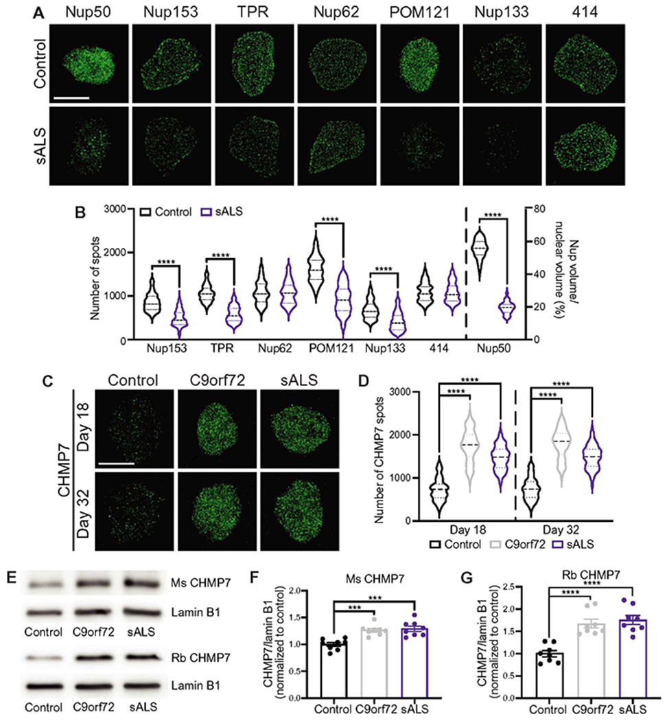
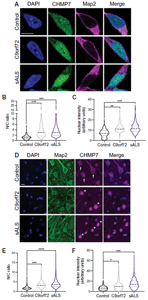
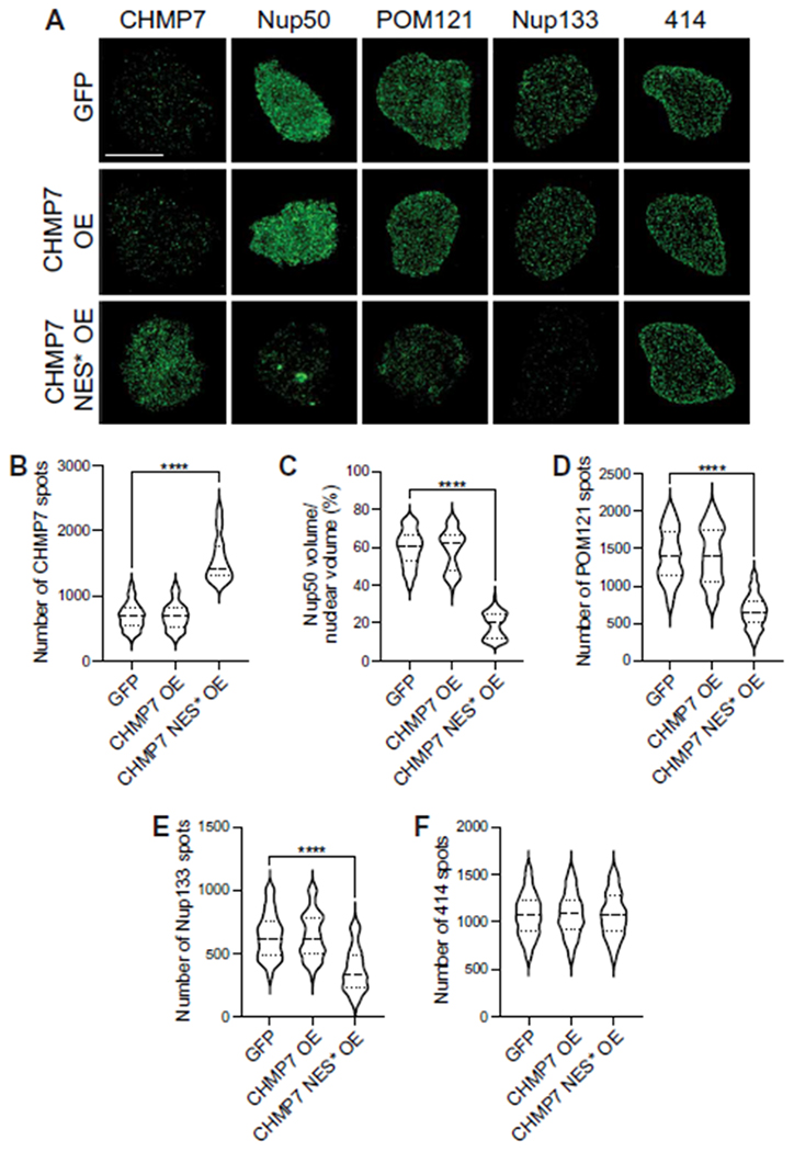
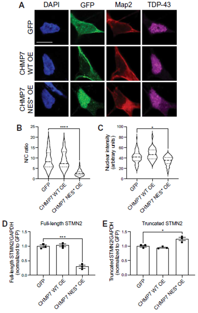
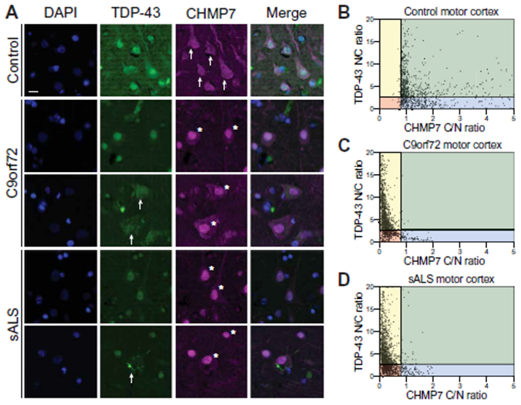
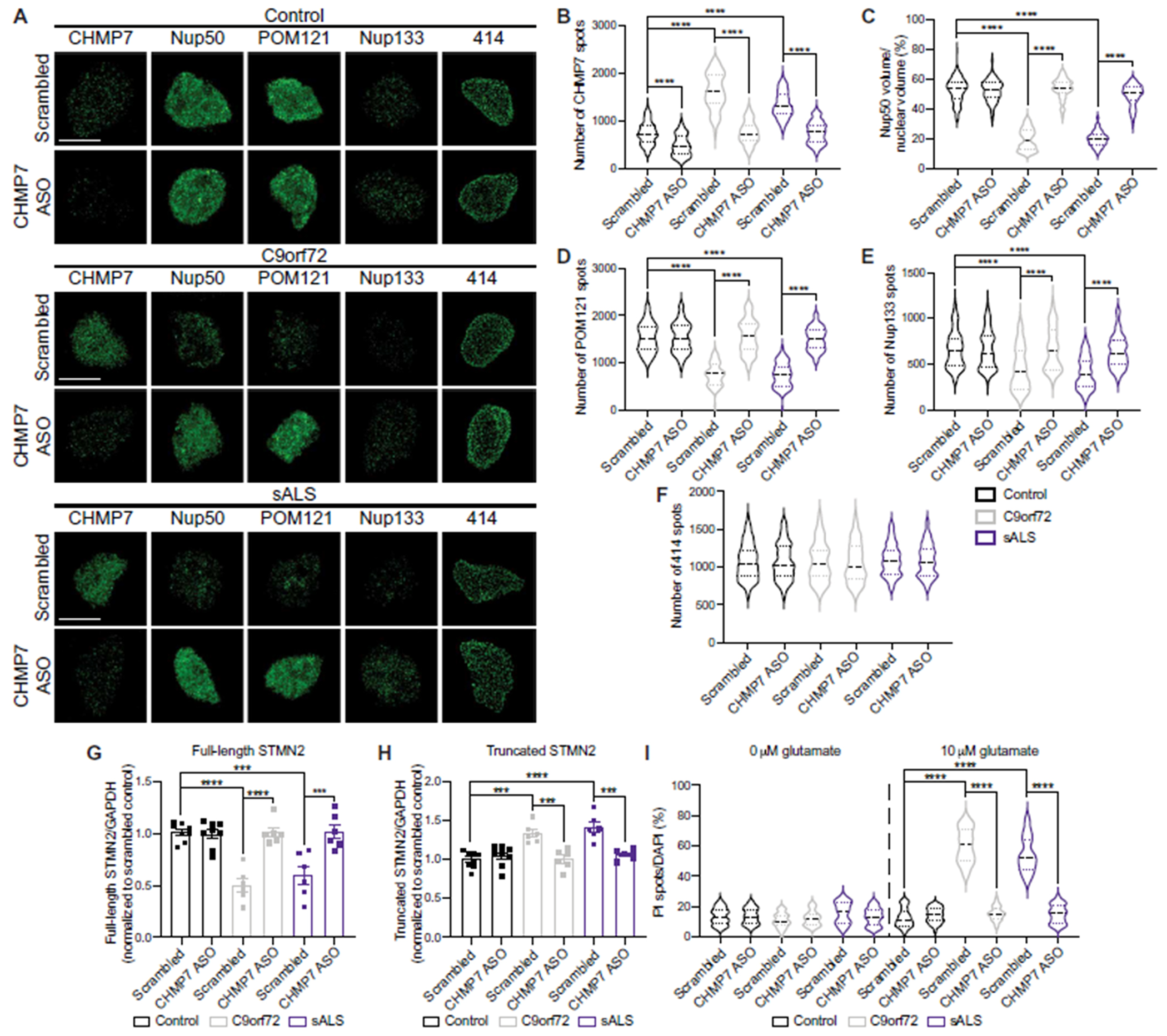
Alterations in the components [nucleoporins (Nups)] and function of the nuclear pore complex (NPC) have been implicated as contributors to the pathogenesis of genetic forms of neurodegeneration including C9orf72 amyotrophic lateral sclerosis/frontotemporal dementia (ALS/FTD). We hypothesized that Nup alterations and the consequential loss of NPC function may lie upstream of TDP-43 dysfunction and mislocalization widely observed in ALS, FTD, and related neurodegenerative diseases. Here, we provide evidence that CHMP7, a critical mediator of NPC quality control, is increased in nuclei of C9orf72 and sporadic ALS induced pluripotent stem cell (iPSC)-derived spinal neurons (iPSNs) and postmortem human motor cortex before the emergence of Nup alterations. Inhibiting the nuclear export of CHMP7 triggered Nup reduction and TDP-43 dysfunction and pathology in human neurons. Knockdown of CHMP7 alleviated disease-associated Nup alterations, deficits in Ran GTPase localization, defects in TDP-43-associated mRNA expression, and downstream glutamate-induced neuronal death. Thus, our data support a role for altered CHMP7-mediated Nup homeostasis as a prominent initiating pathological mechanism for familial and sporadic ALS and highlight the potential for CHMP7 as therapeutic target.


Figures

Fig. 1.
Fig. 1.. Increased nuclear expression of CHMP7 correlates with reduction in specific Nups in C9orf72 and sALS iPSN nuclei.
(A) Maximum intensity projections from SIM imaging of Nups in nuclei isolated from control and sALS iPSNs at day 32 of differentiation. Genotype as indicated on left, antibody as indicated on top. (B) Quantification of Nup spots. n = 10 control and 17 sALS iPSC lines, 50 NeuN+ nuclei per line. Student’s t test was used to calculate statistical significance. ****P < 0.0001. (C) Maximum intensity projections from SIM imaging of CHMP7 in nuclei isolated from control, C9orf72, and sALS iPSNs. Time point as indicated on left, genotype as indicated on top. (D) Quantification of CHMP7 spots. n = 10 control, 8 C9orf72, and 17 sALS iPSC lines, 50 NeuN+ nuclei per line. One-way ANOVA with Tukey’s multiple comparison test was used to calculate statistical significance. ****P < 0.0001. (E to G) Western blot (E) and quantification (F and G) for CHMP7 expression in nuclei isolated from day 18 control, C9orf72, and sALS iPSNs. Antibodies as indicated on right, genotype as indicated on bottom. Note: Two independent CHMP7 antibodies were used. Lamin B1 was used as a loading control. n = 8 control, 8 C9orf72, and 8 sALS iPSC lines. One-way ANOVA with Tukey’s multiple comparison test was used to calculate statistical significance. ***P < 0.001 and ****P < 0.0001. Scale bar, 5 μm.
Fig. 2.
Fig. 2.. The nuclear localization of CHMP7 is increased in C9orf72 and sALS iPSNs and postmortem patient motor cortex.
(A) Immunostaining and confocal imaging for CHMP7 in iPSNs at day 25 of differentiation. Genotype as indicated on left, antibody as indicated on top. (B and C) Quantification of nuclear/cytoplasmic (N/C) ratio (B) and nuclear intensity (C) of CHMP7 immunostaining. n = 7 control, 5 C9orf72, and 7 sALS iPSC lines, at least 50 Map2+ neurons per line. One-way ANOVA with Tukey’s multiple comparison test was used to calculate statistical significance. **P < 0.01 and ***P < 0.001. (D) Immunostaining for CHMP7 in paraffin-embedded postmortem motor cortex. Genotype as indicated on left, antibody as indicated on top. Arrows indicate cytoplasmic CHMP7 immunostaining. Asterisks indicate nuclear CHMP7 immunostaining. (E and F) Quantification of nuclear/cytoplasmic ratio (E) and nuclear intensity (F) of CHMP7 immunostaining. n = 13 control, 17 C9orf72, and 30 sALS cases, at least 100 Map2+ cells per case. One-way ANOVA with Tukey’s multiple comparison test was used to calculate statistical significance. *P < 0.05, ***P < 0.001, and ****P < 0.0001. Scale bars, 10 μm.
Fig. 3.
Fig. 3.. Inhibition of CHMP7 nuclear export in wild-type iPSNs recapitulates disease-associated Nup alterations.
(A) Maximum intensity projections from SIM imaging of nuclei isolated from control iPSNs on day 25 of differentiation after 1 week of GFP-tagged CHMP7 variant overexpression (OE). Overexpression as indicated on left, antibodies as indicated on top. (B to F) Quantification of CHMP7 spots (B), Nup50 volume (C), and POM121 (D), Nup133 (E), and 414 (F) spots. n = 4 control iPSC lines, 50 GFP+ nuclei per line/overexpression. One-way ANOVA with Tukey’s multiple comparison test was used to calculate statistical significance. ****P < 0.0001. Scale bar, 5 μm.
Fig. 4.
Fig. 4.. Inhibition of CHMP7 nuclear export affects TDP-43 localization and function.
(A) Immunostaining and confocal imaging for TDP-43 in control iPSNs on day 32 of differentiation after 2 weeks of GFP-tagged CHMP7 variant overexpression. Overexpression as indicated on left, antibodies as indicated on top. WT, wild type. (B and C) Quantification of nuclear/cytoplasmic ratio (B) and nuclear intensity (C) of TDP-43 immunostaining. n = 3 control iPSC lines, at least 50 Map2+ and GFP+ neurons per line/overexpression. One-way ANOVA with Tukey’s multiple comparison test was used to calculate statistical significance. *P < 0.05 and ****P < 0.0001. (D and E) qRT-PCR for full-length (D) and truncated (E) STMN2 mRNA in control iPSNs on day 32 of differentiation after 2 weeks of GFP-tagged CHMP7 variant overexpression. GAPDH was used for normalization. One-way ANOVA with Tukey’s multiple comparison test was used to calculate statistical significance. *P < 0.05 and ***P < 0.001. Scale bar, 10 μm.
Fig. 5.
Fig. 5.. CHMP7 and TDP-43 co-pathology is present in a subset of neurons in postmortem C9orf72 ALS/FTD and sALS patient motor cortex.
(A) Immunostaining for TDP-43 and CHMP7 in paraffin-embedded postmortem motor cortex. Genotype as indicated on left, antibody as indicated on top. Arrows indicate cytoplasmic CHMP7 or TDP-43 immunostaining. Asterisks indicate nuclear CHMP7 immunostaining. (B to D) Quantification of nuclear/cytoplasmic ratio of TDP-43 versus cytoplasmic/nuclear ratio of CHMP7 in control (B), C9orf72 (C), and sALS (D) motor cortex. Individual data points for each Map2+ neuron analyzed are shown. n = 10 control, 10 C9orf72, and 20 sALS cases. Note: Red indicates high nuclear CHMP7/low nuclear TDP-43, yellow indicates high nuclear CHMP7/high nuclear TDP-43, blue indicates low nuclear CHMP7/low nuclear TDP-43, and green indicates low nuclear CHMP7/high nuclear TDP-43. Scale bar, 10 μm.
Fig. 6.
Fig. 6.. ASO-mediated knockdown of CHMP7 restores the nuclear expression of specific Nups, mitigates TDP-43-mediated splicing defects, and reduces glutamate-induced excitotoxicity in C9orf72 and sALS iPSNs.
(A) Maximum intensity projections from SIM imaging of nuclei isolated from control, C9orf72, and sALS iPSNs on day 40 of differentiation after 2-week exposure to 5 μM scrambled control ASO or CHMP7 ASO 2. Treatment as indicated on left, genotype and antibodies as indicated on top. (B to F) Quantification of CHMP7 spots (B), Nup50 volume (C), and POM121 (D), Nup133 (E), and 414 (F) spots. n = 7 control, 4 C9orf72, and 6 sALS iPSC lines, 50 NeuN+ nuclei per line per treatment. Two-way ANOVA with Tukey’s multiple comparison test was used to calculate statistical significance. ****P < 0.0001. (G and H) qRT-PCR for full-length (G) and truncated (H) STMN2 mRNA in control, C9orf72, and sALS iPSNs on day 46 of differentiation after 3-week exposure to 5 μM scrambled control ASO or CHMP7 ASO 2. GAPDH was used for normalization. n = 8 control, 6 C9orf72, and 6 sALS iPSC lines. Two-way ANOVA with Tukey’s multiple comparison test was used to calculate statistical significance. ***P < 0.001 and ****P < 0.0001. (I) Quantification of percent cell death as measured by propidium iodide (PI) incorporation after exposure to glutamate in control and C9orf72 iPSNs after 2-week exposure to 5 μM scrambled control ASO or CHMP7 ASO 2. n = 7 control, 5 C9orf72, and 6 sALS iPSC lines, five frames per well. Two-way ANOVA with Tukey’s multiple comparison test was used to calculate statistical significance. ****P < 0.0001. Scale bar, 5 μm.

Similar articles

Cited by

References

    1. Ling SC, Polymenidou M, Cleveland DW, Converging mechanisms in ALS and FTD: Disrupted RNA and protein homeostasis. Neuron 79, 416–438 (2013). - PMC - PubMed
    1. Taylor JP, Brown RH Jr., Cleveland DW, Decoding ALS: From genes to mechanism. Nature 539, 197–206 (2016). - PMC - PubMed
    1. Kim G, Gautier O, Tassoni-Tsuchida E, Ma XR, Gitler AD, ALS genetics: Gains, losses, and implications for future therapies. Neuron 108, 822–842 (2020). - PMC - PubMed
    1. Sances S, Bruijn LI, Chandran S, Eggan K, Ho R, Klim JR, Livesey MR, Lowry E, Macklis JD, Rushton D, Sadegh C, Sareen D, Wichterle H, Zhang S-C, Svendsen CN, Modeling ALS with motor neurons derived from human induced pluripotent stem cells. Nat. Neurosci 19, 542–553 (2016). - PMC - PubMed
    1. Zhang X, Hu D, Shang Y, Qi X, Using induced pluripotent stem cell neuronal models to study neurodegenerative diseases. Biochim. Biophys. Acta Mol. Basis Dis 1866, 165431 (2019). - PMC - PubMed

Publication types

MeSH terms

Substances

Supplementary concepts