Case Reports
. 2021 Jul 28;7(1):64.
doi: 10.1038/s41394-021-00429-y.

C1-T2 decompression and fusion for C2 erosive pannus-a case report

Affiliations
Case Reports

C1-T2 decompression and fusion for C2 erosive pannus-a case report

Adan M Omar et al. Spinal Cord Ser Cases. .
No abstract available


Conflict of interest statement

The authors declare no competing interests.

Figures
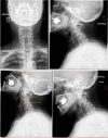
Fig. 1
Fig. 1. Preoperative radiographs.
Preoperative standing cervical spine radiographs, including AP (top left), lateral (top right), extension (bottom left), and flexion (bottom right) views.
Fig. 2
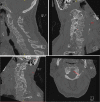
Fig. 2. Preoperative CT Scan.
Preoperative CT scan demonstrating the C2 erosive pannus and diffuse subaxial spondylotic changes.
Fig. 3
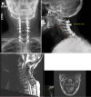
Fig. 3. Preoperative MRI.
Preoperative mid-sagittal T2 (left) and axial T1 MRI sequences demonstrating the C2 erosive pannus, myelomalacia (red arrow), and diffuse subaxial spondylotic changes.
Fig. 4
Fig. 4. Intraoperative Photographs.
Intraoperative photo demonstrating wide decompression of the cervical spine from C1-4 with a C5 dome laminectomy.
Fig. 5
Fig. 5. Early Postoperative Radiographs.
Postoperative radiographs were obtained prior to the patient’s dismissal from the hospital.
Fig. 6
Fig. 6. Two Year Postoperative Radiographs.
Two-year postoperative radiographs (top left and top right) and mid-sagittal (bottom left) and axial (bottom right) T2 MRI demonstrate wide patency of the spinal canal at the decompressed levels, complete regression of the peri-odontoid pannus, and maintained cervical alignment.

Similar articles

References

    1. Radcliff KE, Hussain MM, Moldavsky M, Klocke N, Vaccaro AR, Albert TJ, et al. In vitro biomechanics of the craniocervical junction-a sequential sectioning of its stabilizing structures. Spine J. 2015;15:1618–28. doi: 10.1016/j.spinee.2015.02.004. - DOI - PubMed
    1. Joaquim AF, Appenzeller S. Cervical spine involvement in rheumatoid arthritis-a systematic review. Autoimmun Rev. 2014;13:1195–202. doi: 10.1016/j.autrev.2014.08.014. - DOI - PubMed
    1. Joyce AA, Williams JN, Shi J, Mandell JC, Isaac Z, Ermann J. Atlanto-axial pannus in patients with and without rheumatoid arthritis. J Rheumatol. 2019;46:1431–7. doi: 10.3899/jrheum.181429. - DOI - PubMed
    1. Lagares A, Arrese I, Pascual B, Gòmez PA, Ramos A, Lobato RD. Pannus resolution after occipitocervical fusion in a non-rheumatoid atlanto-axial instability. Eur Spine J. 2006;15:366–9. doi: 10.1007/s00586-005-0969-4. - DOI - PMC - PubMed
    1. Goyal A, Akhras A, Wahood W, Alvi MA, Nassr A, Bydon M. Should multilevel posterior cervical fusions involving C7 cross the cervicothoracic junction? A systematic review and meta-analysis. World Neurosurg. 2019;127:588–95. doi: 10.1016/j.wneu.2019.03.283. - DOI - PubMed

Publication types