

Mapping surgical fields by moving a laser-scanning multimodal scope attached to a robot arm
- PMID: 34321710
- PMCID: PMC8315033
- DOI: 10.1117/12.2044165
Mapping surgical fields by moving a laser-scanning multimodal scope attached to a robot arm
Abstract
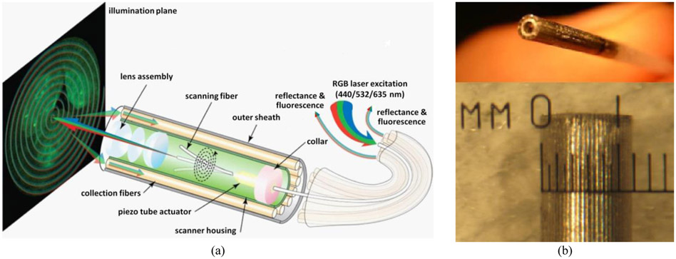
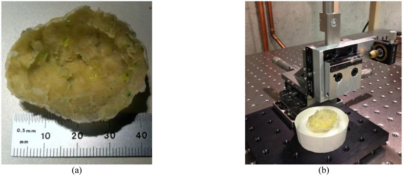
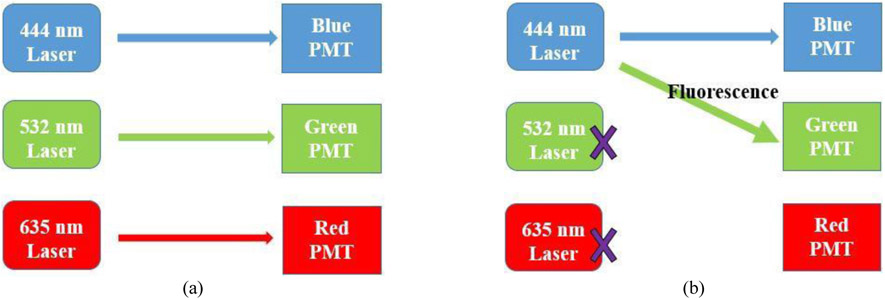
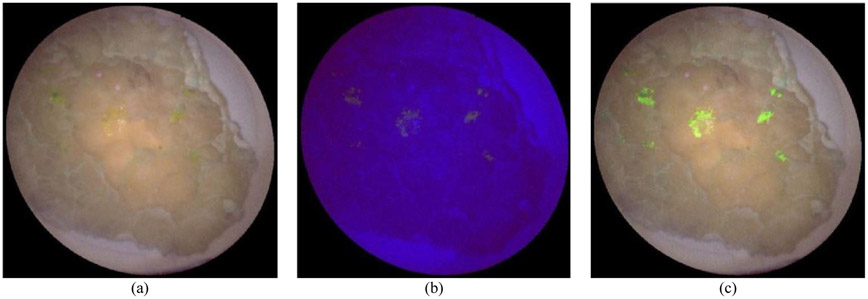
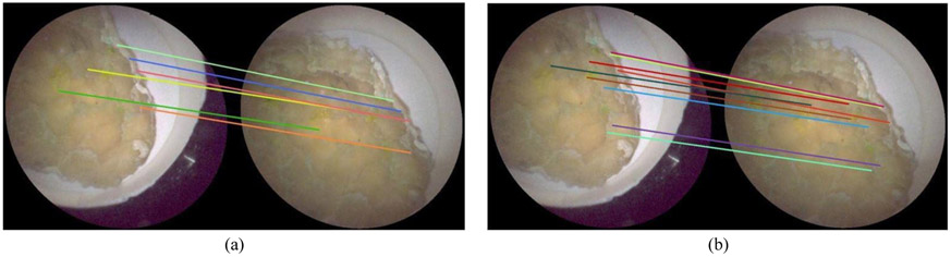
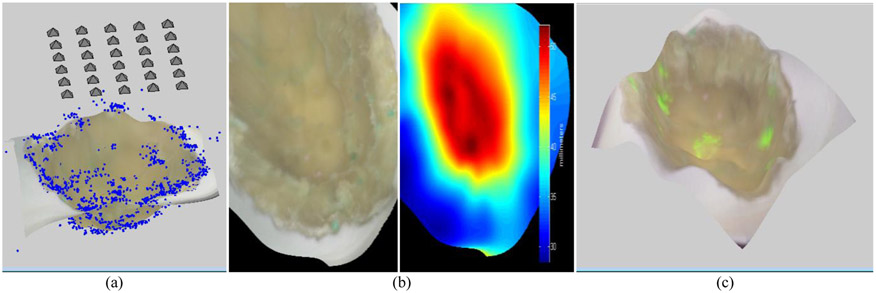
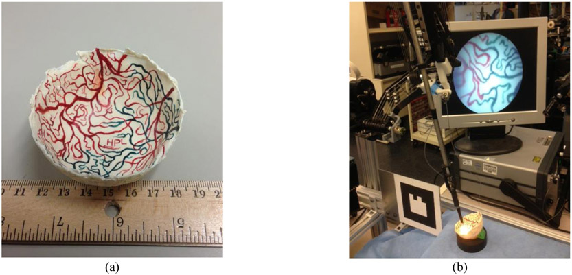
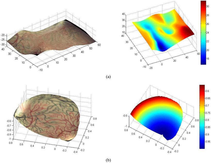
Endoscopic visualization in brain tumor removal is challenging because tumor tissue is often visually indistinguishable from healthy tissue. Fluorescence imaging can improve tumor delineation, though this impairs reflectance-based visualization of gross anatomical features. To accurately navigate and resect tumors, we created an ultrathin/flexible, scanning fiber endoscope (SFE) that acquires reflectance and fluorescence wide-field images at high-resolution. Furthermore, our miniature imaging system is affixed to a robotic arm providing programmable motion of SFE, from which we generate multimodal surface maps of the surgical field. To test this system, synthetic phantoms of debulked tumor from brain are fabricated having spots of fluorescence representing residual tumor. Three-dimension (3D) surface maps of this surgical field are produced by moving the SFE over the phantom during concurrent reflectance and fluorescence imaging (30Hz video). SIFT-based feature matching between reflectance images is implemented to select a subset of key frames, which are reconstructed in 3D by bundle adjustment. The resultant reconstruction yields a multimodal 3D map of the tumor region that can improve visualization and robotic path planning. Efficiency of creating these 3D maps is important as they are generated multiple times during tumor margin clean-up. By using pre-programmed motions of the robot arm holding the SFE, the computer vision algorithms are optimized for efficiency by reducing search times. Preliminary results indicate that the time for creating these multimodal maps of the surgical field can be reduced to one third by using known trajectories of the surgical robot moving the image-guided tool.
Keywords: 3D surface mosaic; fluorescence-guided surgery; image-guided therapy; machine vision; medical robotics.
Figures
References
-
- Soroceanu L, Gillespie Y, Khazaeli MB, and Sontheimer H, “Use of chlorotoxin for targeting of primary brain tumors,” Cancer Res. 58: 4871–9 (1998). - PubMed
-
- Veiseh M, Gabikian P, Bahrami SB, Veiseh O, …, Ellenbogen RG, and Olson JA, “Tumor paint: a chlorotoxin: Cy5.5 bioconjugate for intraoperative visualization of cancer foci.,” Cancer Res. 67:6882–8 (2007). - PubMed
Grants and funding
LinkOut - more resources
Full Text Sources