

Incorporating mucosal-associated invariant T cells into the pathogenesis of chronic liver disease
- PMID: 34321839
- PMCID: PMC8291028
- DOI: 10.3748/wjg.v27.i25.3705
Incorporating mucosal-associated invariant T cells into the pathogenesis of chronic liver disease
Abstract
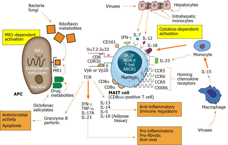
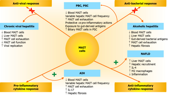
Mucosal-associated invariant T (MAIT) cells have been described in liver and non-liver diseases, and they have been ascribed antimicrobial, immune regulatory, protective, and pathogenic roles. The goals of this review are to describe their biological properties, indicate their involvement in chronic liver disease, and encourage investigations that clarify their actions and therapeutic implications. English abstracts were identified in PubMed by multiple search terms, and bibliographies were developed. MAIT cells are activated by restricted non-peptides of limited diversity and by multiple inflammatory cytokines. Diverse pro-inflammatory, anti-inflammatory, and immune regulatory cytokines are released; infected cells are eliminated; and memory cells emerge. Circulating MAIT cells are hyper-activated, immune exhausted, dysfunctional, and depleted in chronic liver disease. This phenotype lacks disease-specificity, and it does not predict the biological effects. MAIT cells have presumed protective actions in chronic viral hepatitis, alcoholic hepatitis, non-alcoholic fatty liver disease, primary sclerosing cholangitis, and decompensated cirrhosis. They have pathogenic and pro-fibrotic actions in autoimmune hepatitis and mixed actions in primary biliary cholangitis. Local factors in the hepatic microenvironment (cytokines, bile acids, gut-derived bacterial antigens, and metabolic by-products) may modulate their response in individual diseases. Investigational manipulations of function are warranted to establish an association with disease severity and outcome. In conclusion, MAIT cells constitute a disease-nonspecific, immune response to chronic liver inflammation and infection. Their pathological role has been deduced from their deficiencies during active liver disease, and future investigations must clarify this role, link it to outcome, and explore therapeutic interventions.
Keywords: Antimicrobial; Immune regulatory; Innate-like lymphocytes; Mucosal-associated invariant T cell; Pathogenic.
©The Author(s) 2021. Published by Baishideng Publishing Group Inc. All rights reserved.
Conflict of interest statement
Conflict-of-interest statement: The author has no conflict of interest.
Figures
Similar articles
-
Persistent reduction of mucosal-associated invariant T cells in primary biliary cholangitis.J Gastroenterol Hepatol. 2018 Jun;33(6):1286-1294. doi: 10.1111/jgh.14076. Epub 2018 Mar 9. J Gastroenterol Hepatol. 2018. PMID: 29266628
-
Mucosal-Associated Invariant T cell in liver diseases.Int J Biol Sci. 2020 Jan 1;16(3):460-470. doi: 10.7150/ijbs.39016. eCollection 2020. Int J Biol Sci. 2020. PMID: 32015682 Free PMC article. Review.
-
Mucosal-Associated Invariant T Cells Improve Nonalcoholic Fatty Liver Disease Through Regulating Macrophage Polarization.Front Immunol. 2018 Sep 4;9:1994. doi: 10.3389/fimmu.2018.01994. eCollection 2018. Front Immunol. 2018. PMID: 30233587 Free PMC article.
-
Dysregulation and impaired anti-bacterial potential of mucosal-associated invariant T cells in autoimmune liver diseases.Int Immunopharmacol. 2024 Dec 5;142(Pt B):113175. doi: 10.1016/j.intimp.2024.113175. Epub 2024 Sep 21. Int Immunopharmacol. 2024. PMID: 39306887
-
Mucosal-Associated Invariant T Cells: Diplomatic Front-Runners in the Fight against Hepatitis B Virus Infection.Crit Rev Immunol. 2021;41(5):1-16. doi: 10.1615/CritRevImmunol.2021041408. Crit Rev Immunol. 2021. PMID: 35381136 Review.
Cited by
-
Evaluation of mucosal-associated invariant T-cells as a potential biomarker to predict infection risk in liver cirrhosis.PLoS One. 2024 May 1;19(5):e0294695. doi: 10.1371/journal.pone.0294695. eCollection 2024. PLoS One. 2024. PMID: 38691552 Free PMC article.
-
MAIT cells in liver inflammation and fibrosis.Semin Immunopathol. 2022 Jul;44(4):429-444. doi: 10.1007/s00281-022-00949-1. Epub 2022 May 31. Semin Immunopathol. 2022. PMID: 35641678 Free PMC article. Review.
-
Differential Effects of Dietary versus Exercise Intervention on Intrahepatic MAIT Cells and Histological Features of NAFLD.Nutrients. 2022 May 25;14(11):2198. doi: 10.3390/nu14112198. Nutrients. 2022. PMID: 35683998 Free PMC article.
-
Progressive Liver Fibrosis in Non-Alcoholic Fatty Liver Disease.Cells. 2021 Dec 2;10(12):3401. doi: 10.3390/cells10123401. Cells. 2021. PMID: 34943908 Free PMC article. Review.
-
Liver immunology: Biological role and clinical significance.World J Hepatol. 2025 Jul 27;17(7):107541. doi: 10.4254/wjh.v17.i7.107541. World J Hepatol. 2025. PMID: 40747238 Free PMC article. Review.
References
-
- Gold MC, Cerri S, Smyk-Pearson S, Cansler ME, Vogt TM, Delepine J, Winata E, Swarbrick GM, Chua WJ, Yu YY, Lantz O, Cook MS, Null MD, Jacoby DB, Harriff MJ, Lewinsohn DA, Hansen TH, Lewinsohn DM. Human mucosal associated invariant T cells detect bacterially infected cells. PLoS Biol. 2010;8:e1000407. - PMC - PubMed
-
- Montano-Loza AJ, Czaja AJ. Cell mediators of autoimmune hepatitis and their therapeutic implications. Dig Dis Sci. 2015;60:1528–1542. - PubMed
Publication types
MeSH terms
Substances
LinkOut - more resources
Full Text Sources
Medical
