

Case Report: Uroenteric Fistula in a Pediatric-en-bloc Kidney Transplant Manifests as Deceptive Watery Diarrhea and Normal Anion Gap Acidosis
- PMID: 34322462
- PMCID: PMC8310905
- DOI: 10.3389/fped.2021.687396
Case Report: Uroenteric Fistula in a Pediatric-en-bloc Kidney Transplant Manifests as Deceptive Watery Diarrhea and Normal Anion Gap Acidosis
Abstract
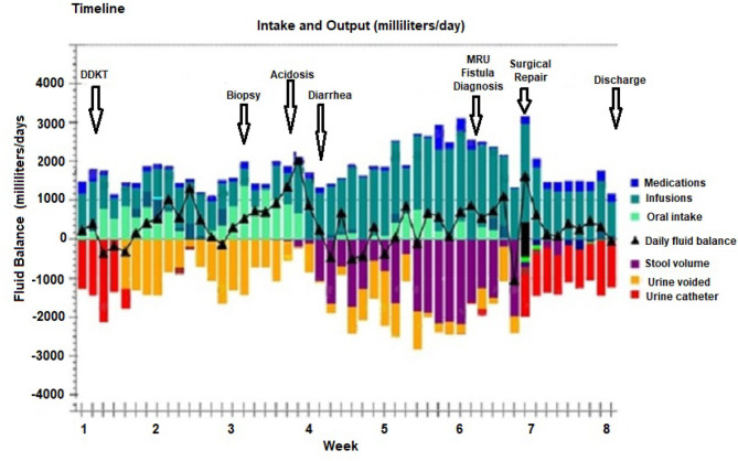
Introduction: The diagnosis of a post-surgical uroenteric fistula can be challenging and may be delayed for months after symptoms begin. A normal anion gap metabolic acidosis has been reported in up to 100% of patients after ureterosigmoidostomy, and bladder substitution using small bowel and/or colonic segments. Here, we describe a rare case of a pediatric patient who developed a uroenteric fistula from the transplant ureters into the small bowel, after an en-bloc kidney transplantation resulting in profound acidosis and deceptive watery diarrhea. Case Presentation: The patient is an 8-year-old girl with end stage kidney disease (ESKD) secondary to focal segmental glomerulosclerosis. Through a right retroperitoneal approach, she underwent a right native nephrectomy and a pediatric deceased donor en-bloc kidney transplant including two separate ureters. One month later, she had a renal allograft biopsy for suspected rejection. During the week after the biopsy, she experienced abdominal pain followed by watery diarrhea and metabolic acidosis requiring continuous bicarbonate/acetate infusions. An extensive gastro-intestinal evaluation for the cause of the diarrhea including endoscopy was inconclusive. The urine output decreased to <500 ml daily; although, the kidney function remained normal. After 2 weeks of unexplained watery diarrhea a magnetic resonance urogram with contrast was performed which demonstrated extravasation of urine from both ureters with fistulization into the small bowel. She underwent corrective surgery which identified the fistulous tract, which was resected and both ureters were re-implanted. The diarrhea and acidosis resolved, and she has maintained normal renal allograft function for over 1 year. Conclusion: An important aspect in the early diagnosis of a uroenteric fistula is the sudden onset of severe hyperchloremic metabolic acidosis that results when urine is diverted into the intestinal tract. The mechanism is similar to that described in cases of urinary diversions and/or bladder augmentation using the intestine. Important diagnostic tools are the measurements of solute excretion and pH in the urine as compared to the "watery diarrhea" or bowel output. Summary: We describe a case of a uroenteric fistula in a pediatric-en-bloc kidney transplant patient that went undiagnosed for almost 3 weeks due to the deceptive nature of the watery diarrhea which was actually urine. A uroenteric fistula should be considered in the differential diagnosis of diarrhea and hyperchloremic metabolic acidosis as a complication of kidney transplant. The simultaneous comparison of stool and urine pH and solute excretions may lead to the diagnosis, appropriate imaging and surgical intervention.
Keywords: CFTR-SLC26; non-anion gap acidosis; pediatric-en-bloc transplant; urinary diarrhea; uroenteric fistula.
Copyright © 2021 Al Barbandi, Defreitas, Infante, Morsi, Arroyo Parejo Drayer, Katsoufis, Seeherunvong, Chandar, Burke and Abitbol.
Conflict of interest statement
The authors declare that the research was conducted in the absence of any commercial or financial relationships that could be construed as a potential conflict of interest.
Figures


Similar articles
-
Case Report: Irreversible Watery Diarrhea, Severe Metabolic Acidosis, Hypokalemia and Achloridria Syndrome Related to Vasoactive Intestinal Peptide Secreting Malignant Pheochromocytoma.Front Endocrinol (Lausanne). 2021 Mar 17;12:652045. doi: 10.3389/fendo.2021.652045. eCollection 2021. Front Endocrinol (Lausanne). 2021. PMID: 33815297 Free PMC article.
-
Severe metabolic acidosis and hypokalemia in a patient with enterovesical fistula.Clin Exp Nephrol. 2007 Sep;11(3):225-229. doi: 10.1007/s10157-007-0475-6. Epub 2007 Sep 28. Clin Exp Nephrol. 2007. PMID: 17891350
-
Follow-up after urinary diversion.Urol Int. 1999;63(1):40-5. doi: 10.1159/000030417. Urol Int. 1999. PMID: 10592489 Review.
-
An unusual case of neuroblastoma presenting with prolonged watery diarrhea in a pediatric patient.Biochem Med (Zagreb). 2025 Jun 15;35(2):020901. doi: 10.11613/BM.2025.020901. Biochem Med (Zagreb). 2025. PMID: 40520658 Free PMC article.
-
Hyperchloremic normal gap metabolic acidosis.Minerva Endocrinol. 2019 Dec;44(4):363-377. doi: 10.23736/S0391-1977.19.03059-1. Epub 2019 Jul 24. Minerva Endocrinol. 2019. PMID: 31347344 Review.
References
Publication types
LinkOut - more resources
Full Text Sources
Research Materials
Miscellaneous