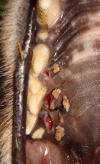
Focal Palatitis (Previously Focal Palatine Erosions) in Captive Cheetahs (Acinonyx jubatus)
- PMID: 34322534
- PMCID: PMC8312244
- DOI: 10.3389/fvets.2021.682150
Focal Palatitis (Previously Focal Palatine Erosions) in Captive Cheetahs (Acinonyx jubatus)
Abstract
Focal palatine erosion (FPE) is a misleading term that is used in the literature to describe inflammatory lesions associated with depressions of the palatal mucosa in cheetah. Cheetahs have large cheek teeth and these depressions are formed to accommodate them. Previously FPE was only described as a mandibular molar tooth malocclusion on the hard palate due to suspected rotation and super eruption of the mandibular molar teeth of cheetahs aged 18 months and older. Two hundred and fifty six cheetahs (135 male, 121 female), originating from two independent facilities, had their oral cavities evaluated as part of an annual health visit over a decade. Ninety-nine cheetahs were seen once, 59 cheetahs were seen twice, 33 were seen three times, 43 on four occasions, 16 on five occasions, 5 on six occasions, and 1 cheetah was seen seven times. Apart from these clinical cases a prospective study on 5 cheetah cubs (3 male and 2 female) was conducted to document their skull development and mandibular molar tooth eruption over a period of 25 months. Of the 261 cheetahs observed none developed rotation or super eruption of their mandibular molar teeth. The term FPE is a misnomer as these inflammatory lesions were found in palatal depressions opposing any of the cusps of all of the cheetah mandibular cheek teeth. It consisted mainly of deep ulcerations, inflammation and oedema and also micro abscess formation. In severe cases oro-nasal fistulas were present. Of all the depressions present on the cheetah's palate, the large one palatal to the 4th maxillary premolar tooth was most commonly affected. In the five cubs evaluated prospectively, focal palatitis was evident from the 7 month evaluation, before all the permanent teeth erupted. Conservative treatment of the inflamed depressions by removing the foreign material through curettage and copious flushing reduced the grade of the inflammation when observed on follow-up. Focal palatine erosion is an incorrect term used to describe focal palatitis that occurs randomly in cheetahs. This focal palatitis is often associated with foreign material trapped in the palatal depressions. Conservative management is sufficient to treat these animals without odontoplasties.
Keywords: cheetah; focal palatine erosions; molar; palatal depressions; palatitis; teeth.
Copyright © 2021 Steenkamp, Boy, van Staden and Bester.
Conflict of interest statement
The authors declare that the research was conducted in the absence of any commercial or financial relationships that could be construed as a potential conflict of interest.
Figures


Similar articles
-
Focal palatine erosion in captive and free-living cheetahs (Acinonyx jubatus) and other felid species.Zoo Biol. 2012 Mar-Apr;31(2):181-8. doi: 10.1002/zoo.20392. Epub 2011 May 3. Zoo Biol. 2012. PMID: 21541986
-
Longitudinal Radiographic Study of Cranial Bone Growth in Young Cheetah.Front Vet Sci. 2019 Jul 30;6:256. doi: 10.3389/fvets.2019.00256. eCollection 2019. Front Vet Sci. 2019. PMID: 31417919 Free PMC article.
-
Oral, Maxillofacial and Dental Diseases in Captive Cheetahs (Acinonyx jubatus).J Comp Pathol. 2018 Jan;158:77-89. doi: 10.1016/j.jcpa.2017.12.004. Epub 2018 Jan 16. J Comp Pathol. 2018. PMID: 29422320
-
Chronic Stress-Related Gastroenteric Pathology in Cheetah: Relation between Intrinsic and Extrinsic Factors.Biology (Basel). 2022 Apr 15;11(4):606. doi: 10.3390/biology11040606. Biology (Basel). 2022. PMID: 35453805 Free PMC article. Review.
-
Anatomy of the Brachycephalic Canine Hard Palate and Treatment of Acquired Palatitis Using CO2 Laser.J Vet Dent. 2019 Sep;36(3):186-197. doi: 10.1177/0898756419893127. J Vet Dent. 2019. PMID: 31928397 Review.
Cited by
-
Menrath ulcers in cats: four cases (2014-2023).J Small Anim Pract. 2025 May;66(5):346-352. doi: 10.1111/jsap.13828. Epub 2025 Jan 12. J Small Anim Pract. 2025. PMID: 39799972 Free PMC article.
-
Getting to the Meat of It: The Effects of a Captive Diet upon the Skull Morphology of the Lion and Tiger.Animals (Basel). 2023 Nov 22;13(23):3616. doi: 10.3390/ani13233616. Animals (Basel). 2023. PMID: 38066967 Free PMC article.
References
-
- Fitch HM, Fagan D. Focal palatine erosion associated with dental malocclusion in captive cheetahs. Zoo Biol. (1982) 1:295–310. 10.1002/zoo.1430010403 - DOI
-
- Fagan DA. Cheetah Focal Palatine Erosion Study Evaluation Protocol. Colyer Institute; (2014). Available online at: http://www.colyerinstitute.org/pdf/fpe-rating-system.pdf (accessed on February 1, 2021).
-
- Scheels JL, Wallace R. editors. Early detection of dental and oral pathology. In: American Association of Zoo Veterinarians, Zoo Dentistry Symposium. Milwaukee, WI: (2002).
-
- Phillips JA, Worley MB, Morsbach D, Williams TM. Relationship among diet, growth and occurrence of focal palatine erosion in wild-caught captive cheetahs. Madoqua. (1993) 18:79–83.
LinkOut - more resources
Full Text Sources

