Review
. 2021 Aug;9(15):e14982.
doi: 10.14814/phy2.14982.

Losing the dogmatic view of cerebral autoregulation

Affiliations
Review

Losing the dogmatic view of cerebral autoregulation

Patrice Brassard et al. Physiol Rep. 2021 Aug.

Abstract

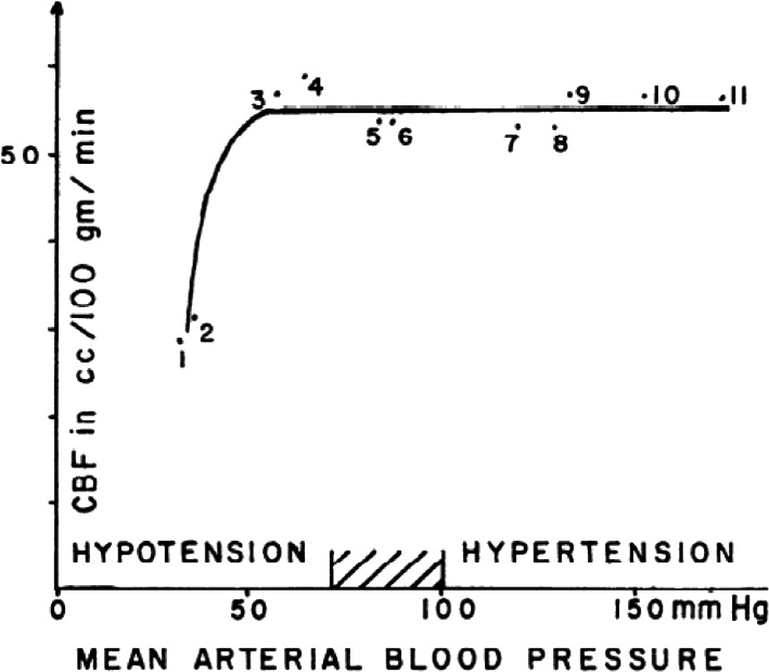
In 1959, Niels Lassen illustrated the cerebral autoregulation curve in the classic review article entitled Cerebral Blood Flow and Oxygen Consumption in Man. This concept suggested a relatively broad mean arterial pressure range (~60-150 mmHg) wherein cerebral blood flow remains constant. However, the assumption that this wide cerebral autoregulation plateau could be applied on a within-individual basis is incorrect and greatly variable between individuals. Indeed, each data point on the autoregulatory curve originated from independent samples of participants and patients and represented interindividual relationships between cerebral blood flow and mean arterial pressure. Nonetheless, this influential concept remains commonly cited and illustrated in various high-impact publications and medical textbooks, and is frequently taught in medical and science education without appropriate nuances and caveats. Herein, we provide the rationale and additional experimental data supporting the notion we need to lose this dogmatic view of cerebral autoregulation.

Keywords: Lassen; arterial blood pressure; autoregulatory curve; cerebral autoregulation; cerebral blood flow.


Conflict of interest statement

The authors declare no conflict of interest.

Figures

FIGURE 1
FIGURE 1
Cerebral blood flow and blood pressure. Mean values of 11 groups of subjects reported in seven studies have been plotted. Various acute and chronic conditions have been selected, characterized by a change in blood pressure. See text for details. Reprinted from Lassen (1959) with permission
FIGURE 2
FIGURE 2
A diagrammatic representation of the relationship of pial arteriolar diameter, cerebral blood flow, and cerebrovascular resistance with blood pressure. It will be seen that pial arteriolar dilatation reaches a maximum well below the lower level of autoregulation. Reprinted from Paulson et al. (1990) with permission
FIGURE 3
FIGURE 3
(a) Finger arterial blood pressure and middle cerebral blood flow velocity over 900 s (presented on log axes) for a human subject in the seated resting position. (b) The corresponding power spectrums, which decompose the time series signals into its various constituent component frequencies. Note that the fundamental frequency (f 0) and its harmonics (f 1, f 2,f 3) correspond to pulsations coincident with the pulse, whereas progressively lower frequency components reflect the longer term oscillations and trends in the time domain. ULF ultra‐low frequency, VLF very low frequency, LF low frequency, HF high frequency. Reprinted from Tzeng & Ainslie (2013) with permission
FIGURE 4
FIGURE 4
Relationship between relative changes in MAP from baseline (Δ%MAP) and concomitant relative changes in CBF (Δ%CBF) when MAP decreases and increases including (left panel) and excluding (right panel) the utilization of cardiovascular drugs to manipulate MAP. These data represent an updated analysis initially published by Numan et al. (2014) Note the consistent pressure‐passive nature of CBF in the pharmacological data (left panel) and the small plateau present in the non‐pharmacological data (right panel)

References

    1. Aaslid, R. , Blaha, M. , Sviri, G. , Douville, C. M. , & Newell, D. W. (2007). Asymmetric dynamic cerebral autoregulatory response to cyclic stimuli. Stroke, 38(5), 1465–1469. 10.1161/STROKEAHA.106.473462 - DOI - PubMed
    1. Aaslid, R. , Lindegaard, K. F. , Sorteberg, W. , & Nornes, H. (1989). Cerebral autoregulation dynamics in humans. Stroke, 20(1), 45–52. 10.1161/01.STR.20.1.45 - DOI - PubMed
    1. Abercrombie, J. (1836). Pathological and practical researches of diseases of the brain and spinal cord, 3rd ed. John Carfrae and Son.
    1. Ainslie, P. N. , & Hoiland, R. L. (2014). Transcranial Doppler ultrasound: Valid, invalid, or both? Journal of Applied Physiology, 117(10), 1081–1083. 10.1152/japplphysiol.00854.2014 - DOI - PubMed
    1. Barrett, K. E. , Barman, S. M. , Boitano, S. , & Brooks, H. (2010). Ganong's review of medical physiology, 23rd ed. McGraw‐Hill.

LinkOut - more resources