. 2021 Sep;100(10):1099-1108.
doi: 10.1177/00220345211026838. Epub 2021 Jul 29.

Biomineralization of Dental Tissues Treated with Silver Diamine Fluoride

Affiliations

Biomineralization of Dental Tissues Treated with Silver Diamine Fluoride

R M Sulyanto et al. J Dent Res. 2021 Sep.

Abstract

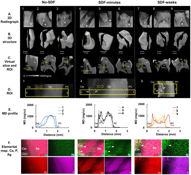
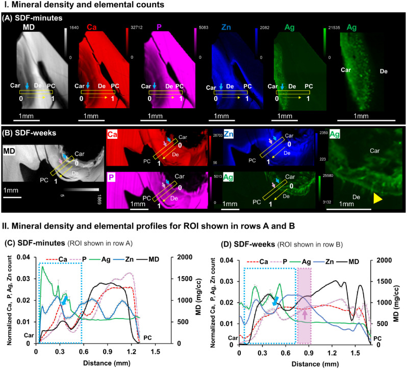
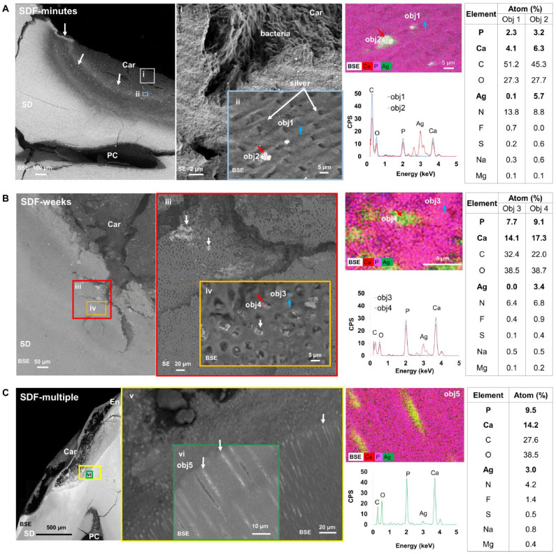
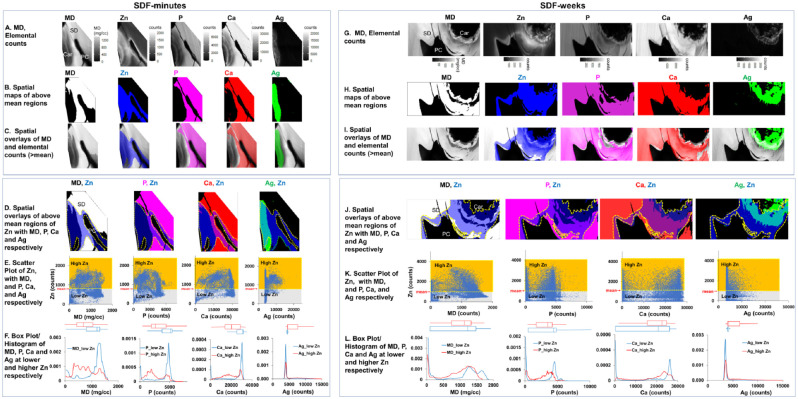
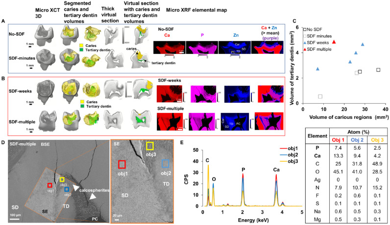
Silver diamine fluoride (SDF) is a dental biomaterial used to arrest dental caries. To better understand SDF's mechanism of action, we examined the localization of silver within the tissues of SDF-treated teeth. Carious primary teeth fixed within 2 min of SDF application (SDF-minutes, n = 3), at 3 wk after SDF application in vivo (SDF-weeks, n = 4), and at 2 y after multiple SDF applications in vivo (SDF-multiple, n = 1) were investigated in this study. Carious primary teeth without SDF application (no-SDF, n = 3) served as controls. Mineral density and structural analyses were performed via micro-X-ray computed tomography and scanning electron microscopy. Elemental analyses were performed through X-ray fluorescence microprobe and energy-dispersive X-ray spectroscopic techniques. SDF-treated teeth revealed higher X-ray-attenuated surface and subsurface regions within carious lesions, and similar regions were not present in no-SDF teeth. Regions of higher mineral density correlated with regions of silver abundance in SDF-treated teeth. The SDF penetration depth was approximated to 0.5 ± 0.02 mm and 0.6 ± 0.05 mm (mean ± SD) for SDF-minutes and SDF-weeks specimens, respectively. A higher percentage of dentin tubular occlusion by silver or calcium phosphate particles was observed in primary teeth treated with SDF-weeks as compared with SDF-minutes. Elemental analysis also revealed zinc abundance in carious lesions and around the pulp chamber. SDF-weeks teeth had significantly increased tertiary dentin than SDF-minutes and no-SDF teeth. These results suggest that SDF treatment on primary teeth affected by caries promotes pathologic biomineralization by altering their physicochemical properties, occluding dentin tubules, and increasing tertiary dentin volume. These seemingly serendipitous effects collectively contribute to the cariostatic activity of SDF.

Keywords: caries; metalloprotease; primary tooth; tertiary dentin; trace metals; zinc.


Conflict of interest statement

Declaration of Conflicting Interests: The authors declared no potential conflicts of interest with respect to the research, authorship, and/or publication of this article.

Figures

Figure 1.
Figure 1.
Mineral density (MD) and elemental maps of silver diamine fluoride (SDF)–treated carious teeth. Nine carious primary teeth in 3 treatment groups were evaluated: no-SDF (teeth 1 to 3), SDF-minutes (teeth 4 to 6), and SDF-weeks (teeth 7 to 9). (A) Two-dimensional (2D) clinical X-ray radiographs, (B) 2D slices within 3-dimensional (3D) volume-rendered structures, and (C) corresponding 2D virtual slices/tomograms in each treatment group. (D) Region of interest (ROI; yellow rectangular box) in each 2D slice for all 9 teeth examined by an energy-dispersive X-ray unit. Representative regions span the carious surface (0) to the pulp chamber (2) for teeth 1, 4, and 9 (yellow arrows, direction; white arrows, Ag). (E) Heterogenous MD profiles along the length of the ROI for each tooth obtained by micro X-ray computed tomography are illustrated. (F) Elemental spatial maps of Ca, P, and Ag (white arrows) of representative ROIs for each treatment group were obtained by energy-dispersive X-ray. B, bone; Car, caries; De, dentin; PC, pulp chamber; T, teeth.
Figure 2.
Figure 2.
Varied distributions of elemental Ca, P, Zn, and Ag “shaped” the mineral density (MD) profiles unique to carious lesions treated with SDF-minutes and SDF-weeks. MD (white) from micro X-ray computed tomography and X-ray fluorescence microprobe elemental maps of Ca (red), P (pink), Zn (blue), and Ag (green) in carious primary teeth treated with a onetime SDF application and extracted (A) 2 min after application (SDF-minutes, tooth 4) and (B) 3 wk after application (SDF-weeks, tooth 9). Right-most panel shows Ag distribution at a higher magnification for both treatment groups and indicates Ag distribution along the length of the dentinal tubules in the SDF-weeks group (yellow arrowhead). MD and elemental profiles for regions of interest (ROIs; yellow boxes) from the carious margin (0) to the pulp chamber (1) in panels A and B are shown for teeth treated with SDF for (C) minutes (tooth 4) and (D) weeks (tooth 9). SDF penetration depth (light blue dotted rectangle) was estimated by spatially correlating the MD profile with that of the Ag and Ca elemental profiles. The penetration depths of SDF-minutes and SDF-weeks were ~0.5 ± 0.02 mm and ~0.6 ± 0.05 mm (mean ± SD), (yellow arrows, direction; white arrows, location of Ag particles) respectively. Ca and P peaks (light blue arrows) within carious lesions indicated potentially remineralized areas. Increased P counts with decreased Ca counts (light purple arrow within purple rectangle) were observed. Car, caries; De, dentin; PC, pulp chamber; SDF, silver diamine fluoride.
Figure 3.
Figure 3.
Morphology and spatial distributions of silver (Ag) in primary teeth affected with caries. Teeth shown are from 3 treatment groups: (A) SDF-minutes, (B) SDF-weeks, and (C) SDF-multiple. Backscattered electron (BSE) and secondary electron (SE) images of a representative tooth in each treatment group were obtained at various magnifications (i–vi). Grayscale differences in BSE micrographs represent variations per the atomic number of elements within regions of varying mineral densities and Ag particles (white arrows). Energy-dispersive X-ray spectra of the same regions with atomic percentages of each element in specific regions (obj 1, 2, 3, 4, 5) are presented in corresponding tables. Obj 1 to 4 were collected at a spot size of 1 µm whereas Obj 5 was obtained from an area of 828 µm2. Objs 1 & 3, blue arrows; Objs 2 & 4, red arrows. Car, caries; En, enamel; PC, pulp chamber; SD, sound dentin; SDF, silver diamine fluoride.
Figure 4.
Figure 4.
Colocalization of zinc (Zn), phosphorus (P), calcium (Ca), and silver (Ag) in regions of known mineral densities (MDs) and spatial correlation between Zn and MD, Zn and Ca, Zn and P, and Zn and Ag in SDF-treated primary teeth. Spatial maps of MD and each element of SDF-minutes (AF) and SDF-weeks (GL) are shown. Regions with MD or elemental counts above mean values were segmented (B, H), and resulting elemental distribution maps were overlaid with MD maps (C, I). Higher Zn counts (above mean represented by yellow dotted line) were observed in carious dentin and around the pulp chamber (D, J). Scatter plots (E, K) illustrate higher (yellow) and lower (gray) Zn regions, and box plots and histograms of MD, Ca, P, and Ag within higher (red line) and lower (blue line) Zn regions are shown (F, L). Car, caries; PC, pulp chamber; SD, sound dentin; SDF, silver diamine fluoride.
Figure 5.
Figure 5.
Tertiary dentin in SDF-treated primary teeth. (A, B) Three-dimensional volume-rendered images and virtual sections overlaid with mineral density segmented volumes illustrate different volumes of carious lesions (yellow) and tertiary dentin (green) inside the pulp chamber. X-ray fluorescence microprobe elemental distribution maps demonstrate Ca (red), P (pink), and Zn (blue) expressions within carious and tertiary dentin regions located around and inside the pulp chamber. (C) Volume estimates of tertiary dentin were plotted against volume estimates of carious lesions for carious primary molars not treated with SDF, treated with SDF for minutes, treated with SDF for weeks, and treated with SDF multiple times. (D) Backscattered electron (BSE) and secondary electron (SE) micrographs of carious lesions treated with SDF multiple times illustrate spherulitic calcified structures within the pulp chamber (orange box). (E) Energy-dispersive X-ray maps show relatively lower Ca and P atomic percentages in less (obj 2) and least (obj 3) mineralized regions as compared with tubular sound dentin (obj 1). Car, caries, PC, pulp chamber; SD, sound dentin; SDF, silver diamine fluoride; TD, tertiary dentin.

References

    1. Barceloux DG. 1999. Zinc. J Toxicol Clin Toxicol. 37(2):279–292. - PubMed
    1. Bimstein E, Damm D. 2018. Human primary tooth histology six months after treatment with silver diamine fluoride. J Clin Pediatr Dent. 42(6):442–444. - PubMed
    1. Chairside guide: silver diamine fluoride in the management of dental caries lesions. 2018. Pediatr Dent. 40(6):492–517. - PubMed
    1. Charadram N, Austin C, Trimby P, Simonian M, Swain MV, Hunter N. 2013. Structural analysis of reactionary dentin formed in response to polymicrobial invasion. J Struct Biol. 181(3):207–222. - PMC - PubMed
    1. Chu CH, Lo ECM, Lin HC. 2002. Effectiveness of silver diamine fluoride and sodium fluoride varnish in arresting dentin caries in Chinese pre-school children. J Dent Res. 81(11):767–770. - PubMed

Publication types