. 2021 Sep 1;131(3):1067-1079.
doi: 10.1152/japplphysiol.00221.2021. Epub 2021 Jul 29.

Characterization of endothelium-dependent and -independent processes in occipital artery of the rat: relevance to control of blood flow to nodose sensory cells

Affiliations

Characterization of endothelium-dependent and -independent processes in occipital artery of the rat: relevance to control of blood flow to nodose sensory cells

Tristan H J Lewis et al. J Appl Physiol (1985). .

Abstract

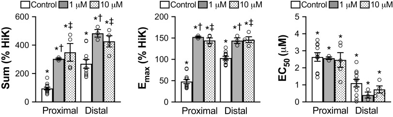
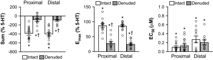
Circulating factors access cell bodies of vagal afferents in nodose ganglia (NG) via the occipital artery (OA). Constrictor responses of OA segments closer in origin from the external carotid artery (ECA) differ from segments closer to NG. Our objective was to determine the role of endothelium in this differential vasoreactivity in rat OA segments. Vasoreactivity of OA segments (proximal segments closer to ECA, distal segments closer to NG) was examined in wire myographs. We evaluated 1) vasoconstrictor effects of 5-hydroxytryptamine (5-HT) in intact and endothelium-denuded OA segments in absence/presence of soluble guanylate cyclase (SGC) inhibitor 1H-[1,2,4]oxadiazolo[4,3-a]quinoxalin-1-one (ODQ), 2) vasodilator responses elicited by the endothelium dependent vasodilator, acetylcholine (ACh), in intact or endothelium-denuded OA segments in absence/presence of ODQ, and 3) vasodilator responses elicited by NO-donor MAHMA NONOate, in intact OA segments in absence/presence of ODQ. Intact distal OA responded more to 5-HT than intact proximal OA. Endothelium denudation increased 5-HT potency in both OA segments, especially proximal OA. ODQ increased maximal responses of 5-HT in both segments, particularly proximal OA. ACh similarly relaxed both OA segments, effects abolished by endothelial denudation and attenuated by ODQ. MAHMA NONOate elicited transient vasodilation in both segments. Effects of ODQ against ACh were segment dependent whereas those against MAHMA NONOate were not. The endothelium regulates OA responsiveness in a segment-dependent fashion. Endothelial cells at the OA-ECA junction more strongly influence vascular tone than those closer to NG. Differential endothelial regulation of OA tone may play a role in controlling blood flow and access of circulating factors to NG.NEW & NOTEWORTHY This study demonstrates that the endothelium-dependent regulation of smooth muscle tone of occipital arteries is segment-dependent. Endothelial cells at the occipital artery-external carotid artery junction (entryway of blood flow to the nodose ganglia) more strongly influence vascular tone than those closer to the nodose ganglia. This differential endothelial regulation of occipital artery tone may control blood flow and access of circulating factors to the nodose ganglia.

Keywords: endothelium-dependent vasodilation; nitric oxide; nodose ganglion; occipital arteries; wire myography.


Conflict of interest statement

No conflicts of interest, financial or otherwise, are declared by the authors.

Figures

Figure 1.
Figure 1.
An in situ photograph of the occipital artery (OA) from an adult Sprague-Dawley rat. The common carotid artery (CCA) branches into the internal (ICA) and external (ECA) carotid arteries. The OA arises from the ECA, and the proximal OA segment (Prox) is closest to the ECA and the distal OA (Dist) is closest to the nodose ganglion (NG). The inferior pharyngeal nerve (IPN) is also shown. Dotted lines indicate approximately where the vessels were cut and removed.
Figure 2.
Figure 2.
Typical examples of the effects of increasing concentrations of 5-hydroxytryptamine (5-HT) on contractile force (mN) of intact (top) and endothelium-denuded (bottom) occipital artery segments bisected into those closest to the external carotid (proximal) or those closest to the nodose ganglion (distal). The term HiK and the time bar below the trace denote the application of 80 mM K+ PSS for 2 min. The numbers 1–9 denote the time of application and concentrations of 5-HT (in µM: 0.001, 0.003, 0.01, 0.03, 0.1, 0.3, 1, 3, 10). The percentages provided at the end of each trace are the maximal contractions elicited by the final concentration of 5-HT compared to the HiK. HiK, high K+; PSS, physiological saline solution.
Figure 3.
Figure 3.
Responses to cumulative addition of 5-hydroxytryptamine (5-HT, 0.001–10 μM) of intact and endothelium-denuded proximal and distal occipital artery segments. The data are presented as mean ± SE as a percentage of the response to 80 mM K+ (HiK). The absolute values of HiK responses used for each group in mN are proximal 2.23 ± 0.18, distal 2.93 ± 0.29, proximal denuded 1.67 ± 0.18, distal denuded 1.99 ± 0.24. n = 12 for each group except distal denuded (n = 11).
Figure 4.
Figure 4.
The responses to cumulative addition of 5-hydroxytryptamine (5-HT, 0.001–10 µM) in intact and endothelium-denuded proximal and distal occipital artery (OA) segments. Left: total percentage change (sum) of the responses. Middle: the maximum change (Emax) in tension. Right: the concentration of 5-HT that elicited 50% of maximal constriction (EC50). The data are presented as means ± SE, cumulative, and Emax as a percentage of the response to 80 mM K+ (HiK). N = 12 for each group except distal denuded (n = 11) and were analyzed using one-way ANOVA. *P < 0.05, significant change from baseline. †P < 0.05, endothelium-denuded versus intact. ‡P < 0.05, intact or endothelium-denuded distal segments versus corresponding proximal segments. HiK, high K+.
Figure 5.
Figure 5.
Responses to cumulative addition of 5-hydroxytryptamine (5-HT, 0.001–10 µM) of intact proximal and distal occipital artery segments (n = 12) and those with 1 µM ODQ (n = 3) or 10 µM ODQ (n = 4). The absolute values of HiK responses used for each group in mN are proximal (control 2.23 ± 0.18, 1 µM ODQ 3.34 ± 0.80, 10 µM ODQ 3.21 ± 0.55) and distal (control 2.93 ± 0.29, 1 µM ODQ 3.34 ± 0.80, 10 µM ODQ 4.65 ± 1.36). The data are presented as mean ± SE as a percentage of the response to 80 mM K+ (HiK). HiK, high K+; ODQ, 1H-[1,2,4]oxadiazolo[4,3,-a]quinoxalin-1-one.
Figure 6.
Figure 6.
The responses to cumulative addition of 5-hydroxytryptamine (5-HT, 0.001–10 µM) on intact proximal and distal occipital artery control segments (n = 12) and those with 1 µM ODQ (n = 3) or 10 µM ODQ (n = 4). Left: total percentage change (sum) of the responses. Middle: the maximum change (Emax) in tension. Right: the concentration of 5-HT that elicited 50% of the maximal constriction (EC50). The data are presented as means ± SE, cumulative, and Emax as a percentage of the response to 80 mM K+ (HiK). *P < 0.05, significant change from 5-HT tone. †P < 0.05, 1 µM ODQ versus control. ‡P < 0.05, 10 µM ODQ versus control. HiK, high K+; ODQ, 1H-[1,2,4]oxadiazolo[4,3,-a]quinoxalin-1-one.
Figure 7.
Figure 7.
Typical examples of the effects of increasing concentrations of acetylcholine (ACh) on contractile force (mN) of intact (top), endothelium-denuded (middle), or intact with 10 µM ODQ (bottom) proximal and distal occipital artery segments. The term HiK and the time bar below the trace denote the application of 80 mM K+ PSS for 2 min. 5-hydroxytryptamine (5-HT) was added to each segment to elicit approximately 50% of the maximal HiK response. The numbers 1–9 denote the time of application and concentrations of ACh (in µM: 0.001, 0.003, 0.01, 0.03, 0.1, 0.3, 1, 3, 10). The percentages provided at the end of each trace represent the maximal dilation elicited by the final concentration of ACh compared to 5-HT tone. HiK, high K+; 5-HT, 5-hydroxytryptamine; ODQ, 1H-[1,2,4]oxadiazolo[4,3,-a]quinoxalin-1-one; PSS, physiological saline solution.
Figure 8.
Figure 8.
Responses to the cumulative addition of acetylcholine (ACh, 0.001–10 µM) of intact and endothelium-denuded proximal and distal occipital artery segments. The data are presented as mean ± SE of the responses expressed as a percentage of agonist-induced constriction (% 5-HT tone). The absolute values of 5-HT tone used for each group in mN are proximal 1.41 ± 0.20, distal 2.54 ± 0.35, proximal denuded 1.75 ± 0.22, distal denuded 2.48 ± 0.24. N = 12 for all groups except distal denuded (n = 11). 5-HT, 5-hydroxytryptamine.
Figure 9.
Figure 9.
The responses to cumulative addition of acetylcholine (ACh, 0.001–10 µM) of intact and endothelium-denuded proximal and distal occipital artery segments. Left: total percentage change (sum) of response. Middle: the maximum change (Emax) in tension. Right: the concentration of ACh that elicited 50% of maximal dilation (EC50). The data are presented as mean ± SE, cumulative, and Emax as a percentage of agonist-induced pretone (% 5-HT). N = 12 for all groups except distal denuded (n = 11). *P < 0.05, significant change from 5-HT pretone. †P < 0.05, endothelium-denuded versus intact. ‡P < 0.05, Intact or endothelium-denuded distal segments versus corresponding proximal segments. 5-HT, 5-hydroxytryptamine.
Figure 10.
Figure 10.
Responses to cumulative addition of acetylcholine (0.001–10 µM) of intact proximal and distal occipital arteries in control segments (n = 14) and those with 1 µM ODQ (n = 4) or 10 µM ODQ (n = 7). The data are presented as means ± SE, expressed as a percentage of agonist-induced constriction (% 5-HT tone). The absolute values of 5-HT tone used for each group in mN are proximal (control 2.23 ± 0.18, 1 µM ODQ 3.45 ± 0.91, 10 µM ODQ 3.78 ± 0.91), and distal (control 2.93 ± 0.29, 1 µM ODQ 3.34 ± 1.11, 10 µM ODQ 4.18 ± 0.88). 5-HT, 5-hydroxytryptamine; ODQ, 1H-[1,2,4]oxadiazolo[4,3,-a]quinoxalin-1-one.
Figure 11.
Figure 11.
The responses to cumulative addition of acetylcholine (ACh, 0.001–10 µM) of intact proximal and distal occipital artery control segments (n = 14) and those with 1 µM ODQ (n = 4) or 10 µM ODQ (n = 7). Left: total percentage change (sum) of the responses. Middle: the maximum change (Emax) in tension. Right: the concentration of ACh that elicited 50% of the maximal dilation (EC50). The data are presented as means ± SE, cumulative, and Emax as a percentage of agonist-induced pretone (% 5-HT). *P < 0.05, significant change from 5-HT pretone. †P < 0.05, 1 µM ODQ versus control. ‡P < 0.05, 10 µM ODQ versus control. 5-HT, 5-hydroxytryptamine; ODQ, 1H-[1,2,4]oxadiazolo[4,3,-a]quinoxalin-1-one.
Figure 12.
Figure 12.
Typical examples of the effects of increasing concentrations of MAHMA NONOate on contractile force (mN) of intact proximal and distal occipital artery segments (top), or intact segments with 10 µM ODQ (bottom). The term HiK and the time bar below the trace denote the application of 80 mM K+ PSS for 2 min. 5-hydroxytryptamine (5-HT pretone) was added to each segment to elicit approximately 50% of the maximal HiK response. The numbers 1–9 denote the time of application and concentrations of MAHMA NONOate (in nM: 0.01, 0.03, 0.1, 0.3, 1, 3, 10, 30, 100). The percentages provided at the end of each trace represent the maximal dilation elicited by the final concentration of MAHMA NONOate compared to 5-HT pretone. HiK, high K+; 5-HT, 5-hydroxytryptamine; MAHMA NONOate, 5-hydroxytryptamine HCl, (Z)-1-[N-Methyl-N-[6-(N-methylammoniohexyl)amino]]diazen-1-ium-1,2-diolate; ODQ, 1H-[1,2,4]oxadiazolo[4,3,-a]quinoxalin-1-one; PSS, physiological saline solution.
Figure 13.
Figure 13.
Top: the maximum responses to cumulative addition of MAHMA NONOate (0.01–100 nM) of intact proximal and distal occipital arteries in control segments (n = 7) and those with 1 µM ODQ (n = 4) or 10 µM ODQ (n = 6). Bottom: the responses after recovery of stable tone to the cumulative addition of MAHMA NONOate (0.01–100 nM) of intact proximal and distal occipital arteries in control segments (n = 7) and those with 1 µM ODQ (n = 4) or 10 µM ODQ (n = 6). The data are presented as means ± SE of the responses expressed as a percentage of agonist-induced constriction (% 5-HT tone). The absolute values of 5-HT tone used for each group in mN are proximal (control 1.54 ± 0.26, 1 µM ODQ 2.18 ± 0.81, 10 µM ODQ 2.53 ± 0.49) and distal (control 1.93 ± 0.20, 1 µM ODQ 2.81 ± 0.97, 10 µM ODQ 3.79 ± 0.82). 5-HT, 5-hydroxytryptamine; MAHMA NONOate, 5-hydroxytryptamine HCl, (Z)-1-[N-Methyl-N-[6-(N-methylammoniohexyl)amino]]diazen-1-ium-1,2-diolate; ODQ, 1H-[1,2,4]oxadiazolo[4,3,-a]quinoxalin-1-one.
Figure 14.
Figure 14.
Top: the maximum responses to cumulative addition of MAHMA NONOate (0.01–100 nM) of intact proximal and distal occipital artery control segments (n = 7) and those with 1 µM ODQ (n = 4) or 10 µM ODQ (n = 6). Bottom: the responses after recovery of stable tone to cumulative addition of MAHMA NONOate (0.01–100 nM) of intact proximal and distal occipital artery control segments (n = 7) and those with 1 µM ODQ (n = 4) or 10 µM ODQ (n = 6). Left: total percentage change (sum) of the responses. Middle: the maximum change (Emax) in tension. Right: the concentration of MAHMA NONOate that elicited 50% of the maximal dilation (EC50). The data are presented as mean ± SE, cumulative, and Emax as a percentage of agonist-induced pretone (% 5-HT). *P < 0.05, significant change from 5-HT pretone. †P < 0.05, 1 µM ODQ versus control. ‡P < 0.05, 10 µM ODQ versus control. 5-HT, 5-hydroxytryptamine; MAHMA NONOate, 5-hydroxytryptamine HCl, (Z)-1-[N-Methyl-N-[6-(N-methylammoniohexyl)amino]]diazen-1-ium-1,2-diolate; ODQ, 1H-[1,2,4]oxadiazolo[4,3,-a]quinoxalin-1-one.

References

    1. Guo Y, Chen H, Chen X, Yu J. Clinical importance of the occipital artery in vascular lesions: a review of the literature. Neuroradiol J 32: 366–375, 2019. doi:10.1177/1971400919857245. - DOI - PMC - PubMed
    1. Lacolley P, Owen JR, Sandock K, Lewis TH, Bates JN, Robertson TP, Lewis SJ. Occipital artery injections of 5-HT may directly activate the cell bodies of vagal and glossopharyngeal afferent cell bodies in the rat. Neuroscience 143: 289–308, 2006. doi:10.1016/j.neuroscience.2006.08.047. - DOI - PubMed
    1. Lacolley P, Owen JR, Sandock K, Lewis TH, Bates JN, Robertson TP, Lewis SJ. 5-HT activates vagal afferent cell bodies in vivo: role of 5-HT2 and 5-HT3 receptors. Neuroscience 143: 273–287, 2006. doi:10.1016/j.neuroscience.2006.07.032. - DOI - PubMed
    1. Mei N, Condamin M, Boyer A. The composition of the vagus nerve of the cat. Cell Tissue Res 209: 423–431, 1980. doi:10.1007/BF00234756. - DOI - PubMed
    1. Pugh WW, Kalia M. Differential uptake of peroxidase (HRP) and peroxidase-lectin (HRP-WGA) conjugate injected in the nodose ganglion of the cat. J Histochem Cytochem 30: 887–894, 1982. doi:10.1177/30.9.6897072. - DOI - PubMed

Publication types

LinkOut - more resources