. 2021 Sep 1;139(9):1015-1021.
doi: 10.1001/jamaophthalmol.2021.2795.

Presumed SARS-CoV-2 Viral Particles in the Human Retina of Patients With COVID-19

Affiliations

Presumed SARS-CoV-2 Viral Particles in the Human Retina of Patients With COVID-19

Carlla A Araujo-Silva et al. JAMA Ophthalmol. .

Abstract

Importance: The presence of the SARS-CoV-2 virus in the retina of deceased patients with COVID-19 has been suggested through real-time reverse polymerase chain reaction and immunological methods to detect its main proteins. The eye has shown abnormalities associated with COVID-19 infection, and retinal changes were presumed to be associated with secondary microvascular and immunological changes.

Objective: To demonstrate the presence of presumed SARS-CoV-2 viral particles and its relevant proteins in the eyes of patients with COVID-19.

Design, setting, and participants: The retina from enucleated eyes of patients with confirmed COVID-19 infection were submitted to immunofluorescence and transmission electron microscopy processing at a hospital in São Paulo, Brazil, from June 23 to July 2, 2020. After obtaining written consent from the patients' families, enucleation was performed in patients deceased with confirmed SARS-CoV-2 infection. All patients were in the intensive care unit, received mechanical ventilation, and had severe pulmonary involvement by COVID-19.

Main outcomes and measures: Presence of presumed SARS-CoV-2 viral particles by immunofluorescence and transmission electron microscopy processing.

Results: Three patients who died of COVID-19 were analyzed. Two patients were men, and 1 was a woman. The age at death ranged from 69 to 78 years. Presumed S and N COVID-19 proteins were seen by immunofluorescence microscopy within endothelial cells close to the capillary flame and cells of the inner and the outer nuclear layers. At the perinuclear region of these cells, it was possible to observe by transmission electron microscopy double-membrane vacuoles that are consistent with the virus, presumably containing COVID-19 viral particles.

Conclusions and relevance: The present observations show presumed SARS-CoV-2 viral particles in various layers of the human retina, suggesting that they may be involved in some of the infection's ocular clinical manifestations.


Conflict of interest statement

Conflict of Interest Disclosures: None reported.

Figures

Figure 1.
Figure 1.. Fundus Picture of Patient 1’s Left Eye Prior to Death
Left eye showing normal optic disc with sharp margins, associated with a temporal subretinal hemorrhage (arrowhead) and increased vessel tortuosity.
Figure 2.
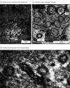
Figure 2.. Visualization of Reticular Changes and Presumed Viral Particles by Transmission Electron Microscopy in Retinal Tissue
A, Sagittal section micrograph of the retinal cells, showing the inner nuclear layer, outer plexiform layer, outer nuclear layer, and the photoreceptor segments. B, Perinuclear region of ganglion cell layer, with reticular changes (inset, white arrowhead) with the presence of presumed viral particles, particles are between 60 and 70 nm (inset). C, Presence of viral particles in the reticulum region, randomly distributed structures presumed to be S1 protein (white arrowheads). Electrodense granularity within the particle was also observed (asterisks).
Figure 3.
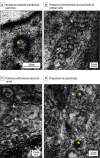
Figure 3.. Presumed Viral Particles by Transmission Electron Microscopy in Retinal Tissue
A, Presence of presumed viral particles in retinal cells, particles between 60 and 70 nm (white arrowhead), with dense electron granulation inside, indicating packaging of genetic material (asterisk). B, Numerous double membrane particles (white arrowhead). C, Presumed viral particles are present in the lumen of the endoplasmic reticulum in the outer nuclear layer of the cells (white arrowhead). D, Particles with dense electron grain inside (arrowhead and asterisk).
Figure 4.
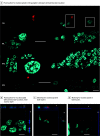
Figure 4.. Presence of Labeling for Nucleocapsid Protein in Neurosensory Retina
A, Punctuation for nucleocapsid (red) in the ganglion cell layer (CGL) with perinuclear location (inset). B, Punctuation (red) for nucleocapsid with perinuclear location in the inner nuclear layer (INL) (inset, x- and y-axis). C and D, Marking for nucleocapsid (red) in both layers, visibly on the x- and y-axis present inside the perinuclear region layer with point marking. Nuclei was stained with Hoescht in cyan. Scale bar: A, 10 nm; inset, 5 nm; B-D, 5 nm. ONL indicates outer nuclear layer.
Figure 5.
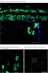
Figure 5.. Presence of Labeling for S1 Protein in the Neurosensory Retina
A, Presence of S1 protein (red) with ganglion cell layer (GCL) distribution, inner plexiform layer, inner nuclear layer (inset, x- and y-axis), outer plexiform layer (OPL), outer nuclear layer (ONL), and both the photoreceptor segments, retinal pigment epithelium (RPE), and choroid (Ch). B, Presence of the S1 protein (red) with distribution in the inner nuclear layer (INL) in the perinuclear region (inset, x- and y-axis). C, There is a positive presence for protein S1 in both INL and ONL, with a more diffuse marking pattern (red) and perinuclear (inset, x- and y-axis). Nuclei was stained with Hoescht in cyan. Scale bar: A, 10 nm; inset, 5 nm; B-C, 5 nm.

Comment in

References

    1. Ke Z, Oton J, Qu K, et al. . Structures and distributions of SARS-CoV-2 spike proteins on intact virions. Nature. 2020;588(7838):498-502. doi:10.1038/s41586-020-2665-2 - DOI - PMC - PubMed
    1. Yao H, Song Y, Chen Y, et al. . Molecular architecture of the SARS-CoV-2 virus. Cell. 2020;183(3):730-738.e13. doi:10.1016/j.cell.2020.09.018 - DOI - PMC - PubMed
    1. Walls AC, Park YJ, Tortorici MA, Wall A, McGuire AT, Veesler D. Structure, function, and antigenicity of the SARS-CoV-2 spike glycoprotein. Cell. 2020;181(2):281-292.e6. doi:10.1016/j.cell.2020.02.058 - DOI - PMC - PubMed
    1. Zhou P, Yang XL, Wang XG, et al. . A pneumonia outbreak associated with a new coronavirus of probable bat origin. Nature. 2020;579(7798):270-273. doi:10.1038/s41586-020-2012-7 - DOI - PMC - PubMed
    1. Kaya H, Çalışkan A, Okul M, Sarı T, Akbudak İH. Detection of SARS-CoV-2 in the tears and conjunctival secretions of coronavirus disease 2019 patients. J Infect Dev Ctries. 2020;14(9):977-981. doi:10.3855/jidc.13224 - DOI - PubMed

Publication types