. 2021 Jul 29;138(4):318-330.
doi: 10.1182/blood.2020008221.

CD70-specific CAR T cells have potent activity against acute myeloid leukemia without HSC toxicity

Affiliations

CD70-specific CAR T cells have potent activity against acute myeloid leukemia without HSC toxicity

Tim Sauer et al. Blood. .

Abstract

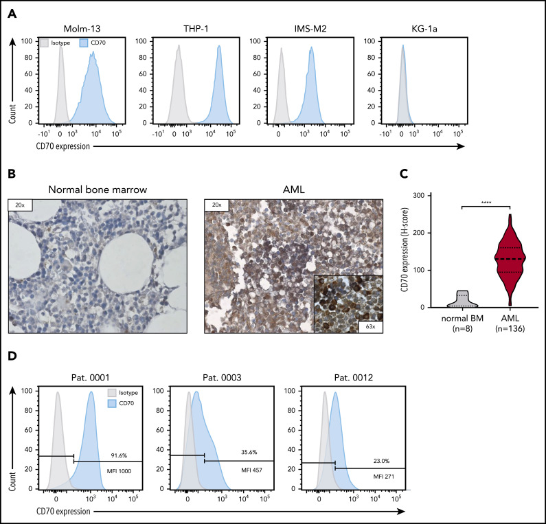
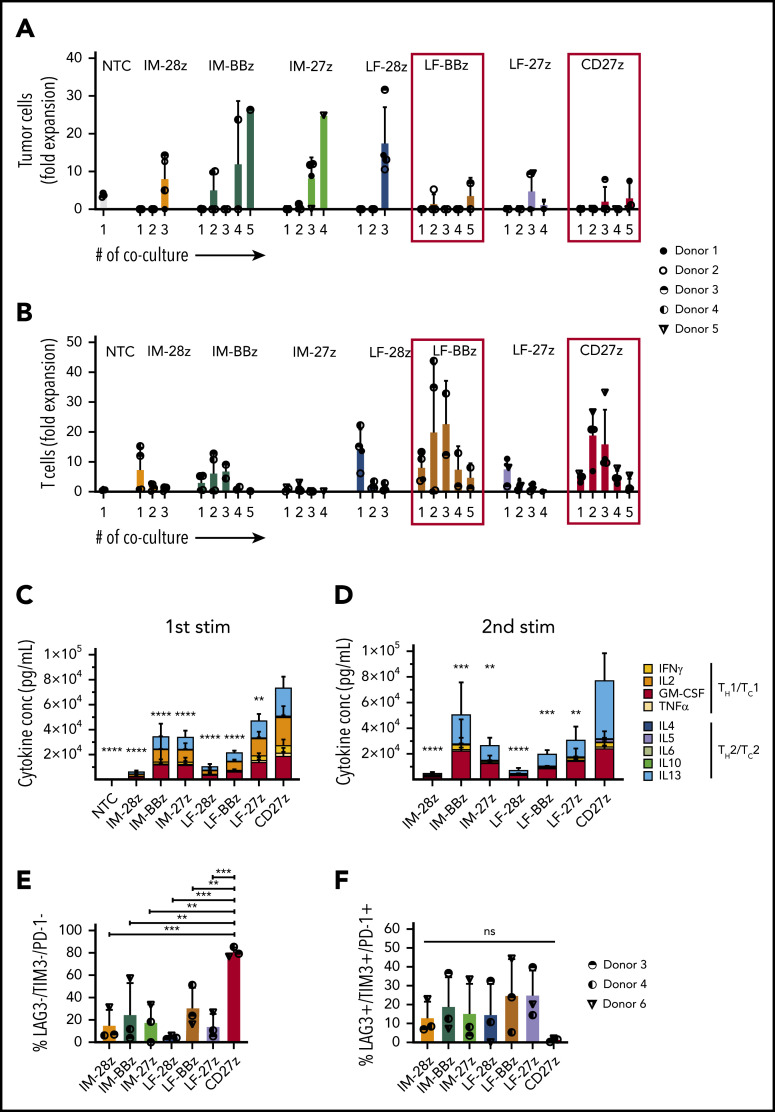
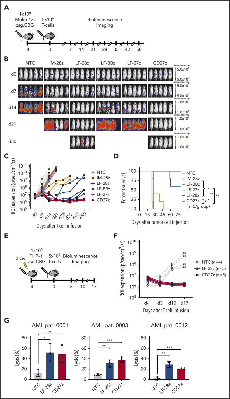
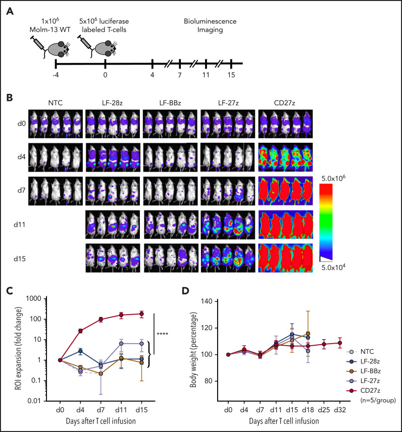
The prognosis of patients with acute myeloid leukemia (AML) remains dismal, highlighting the need for novel innovative treatment strategies. The application of chimeric antigen receptor (CAR) T-cell therapy to patients with AML has been limited, in particular by the lack of a tumor-specific target antigen. CD70 is a promising antigen to target AML, as it is expressed on most leukemic blasts, whereas little or no expression is detectable in normal bone marrow samples. To target CD70 on AML cells, we generated a panel of CD70-CAR T cells that contained a common single-chain variable fragment (scFv) for antigen detection, but differed in size and flexibility of the extracellular spacer and in the transmembrane and the costimulatory domains. These CD70scFv CAR T cells were compared with a CAR construct that contained human CD27, the ligand of CD70 fused to the CD3ζ chain (CD27z). The structural composition of the CAR strongly influenced expression levels, viability, expansion, and cytotoxic capacities of CD70scFv-based CAR T cells, but CD27z-CAR T cells demonstrated superior proliferation and antitumor activity in vitro and in vivo, compared with all CD70scFv-CAR T cells. Although CD70-CAR T cells recognized activated virus-specific T cells (VSTs) that expressed CD70, they did not prevent colony formation by normal hematopoietic stem cells. Thus, CD70-targeted immunotherapy is a promising new treatment strategy for patients with CD70-positive AML that does not affect normal hematopoiesis but will require monitoring of virus-specific T-cell responses.


Figures

None
Graphical abstract
Figure 1.
Figure 1.
CD70 expression of AML cell lines and primary bone marrow samples from patients with AML. (A) Evaluation of CD70 expression in several primary AML cell lines by flow cytometry. (B) Representative images of immunohistochemical staining for CD70 expression of bone marrow samples derived from patients without AML (normal bone marrow) and patients with newly diagnosed AML. Leukemic blasts in patients with AML were identified by their morphology. (C) Comparison of CD70 expression in bone marrow samples of 136 patients with AML and 8 individuals with normal bone marrow findings by immunohistochemical staining. The H-score was used to determine the staining intensity. ****P < .0001, by unpaired Student t test. (D) Analysis of CD70 expression in bone marrow samples of patients with newly diagnosed AML by flow cytometry. Percentage of CD70-expressing cells and median fluorescence intensity (MFI) of the CD70-positive cell population are depicted.
Figure 2.
Figure 2.
Generation and characterization of CD70-CAR T cells. (A) Representative histograms of CD70-CAR expression levels after transduction of activated T cells. CD70scFv-CAR T cells were detected by staining with biotin-labeled protein L, followed by staining with APC-conjugated streptavidin. CD27z-CAR expression was confirmed by detection of a truncated CD19 using an APC-conjugated CD19 specific antibody. (B) The frequency of CD70-CAR expressing T cells of 4 different donors. (C) The frequency of viable cells for CD70-CAR and NT T cells of 4 different donors, as determined by forward vs side scatter gating. (D) Fold change in the number of CD70-CAR T cells in culture during the manufacturing process without antigen stimulation. CAR T cells from 4 different donors were counted in weekly intervals. (E) Phenotypical characterization of CD70-CAR T cells by flow cytometry. The frequency of CD70-CAR T cells with a less (naive/CM T cells; left) and a more (effector memory/terminally differentiated effector memory cells reexpressing CD45RA [EM/EMRA] T cells; right) differentiated phenotype, as determined by the expression of CD45RO and CCR7 in 4 different donors. The colored bars represent the mean of results from 4 different donors, and the error bars indicate the standard deviation (SD). *P < .05; **P < .01; ***P < .001; ****P < .0001; ns, not significant, by unpaired Student t test. (F-G) A chromium-51 release assay was used to determine antigen-dependent lysis of CD70-CAR T cells against CD70-positive AML cell lines (Molm-13, THP-1) (F) and KG-1a (CD70) cells (G) as the control. The graph shows the mean results of 3 technical replicates from 4 different donors, and the error bars indicate the SD. P values were calculated by 2-way ANOVA and are shown in the table below the graphs. *P < .05; **P < .01; ***P < .001; ****P < .0001; ns, not significant.
Figure 3.
Figure 3.
Functional characterization of CD70-CAR T cells after repeated antigen stimulation in vitro. CD70-CAR/NT T cells and AML cells (Molm-13) were cocultured for 5 days, and fresh Molm-13 cells were repeatedly added every 5 days thereafter. The absolute cell count of tumor and T cells at the end of each coculture was determined by flow cytometry with CountBright counting beads. (A) The absolute cell count of Molm-13 cells at the end of each coculture. (B) The absolute cell count of CD70-CAR/NT T cells at the end of each coculture. (C-D) Cytokine production by CD70-CAR and NT T cells after antigen stimulation was determined with the multiplex detection assay. Data show the quantitative determination of TH1/TC1– and TH2/TC2–associated cytokines 24 hours after the first (C) and second (D) antigen stimulation. Data are the mean of results from 4 different donors. P values indicating the statistical significance for secreted cytokines of CD27z-CAR-T cells in comparison with all other constructs and NT T cells were calculated by 2-way ANOVA. (E-F) Expression of LAG-3, TIM-3 and PD-1, markers associated with T-cell exhaustion on CD70-CAR T cells after antigen stimulation. Data are the mean frequency ± standard deviation (SD) of triple-negative (E) and triple-positive (F) T cells from 3 different donors 72 hours after the third antigen stimulation. The error bars indicate the SD. **P < .01; ***P < .001; ****P < .0001; ns, not significant, by unpaired Student t test.
Figure 4.
Figure 4.
Antileukemic activity of CD70-CAR T cells in 2 murine AML xenograft models and against primary AML blast cells. (A) The experimental setup of the Molm-13 xenograft model. CD70-CAR T cells (5 × 10⁶) were injected 4 days after engraftment of luciferase-labeled Molm-13 cells (1 × 10⁶). BLI was performed before T-cell injection on day 0 and weekly thereafter (n = 5 for each treatment group). (B) BLI on the indicated days (after T-cell injection) of mice with Molm-13 cell engraftment and treatment with CD70-CAR or NT T cells. (C) Quantitative analysis of bioluminescence signals for individual mice from each treatment group. (D) Kaplan-Meier survival plot of mice treated with CD70-CAR or NT T cells. The log-rank (Mantel-Cox) test was used to perform statistical analyses of survival between the treatment groups. (E) The experimental setup of the THP-1 xenograft model. After sublethal irradiation, NSG mice underwent injection of 1 × 10⁶ luciferase-labeled THP-1 tumor cells. 4 days later, after engraftment, the mice then received an injection of 5 × 10⁶ CD70-CAR or NT T cells and were subsequently monitored by BLI. (F) Quantitative analysis of bioluminescence signals for individual mice with THP-1 tumor cell engraftment and treatment with LF-28z-, CD27z-CAR, or NT T cells. (G) Cytotoxicity of LF-28z- and CD27z-CAR T cells Ts against AML blasts from 3 patients with AML and various levels of CD70 expression was determined with a chromium-51 release assay. Data are the mean percentage of lysis ± standard deviation at an E:T target ratio of 40:1 from 3 different donors, with 3 technical replicates. *P < .05; **P < .01; ***P < .001, by unpaired Student t test.
Figure 5.
Figure 5.
Expansion and trafficking of CD70-CAR T cells in vivo. (A) The experimental setup. NSG mice underwent AML engraftment with 1 × 10⁶ Molm-13 cells and injection of 5 × 10⁶ CD70-CAR or NT T cells genetically modified to express a luciferase-GFP fusion protein 4 days later. T-cell expansion and trafficking were monitored by BLI on days 4, 7, 11, and 15 (n = 5 for each treatment group). (B) BLI of mice on the indicated days after infusion of CD70-CAR or NT T cells. (C) Quantitative analysis of BLI for each treatment group. Data are the mean ± standard deviation (SD) of 5 animals per treatment group. The area under the curve was calculated for each treatment group and compared by unpaired Student t test; ****P < .0001. (D) The body weight of treated mice was measured as an indicator of their overall condition. The change in body weight compared with the day of T-cell injection for each treatment group is depicted. Data represent the mean of each group ± SD. Results represent pooled data from 5 animals.
Figure 6.
Figure 6.
CD70-CAR T cells eliminate multivirus-specific T cells, but spare normal HSCs. (A-B) CD70 expression on MVSTs was determined by flow cytometry at different time points after stimulation. Bar graphs representing the percentage of CD70-positive cells (A) and the mean fluorescence intensity (MFI) of CD70 expression of MVSTs (B) at the indicated time points. CellTrace Violet–labeled MVSTs were cocultured with autologous CD70-CAR or NT T cells and harvested after 72 hours. The absolute cell count of both T-cell populations was determined by flow cytometry with CountBright counting beads. (C) Representative dot plots for the different T-cell groups are shown, in which autologous MVSTs can be distinguished from CAR or NT T cells by their CellTrace Violet labeling. (D) The total number of MVTSs (left graph) and CAR/NT T cells (right graph) after 3 days of coculture. (E) The percentage of MVSTs with intracellular IFN-γ and TNF-α expression after 3 days of coculture with CD70-CAR or NT T cells, followed by stimulation with EBV-, adenovirus-, and CMV-specific pepmixes. Data are the mean ± standard deviation (SD) of results from 4 different donors. (F) CD70-CAR, CD33-CAR, and NT T cells were cocultured with normal CD34-positive HSCs for 6 hours, and the cells were seeded in a standardized medium for CFU assays. The total number of colonies after the first plating were determined after 2 weeks of incubation. Data are the mean number of colonies formed by HSCs from 2 different donors after incubation with CAR or NT T cells from 3 different donors. The error bars indicate the SD. Two independent investigators counted colonies from 2 technical replicates for each condition. *P < .05; **P < .01; ***P < .001; ****P < .0001; ns, not significant, by unpaired Student t test.

References

    1. Dombret H, Gardin C. An update of current treatments for adult acute myeloid leukemia. Blood. 2016;127(1):53-61. - PMC - PubMed
    1. Dinmohamed AG, Visser O, van Norden Y, et al. . Treatment, trial participation and survival in adult acute myeloid leukemia: a population-based study in the Netherlands, 1989-2012. Leukemia. 2016;30(1):24-31. - PubMed
    1. Thomas D, Majeti R. Biology and relevance of human acute myeloid leukemia stem cells. Blood. 2017;129(12):1577-1585. - PMC - PubMed
    1. Ishikawa F, Yoshida S, Saito Y, et al. . Chemotherapy-resistant human AML stem cells home to and engraft within the bone-marrow endosteal region. Nat Biotechnol. 2007;25(11):1315-1321. - PubMed
    1. Ritchie DS, Neeson PJ, Khot A, et al. . Persistence and efficacy of second generation CAR T cell against the LeY antigen in acute myeloid leukemia. Mol Ther. 2013;21(11):2122-2129. - PMC - PubMed

Publication types

MeSH terms

LinkOut - more resources