. 2021 Oct 27;38(11):4962-4976.
doi: 10.1093/molbev/msab220.

Evolution of Renal-Disease Factor APOL1 Results in Cis and Trans Orientations at the Endoplasmic Reticulum That Both Show Cytotoxic Effects

Affiliations

Evolution of Renal-Disease Factor APOL1 Results in Cis and Trans Orientations at the Endoplasmic Reticulum That Both Show Cytotoxic Effects

Daria Müller et al. Mol Biol Evol. .

Abstract

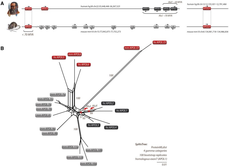
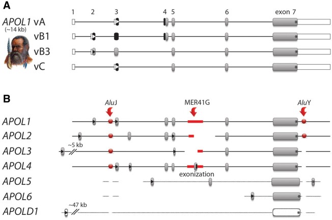
The recent and exclusively in humans and a few other higher primates expressed APOL1 (apolipoprotein L1) gene is linked to African human trypanosomiasis (also known as African sleeping sickness) as well as to different forms of kidney diseases. Whereas APOL1's role as a trypanolytic factor is well established, pathobiological mechanisms explaining its cytotoxicity in renal cells remain unclear. In this study, we compared the APOL family members using a combination of evolutionary studies and cell biological experiments to detect unique features causal for APOL1 nephrotoxic effects. We investigated available primate and mouse genome and transcriptome data to apply comparative phylogenetic and maximum likelihood selection analyses. We suggest that the APOL gene family evolved early in vertebrates and initial splitting occurred in ancestral mammals. Diversification and differentiation of functional domains continued in primates, including developing the two members APOL1 and APOL2. Their close relationship could be diagnosed by sequence similarity and a shared ancestral insertion of an AluY transposable element. Live-cell imaging analyses showed that both expressed proteins show a strong preference to localize at the endoplasmic reticulum (ER). However, glycosylation and secretion assays revealed that-unlike APOL2-APOL1 membrane insertion or association occurs in different orientations at the ER, with the disease-associated mutants facing either the luminal (cis) or cytoplasmic (trans) side of the ER. The various pools of APOL1 at the ER offer a novel perspective in explaining the broad spectrum of its observed toxic effects.

Keywords: APOL gene family, APOL phylogeny, APOL selection analyses; APOL1; APOL2; endoplasmic reticulum; evolutionary medicine; kidney disease.


Figures

Fig. 1.
Fig. 1.
Local APOL gene family and SplitsTree reconstructions for humans and mice. (A) Genomic location of APOL genes from human and mouse with genomic coordinates. Arrows indicate gene orientations. APOLD1 evolved early in vertebrates about 300 Ma, whereas APOL6 was probably inherited by a human–mouse ancestor that lived about 70 Ma. (B) SplitsTree reconstruction of APOL genes based on protein sequences. A similar tree topology was derived by maximum likelihood and Bayesian tree reconstructions (supplementary data SD1, Supplementary Material online). The APOL genes of mice (mm for Mus musculus) are indicated in gray boxes. Black boxes represent human APOL genes (hs for Homo sapiens). The central parallelograms of the reconstruction represent conflicting phylogenetic signals. The tree reconstruction reveals a common origin of mm and hs APOL6 (red boxes). Bootstrap values are shown for representative branches. Balls indicate the clade-supporting integrations of AluJ and AluY elements. The human APOL2 and APOL1 genes share an orthologous AluY element. The orthologous AluY transposons were already present in Catharrine primates (diverged about 30 Ma). The diagnostic AluJ elements were detected at orthologous positions in APOL1-4 in all investigated primates and were probably inherited from a common primate ancestor of about 70 Ma. The double-slash in the mouse locus indicates the exclusion of a large genomic region. Human and mouse APOLD1 are located on chromosomes 12 and 6, respectively, and diverged significantly from other APOL genes.
Fig. 2.
Fig. 2.
APOL1 isoforms and related APOL gene members. (A) Gene structures of the four well-characterized APOL1 isoforms vA, vB1, vB3, and vC (Khatua et al. 2015). Exons for APOL1 vA (gene size ∼14 kb) are labeled 1–7. White frames indicate UTRs. Black boxes represent leading SPs, gray boxes the remaining protein-coding exons, and black lines the intronic regions. Start codons are indicated by triangles and stop codons by asterisks. (B) Cumulative gene structures of all human APOL gene members and isoforms. Red arrows indicate the insertion points of three diagnostic transposable elements (AluJ merging APOL1-4, MER41G representing alternative exon insertion/exonization in APOL4, AluY indicating a common ancestry of APOL1-2). Highly diverged sequences are shown by dotted lines (annotated alignments are provided as supplementary data SD4, Supplementary Material online).
Fig. 3.
Fig. 3.
APOL1–APOL2 gene diversification is accompanied by organ-specific expression. (A) Expression levels of APOL1 (L1, green) and APOL2 (L2, orange) transcriptomes (expressed as RPKM-normalized counts) during human organ development derived from the Evo-devo database. White background indicates embryonic or prenatal stages and gray background postnatal stages of mammalian organ development (supplementary data SD20, Supplementary Material online).
Fig. 4.
Fig. 4.
APOL1 splice variants and APOL2 are targeted to the ER. (A) Schemes of C-terminally GFP-tagged APOL1 splice variants, GFP-APOL1 (lacking the SP, ΔSP, aa1-27), and N-terminally GFP- or RFP-tagged APOL2. Live-cell imaging of AB8 podocytes, expressing APOL1-GFP splice variants vA, vB1, vB3 and vC (BE), GFP-APOL2 (F), or GFP-tagged APOL1 together with RFP-APOL2 (G). ER membranes are visualized with live-cell imaging dye ER Tacker. Scale bars: 20 µm.
Fig. 5.
Fig. 5.
Different ER insertion orientations accompany APOL1–APOL2 diversification. (A) The endogenous SP of the reporter enzyme SEAP (secreted alkaline phosphatase, gray box) was replaced by the N-termini of the APOL1 splice variants (vA, vB1, vB3, vC). The endogenous SP of SEAP (SPSEAP) and the well-known SP of human serum albumin (SPHSA) served as positive, and SEAP lacking its SP (ΔSP) as negative control. (B) SEAP secretion assay in HEK293T cells. Fluorescent SEAP secretion was detectable for positive controls (SPSEAP and SPHSA) and splice variants vA, vB1, and vC. SEAP with the N-termini of APOL1 vB3 and without the SEAP SP (ΔSP) were not secreted (***P < 0.0001). (C) Scheme: APOL1 splice variants vA, vB1, vB3, vC, a mutant lacking the complete APOL1 N-terminus (aa1-59; ΔN59), and APOL2 were fused with an artificial C-terminal N-glycosylation tag (GT). (D) Western blot analyses of Glyco-tag (GT) APOL proteins (shown in C) before and after PNGaseF glycosidase digestion showed N-glycosylation and therefore ER luminal localization only for APOL1 vA, vB1, and vC (marked by black asterisks). (E) Scheme: C-terminally GFP-tagged APOL1 (G0 and renal risk variants G1 and G2), APOL1 lacking C-terminal aa305–398 (ΔC305), and APOL2. (F) Western blot analysis of these GFP-GT-tagged proteins (shown in E) before and after PNGaseF glycosidase digestion demonstrates luminal ER localization for these proteins (asterisks), except for APOL2-GFP-GT.
Fig. 6.
Fig. 6.
APOL1 cytotoxicity is caused by cis- and trans-orientated APOL1 pools. (A) Schemes of APOL1 variants used for cell viability assays. Left panel: We used APOL1 G0, and renal risk variants G1 and G2 combined with C-terminal GFP (APOL1-GFP, vA aa1–398), and APOL1 fusion proteins (G0, G1, and G2) in which the APOL1 SP was replaced by an N-terminal GFP-tag (GFP-APOL1 ΔSP; aa28–398). Right panel: To analyze the impact of the putative pro-apoptotic “BH3-only motif” (BSM) in APOL1 pools at the outer ER membrane (trans orientation), we replaced the BSM of GFP-APOL1 by either an Alanine stretch (B6xAla) or the BSM of APOL2 (B2). GFP-tagged APOL2 was used as a control. (B) Cell viability assay of stable HEK293T cell lines expressing proteins summarized in A: APOL1-expressing cell lines, particularly those with RRVs, exhibited cytotoxic effects in cis and trans orientations. Toxic GFP-APOL1 (ΔSP, G2) in which the BSM was replaced by Alanine (GFP-APOL1B6xAla) or the BSM of APOL2 (GFP-APOL1 B2) and GFP-APOL2 showed no cytotoxicity (***P < 0.0001).
Fig. 7.
Fig. 7.
APOL1 vA gene structure and regions with positively selected sites. (A) Amino acids (single letter code) and coordinates mark positively selected sites for APOL1–4 derived from at least two of three PAML models (M2/M8/BS) as large stars. The positively selected sites found by Smith and Malik (2009) are indicated for APOL1–2, APOL3–4, and APOL6. Red labeled letters show overlapping sites of different studies. The APOL1 variation of alternative amino acids or deletion sites (Δ) are shown above the exon structure (gray). APOL1 was previously divided into four functional regions, an SP, a PFD, an MAD, and the SID (green boxes). The PFD also contains a putative pro-apoptotic BH3-only domain sequence motif (BSM). More recent conceptions suggest an SP (gray bar) and up to four TMs (SP, gray and TM1-4, black bars with their coordinates indicated as numbers). The first two TMs are also referred to as membrane insertion domain (MID) or helix–turn–helix region (H-L-H). The TM4 overlaps with a pore-lining region (PLR), followed by leucine-zipper domain (ZIP). (B) Model of possible orientations of APOL1 (and APOL2) at the ER. The presence or absence of the SP determines the orientation of APOL1 splice variants and of APOL2 at the ER. Functional SPs result in a luminal (cis) ER localization of the APOL1 C-terminus. If APOL1 functions as an ion pore, this most likely requires two (I), “two and a half” (II), or four (III) TMs (black boxes). During evolution, positively selected amino acids accumulate in regions within, or close to, the putative TM3 and TM4 (red arrows). APOL1 vB3 and APOL2, that lack functional SPs (IV), are most likely bound to the cytoplasmic leaflet of the ER membrane (trans). Since cis as well as trans pools of APOL1 show cytotoxic effects—that are pronounced in the case of renal risk variants—our data suggest the presence of different cellular pathomechanisms.

References

    1. Adams JM, Cory S. 2018. The BCL-2 arbiters of apoptosis and their growing role as cancer targets. Cell Death Differ. 25(1):27–36. - PMC - PubMed
    1. Almagro Armenteros JJ, Tsirigos KD, S�by CK, Petersen T N, Winther O, Brunak S, von Heijne G, Nielsen H. 2019. SignalP 5.0 improves signal peptide predictions using deep neural networks. Nat Biotechnol. 37(4):420–423. - PubMed
    1. Beckerman P, Bi-Karchin J, Park ASD, Qiu C, Dummer PD, Soomro I, Boustany-Kari CM, Pullen SS, Miner JH, Hu C-AA, et al.2017. Transgenic expression of human APOL1 risk variants in podocytes induces kidney disease in mice. Nat Med. 23(4):429–438. - PMC - PubMed
    1. Bruggeman LA, O’Toole JF, Ross MD, Madhavan SM, Smurzynski M, Wu K, Bosch RJ, Gupta S, Pollak MR, Sedor JR, et al.2014. Plasma apolipoprotein L1 levels do not correlate with CKD. J Am Soc Nephrol. 25(3):634–644. - PMC - PubMed
    1. Bruno J, Pozzi N, Oliva J, Edwards JC.. 2017. Apolipoprotein L1 confers pH-switchable ion permeability to phospholipid vesicles. J Biol Chem. 292(44):18344–18353. - PMC - PubMed

Publication types