. 2022 Feb 17;29(2):339-350.e10.
doi: 10.1016/j.chembiol.2021.07.005. Epub 2021 Jul 28.

DogCatcher allows loop-friendly protein-protein ligation

Affiliations

DogCatcher allows loop-friendly protein-protein ligation

Anthony H Keeble et al. Cell Chem Biol. .

Abstract

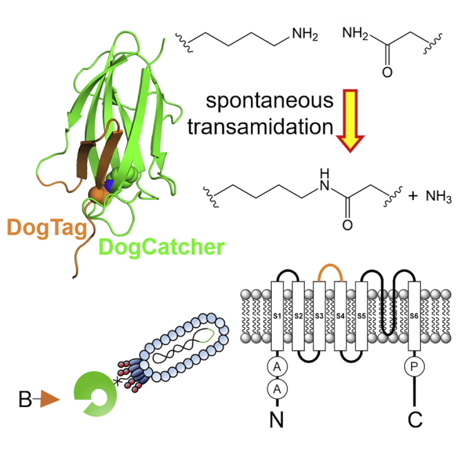
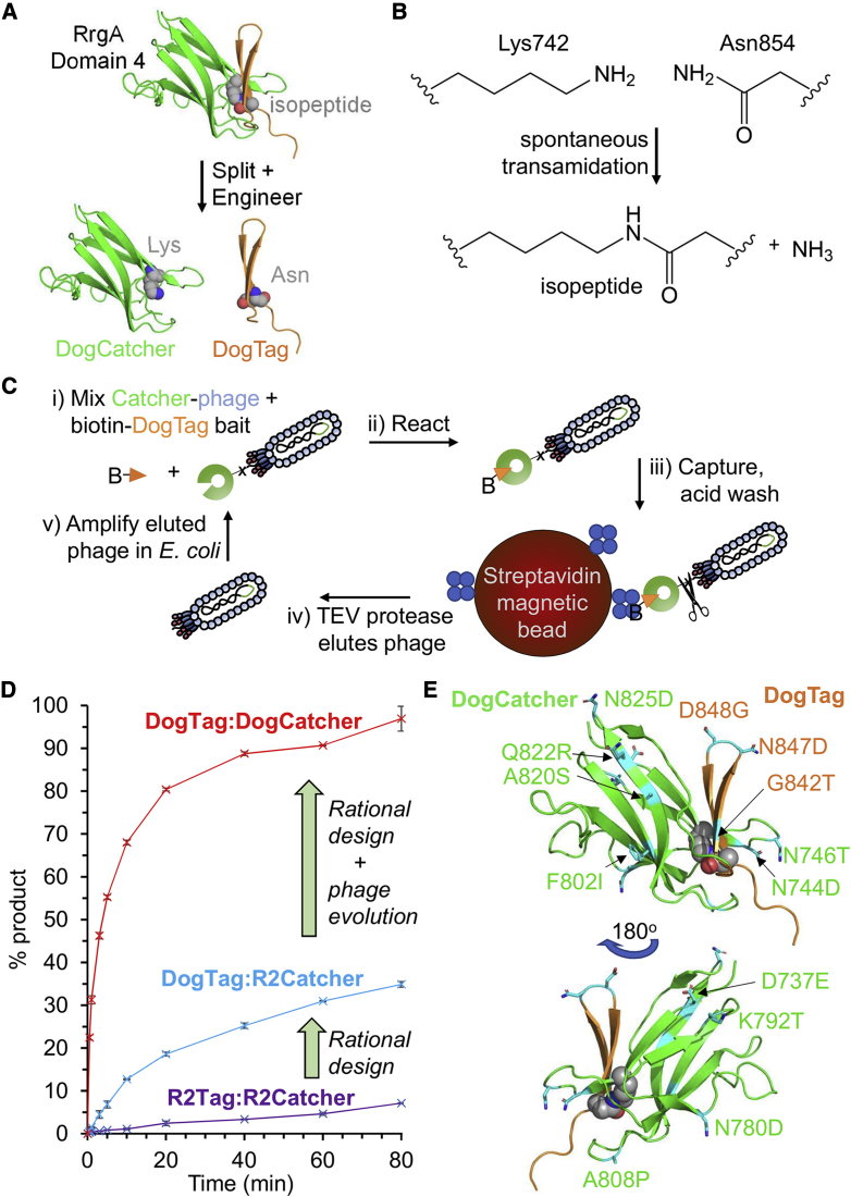
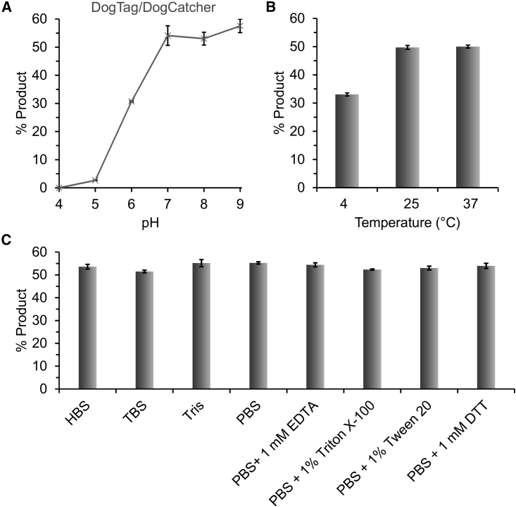
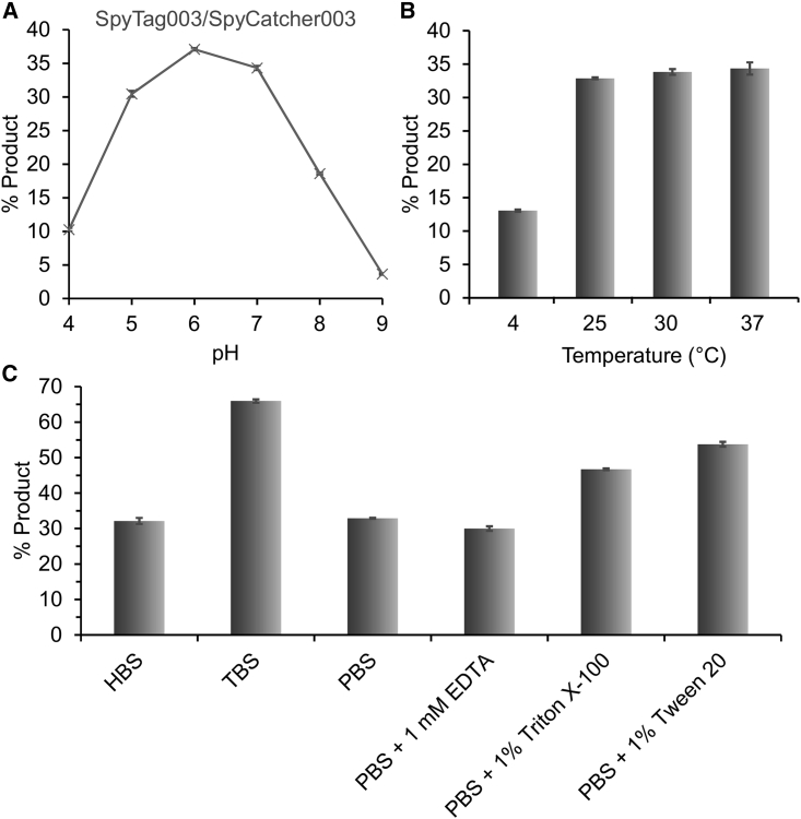
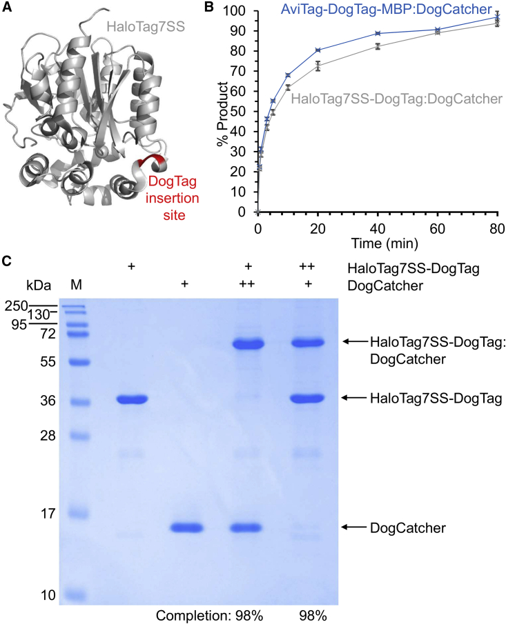
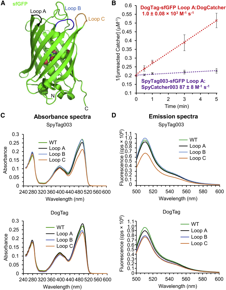
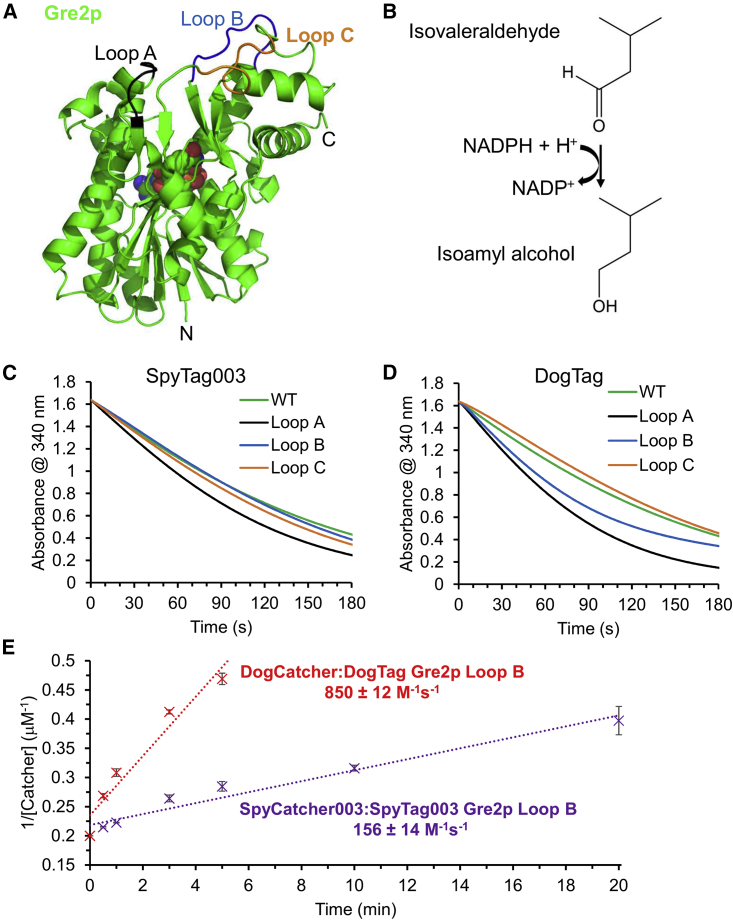
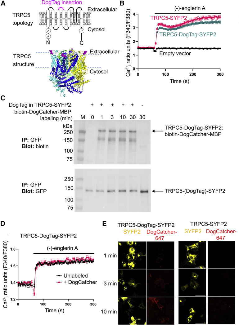
There are many efficient ways to connect proteins at termini. However, connecting at a loop is difficult because of lower flexibility and variable environment. Here, we have developed DogCatcher, a protein that forms a spontaneous isopeptide bond with DogTag peptide. DogTag/DogCatcher was generated initially by splitting a Streptococcus pneumoniae adhesin. We optimized DogTag/DogCatcher through rational design and evolution, increasing reaction rate by 250-fold and establishing millimolar solubility of DogCatcher. When fused to a protein terminus, DogTag/DogCatcher reacts slower than SpyTag003/SpyCatcher003. However, inserted in loops of a fluorescent protein or enzyme, DogTag reacts much faster than SpyTag003. Like many membrane proteins, the ion channel TRPC5 has no surface-exposed termini. DogTag in a TRPC5 extracellular loop allowed normal calcium flux and specific covalent labeling on cells in 1 min. DogTag/DogCatcher reacts under diverse conditions, at nanomolar concentrations, and to 98% conversion. Loop-friendly ligation should expand the toolbox for creating protein architectures.

Keywords: SpyTag; TRPC; bioconjugation; chemical biology; epitope tag; ion channel; protein design; protein engineering; split protein; synthetic biology.


Conflict of interest statement

Declaration of interests M.H. and J.H. are authors on a patent covering RrgA splitting (UK Intellectual Property Office 1509782.7). M.H. is an author on a patent covering DogTag and SnoopLigase (UK Intellectual Property Office 1705750.6). M.H. and A.H.K. are authors on a patent application for SpyTag003/SpyCatcher003 (UK Intellectual Property Office, 1903479.2). M.H., A.H.K., M.P.F., and V.K.Y. are authors on a patent application related to DogCatcher (UK Intellectual Property Office 2104999.4.). M.H. is a SpyBiotech co-founder, shareholder, and consultant. All other authors have no conflicts of interest.

Figures

None
Graphical abstract
Figure 1
Figure 1
Splitting and engineering to create DogTag/DogCatcher (A) Domain splitting. RrgA domain 4 contains an intramolecular isopeptide bond (shown as spheres; schematic based on PDB: 2WW8). The domain was genetically split to create R2Catcher and a β hairpin called R2Tag, which, after further engineering, became DogCatcher and DogTag. (B) Chemistry of amide bond formation between Lys742 of DogCatcher and Asn854 of DogTag. (C) Phage display evolution of DogCatcher. A library of error-prone Catcher variants was displayed on M13 phage pIII and panned for rapid covalent bond formation to DogTag bait linked to biotin (B). (D) Enhancement of reaction speed. Amide bond formation rate for R2Tag/R2Catcher (purple trace), DogTag/R2Catcher (cyan trace), or DogTag/DogCatcher (red trace) in PBS (pH 7.5) at 25°C with 5 μM of each protein. Mean ± 1 SD, n = 3 based on SDS-PAGE densitometry. Some error bars are too small to be visible. (E) Mapping of the mutations (cyan) engineered into RrgA domain 4 (PDB: 2WW8) to create DogCatcher (green) and DogTag (orange). A second view is shown with 180° rotation to illustrate residues on the opposite face.
Figure 2
Figure 2
Condition dependence of DogTag/DogCatcher reactivity (A) pH dependence: 2 μM AviTag-DogTag-MBP and 2 μM DogCatcher were reacted for 30 min at 25°C in SPG buffer at the indicated pH. (B) Temperature dependence: 2 μM AviTag-DogTag-MBP and 2 μM DogCatcher were reacted for 30 min in SPG, pH 7.0, at the indicated temperature. (C) Buffer dependence: 5 μM AviTag-DogTag-MBP and 5 μM DogCatcher were reacted for 5 min at 25°C, pH 7.5, in the indicated buffer. HBS, HEPES-buffered saline; TBS, Tris-buffered saline. Mean ± 1 SD, n = 3; some error bars are too small to be visible.
Figure 3
Figure 3
Condition dependence of SpyTag003/SpyCatcher003 reactivity (A) pH dependence: 1 μM SpyTag003-MBP and 1 μM SpyCatcher003 were reacted for 15 s at 25°C in SPG buffer at the indicated pH. (B) Temperature dependence: 100 nM SpyTag003-MBP was reacted with 100 nM SpyCatcher003-sfGFP for 2 min, pH 7.4, at the indicated temperature. (C) Buffer dependence: 100 nM SpyTag003-MBP was reacted with 100 nM SpyCatcher003-sfGFP for 2 min in the indicated buffer, pH 7.5, at 25°C. HBS, HEPES-buffered saline; TBS, Tris-buffered saline. Mean ± 1 SD, n = 3; some error bars are too small to be visible.
Figure 4
Figure 4
DogTag/DogCatcher reacted close to completion when DogTag was internal (A) Site of DogTag insertion in red in HaloTag7 (gray, PDB: 5Y2Y). (B) DogCatcher reaction rate with the internal DogTag in HaloTag7SS (gray trace) was similar to the unconstrained DogTag in AviTag-DogTag-MBP (blue trace). Each protein was at 5 μM in PBS, pH 7.5, at 25°C. Mean ± 1 SD, n = 3; some error bars are too small to be visible. (C) Testing DogTag/DogCatcher reaction to completion. DogCatcher was incubated with HaloTag7SS-DogTag in PBS, pH 7.5, for 200 min at 25°C, before SDS-PAGE with Coomassie staining. +, 10 μM; ++, 20 μM; M, molecular weight markers. 98% loss was seen for HaloTag7SS-DogTag in the presence of excess DogCatcher, based on densitometry, or for DogCatcher in the presence of excess HaloTag7SS-DogTag.
Figure 5
Figure 5
DogTag functioned well within the β barrel domain of sfGFP and reacted faster than SpyTag003 (A) Structure of sfGFP (PDB: 2B3P) showing the three loops chosen for tag insertion. (B) Second-order reaction plot comparing the reaction speed of DogCatcher with DogTag in sfGFP loop A (red trace), relative to SpyCatcher003 reaction with SpyTag003 (purple trace) in PBS, pH 7.5, at 25°C. Mean ± 1 SD, n = 3. Some error bars are too small to be visible. (C) Comparison of the absorbance spectra of sfGFP (WT) or variants with SpyTag003 or DogTag at the indicated loop. (D) Comparison of the fluorescence emission of sfGFP (WT) or variants with SpyTag003 or DogTag at the indicated loop upon excitation at 488 nm. cps, counts per second.
Figure 6
Figure 6
Tag reactivity and enzyme activity after loop insertion (A) Structure of Gre2p showing the three loops chosen for tag insertion (PDB: 4PVD). NADPH is shown as spheres. (B) Schematic of the reaction catalyzed by Gre2p. (C) SpyTag003 loop insertion had little effect on enzyme activity. Comparison of isovaleraldehyde reductase activity of the Gre2p variants, assayed by the decrease in absorbance at 340 nm as NADPH is converted into NADP+. Data represent the mean of three biological replicates. (D) DogTag loop insertion had little effect on enzyme activity, assayed as in (C). Data represent the mean of three biological replicates. (E) DogTag/DogCatcher (red trace) reacted faster than SpyTag003/SpyCatcher003 (purple trace) in loop B of Gre2p. Second-order reaction plot in PBS, pH 7.5, at 25°C. Mean ± 1 SD, n = 3. Some error bars are too small to be visible.
Figure 7
Figure 7
Specific targeting of an ion channel using DogTag/DogCatcher (A) Schematic of TRPC5, with the insertion site (pink) of DogTag in the second extracellular loop marked on a topology diagram and a crystal structure (PDB: 6YSN, each chain of the tetramer in a different color). A, ankyrin repeat domain; P, TRP domain. (B) DogTag insertion had minimal effect on ion channel opening. Representative intracellular calcium measurements (Ca2+i) from one 96-well plate (mean ± 1 SE, n = 4) showing activation of TRPC5-SYFP2 (red trace) or TRPC5-DogTag-SYFP2 (teal trace) in HEK 293 cells by 30 nM (−)-englerin A (present during the period marked with a horizontal line). No calcium response was induced by (−)-englerin A in empty vector-transfected cells (black trace). (C) Rapid labeling by DogCatcher at the cell surface. COS-7 cells expressing TRPC5-DogTag-SYFP2 or TRPC5-SYFP2 control were incubated with 5 μM biotin-DogCatcher-MBP for the indicated time at 25°C. Cell lysates were immunoprecipitated with GFP-Trap before blotting for either biotin (top panel) or fluorescent protein (bottom panel). (D) DogCatcher reaction had minimal effect on ion channel opening. Representative intracellular calcium measurements (Ca2+i) from one 96-well plate (mean ± 1 SE, n = 6) showing activation of TRPC5-DogTag-SYFP2 in HEK293 cells by 10 nM (−)-englerin A (present during the period marked with a horizontal line), with (red trace) or without (black trace) 30 min pre-treatment with 5 μM biotin-DogCatcher-MBP. (E) DogCatcher labeled specifically at the cell surface: 5 μM DogCatcher-647 was incubated for varying times at 25°C with live COS-7 cells expressing TRPC5-DogTag-SYFP2 or TRPC5-SYFP2, before fixation and confocal microscopy. Images represent confocal slices, with SYFP2 in yellow and DogCatcher-647 in red. Scale bar, 50 μm.

References

    1. Akbulut Y., Gaunt H.J., Muraki K., Ludlow M.J., Amer M.S., Bruns A., Vasudev N.S., Radtke L., Willot M., Hahn S., et al. (–)-Englerin A is a potent and selective activator of TRPC4 and TRPC5 calcium channels. Angew. Chem. 2015;54:3787–3791. - PMC - PubMed
    1. Akiba H., Takayanagi K., Kusano-Arai O., Iwanari H., Hamakubo T., Tsumoto K. Generation of biparatopic antibody through two-step targeting of fragment antibodies on antigen using SpyTag and SpyCatcher. Biotechnol. Rep. 2020;25:e00418. - PMC - PubMed
    1. Anderson P.A., Greenberg R.M. Phylogeny of ion channels: clues to structure and function. Comp. Biochem. Physiol. B Biochem. Mol. Biol. 2001;129:17–28. - PubMed
    1. Bae Y., Lee S.K., Chae Y.C., Park C.Y., Kang S. Accessibility-dependent topology studies of membrane proteins using a SpyTag/SpyCatcher protein-ligation system. Int. J. Biol. Macromol. 2021;175:171–178. - PubMed
    1. Banerjee A., Howarth M. Nanoteamwork: covalent protein assembly beyond duets towards protein ensembles and orchestras. Curr. Opin. Biotechnol. 2018;51:16–23. - PubMed

Publication types