. 2021 Sep 15;41(37):7712-7726.
doi: 10.1523/JNEUROSCI.0440-21.2021. Epub 2021 Jul 29.

Intercellular Arc Signaling Regulates Vasodilation

Affiliations

Intercellular Arc Signaling Regulates Vasodilation

June Bryan de la Peña et al. J Neurosci. .

Abstract

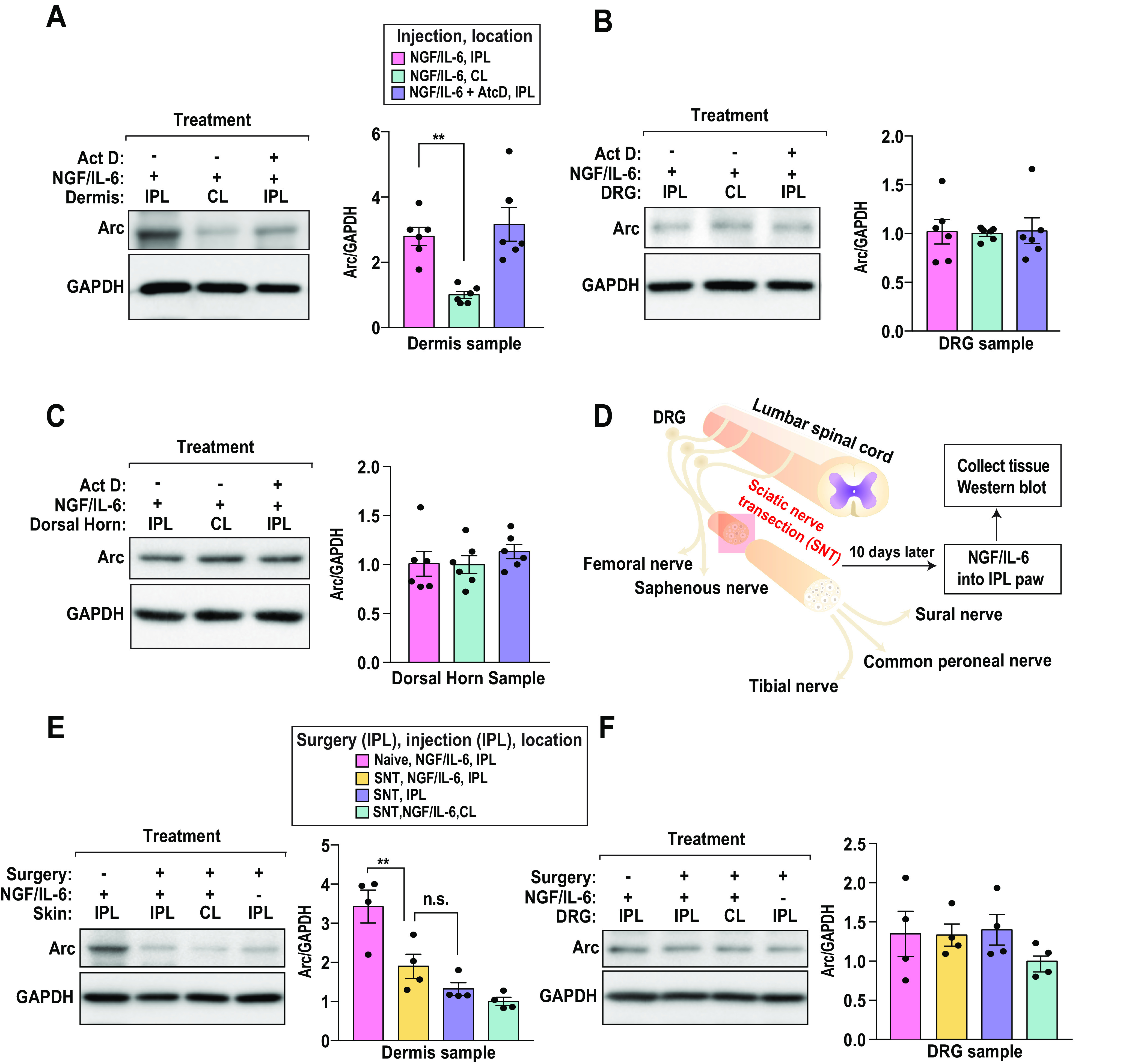
Injury responses require communication between different cell types in the skin. Sensory neurons contribute to inflammation and can secrete signaling molecules that affect non-neuronal cells. Despite the pervasive role of translational regulation in nociception, the contribution of activity-dependent protein synthesis to inflammation is not well understood. To address this problem, we examined the landscape of nascent translation in murine dorsal root ganglion (DRG) neurons treated with inflammatory mediators using ribosome profiling. We identified the activity-dependent gene, Arc, as a target of translation in vitro and in vivo Inflammatory cues promote local translation of Arc in the skin. Arc-deficient male mice display exaggerated paw temperatures and vasodilation in response to an inflammatory challenge. Since Arc has recently been shown to be released from neurons in extracellular vesicles (EVs), we hypothesized that intercellular Arc signaling regulates the inflammatory response in skin. We found that the excessive thermal responses and vasodilation observed in Arc defective mice are rescued by injection of Arc-containing EVs into the skin. Our findings suggest that activity-dependent production of Arc in afferent fibers regulates neurogenic inflammation potentially through intercellular signaling.SIGNIFICANCE STATEMENT Nociceptors play prominent roles in pain and inflammation. We examined rapid changes in the landscape of nascent translation in cultured dorsal root ganglia (DRGs) treated with a combination of inflammatory mediators using ribosome profiling. We identified several hundred transcripts subject to rapid preferential translation. Among them is the immediate early gene (IEG) Arc. We provide evidence that Arc is translated in afferent fibers in the skin. Arc-deficient mice display several signs of exaggerated inflammation which is normalized on injection of Arc containing extracellular vesicles (EVs). Our work suggests that noxious cues can trigger Arc production by nociceptors which in turn constrains neurogenic inflammation in the skin.

Keywords: Arc; DRG; neuroinflamation; nociceptors; translational control.


Figures

Figure 1.
Figure 1.
Ribosome profiling identifies Arc as a rapidly-translated gene. A, Schematic representation of the ribosome profiling approach applied to DRG neurons. B, Intraexperimental correlations for ribosome profiling in the inflammatory mediator treatment group. A list of genes with significant differences is translation is provided in Extended Data Figure 1-1. C, Intraexperimental correlations for RNA-Seq in the inflammatory mediator treatment group. R values correspond to Pearson's correlations in B, C. Transcripts with significant changes in abundance are indicated in Extended Data Figure 1-2. D, Aggregate sites of ribosome protected footprints in 5′ UTRs, coding sequences, and 3′ UTRs. E, A volcano plot depicting changes in translation (log2 ≥ –1.5; false discovery rate < 0.05) across the transcriptome. The ratio of ribosome density following treatment with the inflammatory mediators divided by the vehicle levels is plotted against sample dispersion. Gene density is shaded according to the inset bar; p values for Arc were determined using a two-tailed Student's t test. F, Gene ontology analysis of genes with significant translational enhancement was conducted with Enrichr (Kuleshov et al., 2016). G, Traces of the 5′ end of Arc. Scales are indicated with bars. H, Application of NGF/IL-6 to primary culture DRG neurons upregulated Arc protein in a time course of 0–120 min; n = 9 for 0- and 20 min groups, n = 3 for 60- and 120 min groups. Ordinary one-way ANOVA: F(3,20) = 95.79, p < 0.0001. Dunnett's multiple comparisons test: 0 versus 20 min, ***p < 0.001; 0 versus 120 min, ***p < 0.001.
Figure 2.
Figure 2.
Bioinformatic analysis of differentially translated genes. A, Phase of ribosome protected footprints. B, To be considered a gene with a significant change in abundance or translation, a 1.5-fold (50% upregulated, or 33% downregulated) change and a significance value of <0.05 was required (Student's t test, two-tailed, assuming unequal variances between the two groups tested). C, Comparative analysis of UTR length. D, GC bias in 5′ UTRs. E, A pyrimidine-rich translational element (PRTE) was detected with MEME (E value 4.1 × 10−58; 1). F, A core PRTE is significantly enriched in preferentially translated 5′ UTRs; p value estimate from Fisher's exact test.
Figure 3.
Figure 3.
Arc is constitutively expressed in peripheral and spinal sites important for nociceptive signaling. A, Single DRG neuron-sequencing tSNE clusters showing expression of Arc with the light neurofilament expressed in large diameter neurons (Nefl) and across DRG sensory neurons of peptidergic; Calca (CGRP) and non-peptidergic [P2x3 (P2X3)] subclusters (Li et al., 2016). B, In situ hybridizations of Arc and markers for L4–L5 DRGs together with large diameter neurons expressing neurofilament protein NF200 (NF200, blue), Calca (CGRP, green), and neurons expressing P2x3 (P2X3 red). Validation for negative control probes is provided in Extended Data Figure 3-1. C, Quantification of Arc expression in the DRG. Expression of Arc is greatest in NF200 expressing large diameter neurons. Intraplantar administration of the inflammatory mediators increased Arc mRNA in ipsilateral (IPL) DRGs expressing Calca (CGRP) mRNA; n = 3 animals per group; multiple slices from L4–L5 DRGs. Two-way ANOVA: cell-type factor, F(2,55) = 35.80, p < 0.0001; treatment factor, F(1,55) = 4.545, p = 0.0375. Bonferroni's multiple comparisons test: NF200 versus CGRP, ***p < 0.0001; NF200 versus P2XR, ***p < 0.0001; CGRP-IPL versus CGRP-CL, *p = 0.0113. D, In situ hybridization of Arc in the dorsal horn of the spinal cord (Lamina I–VI). E, Quantification of D. Arc expression is highest in Laminal Layer II. Intraplantar administration of inflammatory mediators did not significantly impact arc mRNA expression.
Figure 4.
Figure 4.
Expression of eGFP-Arc. A, eGFP-Arc is ubiquitously expressed in the DRG and co-localizes with peripherin expressing neurons. B, eGFP-Arc is expressed in large diameter DRG neurons that express NF200 in the DRG. C, Expression patterns in the sciatic nerve for eGFP-Arc and NF200. D, eGFP-Arc is expressed in the glabrous skin and co-localizes with CGRP-expressing fibers. E, eGFP-Arc is expressed in the glabrous skin and co-localizes with β-III tubulin-expressing fibers. Arrowheads indicate colocalization.
Figure 5.
Figure 5.
Induced biosynthesis of Arc in the skin depends on the presence of intact primary afferents. A, Intraplantar administration of inflammatory mediators in the ipsilateral (IPL) paw of mice triggered a rapid translation of arc in skin that is not modified by the transcription inhibitor Act D (300 ng/25 μl); n = 6 animals per group. Ordinary one-way ANOVA: F(2,15) = 11.33, p = 0.0010. Bonferroni's multiple comparisons test: NGF/IL-6, IPL versus NGF/IL-6, CL; **p = 0.0022. IPL, ipsilateral or injected side; CL, contralateral or uninjected side. B, Intraplantar administration of inflammatory mediators, in the presence or absence of Act D (300 ng/25 μl), did not alter Arc abundance in the DRG. C, Intraplantar administration of inflammatory mediators, in the presence or absence of Act D (300 ng/25 μl), did not modify Arc abundance in the spinal dorsal horn (SDH). D, A cartoon detailing the experimental protocol for sciatic nerve transection (SNT). Sciatic nerve was axotomized, and 10 d later, animals received an intraplantar injection of the inflammatory mediators. One hour after injection, Arc protein expression was analyzed in DRG and glabrous skin from injected (ipsilateral, IPL) and noninjected (contralateral, CL) paws. E, Increase in Arc abundance in the skin caused by intraplantar administration of inflammatory mediators requires intact primary afferent fibers; n = 4 animals per group. Ordinary one-way ANOVA: F(3,12) = 15.14, p = 0.0002. Tukey's multiple comparisons test: naive, NGF/IL-6, IPL versus SNT, NGF/IL-6, IPL; **p = 0.0094. n.s., non-significant. F, SNT surgery transection did not alter Arc abundance in the DRG.
Figure 6.
Figure 6.
Sensory neurons produce Arc-containing EVs. A, left panel, Cryo-EM image of EVs purified from untransfected cells. Right panel, Closeup of region with a membrane bilayer bound vesicle (white arrow). B, left panel, Cryo-EM image of EVs purified from cells transfected with Arc. Right panel, Closeup on three types of vesicles that increase in abundance. After transfection the most abundant vesicle type are single membrane vesicles (gray arrow). Low-density lipoprotein (LDL)-like vesicles (black arrow), which have a characteristic striped pattern because of stacked cholesteryl esters, and bilayered vesicles (white arrow) increase in abundance as well. C, left panel, Average number of observed vesicles per image. A total of 20 consecutive images for each sample were evaluated. The number of EVs/image increases ∼3.9-fold for the Arc transfected sample. Unpaired t test with Welch's correction; ***p = 0.0003. Size distributions are provided in Extended Data Figure 6-1. D, Immunoblot analysis of vesicles isolated from cells transfected with Arc are enriched for Arc, Actin, GAPDH, and vesicular markers. E, qPCR quantification of Arc mRNA reveals that Arc overexpression results in significant enrichment of the transcript (n = 7–9). Unpaired t test with Welch's correction; **p = 0.0012. F, A schematic depicting the model for Arc-mediated RNA transfer. Media from cells that overexpress Arc may mediate transfer of abundant mRNA species such as firefly luciferase (Fluc). Transfer of media from donor cells to recipient cells may result in translation of encapsulated RNAs. G, Firefly luciferase assay data for donor cultures normalized to a mock transfection sample. Ordinary one-way ANOVA: F(4,10) = 23, p ≤ 0.0001. Tukey's multiple comparisons test: mock versus Arc (not significant, n.s.), p ≥ 0.999; mock versus Fluc, **p = 0.0020; mock versus Arc + Fluc (1), ***p = 0.0003; mock versus Arc + Fluc (2), ***p = 0.0003. H, Firefly luciferase assay data for recipient cultures normalized to a mock transfection sample. Ordinary one-way ANOVA: F(4,10) = 1220, p ≤ 0.0001. Tukey's multiple comparisons test: mock versus Arc (n.s.), p = 0.1717; mock versus Fluc (n.s.), p = 0.6851; mock versus Arc + Fluc, ****p ≤ 0.0001; mock versus Arc + Fluc + dynasore (n.s.), p = 0.2012; Arc + Fluc versus Arc + Fluc + dynasore, ****p ≤ 0.0001.
Figure 7.
Figure 7.
Arc-containing EVs reduce thermal responses and vascularity in the skin following intraplantar CFA injection. A, FLIR images of the paw of wild-type (WT) and ARC KO mice after injection of 5 µg of CFA, with and without pretreatment of purified Arc vesicles (encircled paw). Arc expression in the DRG and skin of Arc KO mice and WT mice is provided in Extended Data Figure 7-1. B, Arc KO mice have increased ΔTemperature than WT mice, indicating exaggerated vasodilation. Two-way ANOVA: genotype effect, F(1,88) = 25.62, p < 0.0001; time effect, F(3,88) = 2.772, p = 0.0462. Bonferroni's multiple comparison test: WT versus Arc KO, 3 h **p = 0.0015, 24 h ***p = 0.0005. Data are the difference in temperature between the injected (IPL) versus the uninjected (CL) paw of each mouse (ΔTemperature, °C). The thick line connects mean values and the flanking shaded areas denote SEM, n = 12 animals per group. C, Pretreatment with purified Arc containing EVs rescues the exaggerated vasodilation in the Arc KO mice. Two-way ANOVA: vesicle effect, F(1,88) = 26.75, p < 0.0001; time effect, F(3,88) = 2.720, p = 0.0493. Bonferroni's multiple comparison test: WT versus Arc KO, 3 h **p = 0.0057, 24 h **p = 0.0027, 48 h *p = 0.0458. As in panel B, the data represent change in temperature between IPL and CL paws. The thick line connects mean values and the flanking shaded areas denote SEM, n = 12 animals per group. D, Doppler ultrasound images of the paw of WT and ARC KO mice after injection of 5 µg of CFA. Vesicle treatments were either from cells expressing Arc or Arc vesicles isolated from a transfected negative control. E, Paw volumes for WT and Arc KO mice injected with CFA. Injection of EVs did not result in significant changes. Each bar represents the mean ± SEM of the change in volume between the CFA-injected and uninjected paw (ΔPaw volume); n = 6 animals per group. F, Arc KO mice have increased vascularity than WT mice after CFA-injection, indicating exaggerated vasodilation. Pretreatment with purified Arc vesicles, but not of Neg vesicles, rescues the exaggerated vasodilation in the Arc KO mice. Unpaired t test, WT versus Arc KO *p = 0.0432, WT versus Arc KO + Neg EV *p = 0.0244, ARC KO versus ARC KO + Arc EV *p = 0.0258, Arc KO + Neg EV versus Arc KO + Arc EV *p = 0.0276. Each bar represents the mean ± SEM of the relative change in percent vascularity after CFA injection; n = 6 animals per group.

References

    1. Afanasyev P, Ravelli RB, Matadeen R, De Carlo S, van Duinen G, Alewijnse B, Peters PJ, Abrahams JP, Portugal RV, Schatz M, van Heel M (2015) A posteriori correction of camera characteristics from large image data sets. Sci Rep 5:10317. 10.1038/srep10317 - DOI - PMC - PubMed
    1. Andreu Z, Yáñez-Mó M (2014) Tetraspanins in extracellular vesicle formation and function. Front Immunol 5:442. 10.3389/fimmu.2014.00442 - DOI - PMC - PubMed
    1. Ashley J, Cordy B, Lucia D, Fradkin LG, Budnik V, Thomson T (2018) Retrovirus-like Gag protein Arc1 binds RNA and traffics across synaptic boutons. Cell 172:262–274.e11. 10.1016/j.cell.2017.12.022 - DOI - PMC - PubMed
    1. Avona A, Burgos-Vega C, Burton MD, Akopian AN, Price TJ, Dussor G (2019) Dural calcitonin gene-related peptide produces female-specific responses in rodent migraine models. J Neurosci 39:4323–4331. 10.1523/JNEUROSCI.0364-19.2019 - DOI - PMC - PubMed
    1. Bailey TL, Boden M, Buske FA, Frith M, Grant CE, Clementi L, Ren J, Li WW, Noble WS (2009) MEME SUITE: tools for motif discovery and searching. Nucleic acids Res 37:W202–W208. 10.1093/nar/gkp335 - DOI - PMC - PubMed

Publication types

MeSH terms

Substances