. 2021 Aug 3;118(31):e2100178118.
doi: 10.1073/pnas.2100178118.

Replication-dependent histone biosynthesis is coupled to cell-cycle commitment

Affiliations

Replication-dependent histone biosynthesis is coupled to cell-cycle commitment

Claire Armstrong et al. Proc Natl Acad Sci U S A. .

Abstract

The current model of replication-dependent (RD) histone biosynthesis posits that RD histone gene expression is coupled to DNA replication, occurring only in S phase of the cell cycle once DNA synthesis has begun. However, several key factors in the RD histone biosynthesis pathway are up-regulated by E2F or phosphorylated by CDK2, suggesting these processes may instead begin much earlier, at the point of cell-cycle commitment. In this study, we use both fixed- and live-cell imaging of human cells to address this question, revealing a hybrid model in which RD histone biosynthesis is first initiated in G1, followed by a strong increase in histone production in S phase of the cell cycle. This suggests a mechanism by which cells that have committed to the cell cycle build up an initial small pool of RD histones to be available for the start of DNA replication, before producing most of the necessary histones required in S phase. Thus, a clear distinction exists at completion of mitosis between cells that are born with the intention of proceeding through the cell cycle and replicating their DNA and cells that have chosen to exit the cell cycle and have no immediate need for histone synthesis.

Keywords: NPAT; SLBP; histone locus body; replication-dependent histone; restriction point.


Conflict of interest statement

The authors declare no competing interest.

Figures

Fig. 1.
Fig. 1.
Connection of RD histone biosynthesis to cell-cycle progression. (A) Schematic of the influence of cell-cycle entry on RD histone biosynthesis. Cells in the presence of mitogens will up-regulate Cyclin D/CDK4/CDK6 activity, leading to an initial phosphorylation of Rb, release of E2F, and up-regulation of Cyclin E/CDK2 that will further phosphorylate Rb and push cells through the restriction point (the point of cell-cycle commitment). Liberated E2F causes transcriptional up-regulation (red arrows) of two key factors in RD histone mRNA biosynthesis, NPAT and SLBP, responsible for RD histone mRNA transcription and stability, respectively. CDK2/Cyclin E also phosphorylates NPAT to promote the activation of RD histone transcription. (B) Two hypothetical models of RD histone biosynthesis during cell-cycle progression. The canonical model of RD histone biosynthesis has histone production solely coupled to DNA replication in S phase. An alternative model is that in cells born committed to the cell cycle (CDK2inc cells), RD histone production is already high at birth. (C) Log2 fold change of RD histone mRNA (red) in CDK2low versus CDK2inc cells, versus all mRNAs measured (black); RNA sequencing data obtained from ref. . With only a twofold down-regulation of RD histone mRNAs in CDK2low versus CDK2inc cells, these data do not support either model presented in B, but rather imply an intermediate model.
Fig. 2.
Fig. 2.
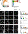
RD histone genes and synthesis factors are up-regulated in G1 versus G0 MCF10A cells. (A) Scatter plot of EdU versus DNA content and phospho-Rb versus DNA content used to define G0 (blue, 5% of cells), G1 (green, 37% of cells), S (orange, 38% of cells), and G2/M (red, 20% of cells). (B) Representative images of cells stained with Hoechst to measure DNA content, phospho-Rb S807/811 or S780, and EdU, along with either NPAT, SLBP, or H1.1 (HIST1H1A), H3.1 (HIST1H3A) or H4.2 (HIST2H4A) mRNA. Arrows indicate example cells of each cell-cycle phase; G0 (blue), G1 (green), S (orange), and G2/M (red). Cutoffs for cell-cycle phases can be found in SI Appendix, Fig. S1. (C) Violin plots by cell-cycle phase of whole-cell median intensity for H1.1, H3.1, and H4.2 mRNA, HLB total intensity (defined in Materials and Methods), and median nuclear intensity for SLBP. Axes were determined by maximum and minimum signal for each protein and mRNA. Cell counts and P values from two-sample Student’s t test for each group can be found in SI Appendix. P values are indicated as ****P ≤ 0.0001, **P ≤ 0.01, *P ≤ 0.05, and ns, P > 0.05.
Fig. 3.
Fig. 3.
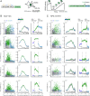
Dynamics of RD histone biosynthesis during cell-cycle progression. (A) Schematic of CDK2 sensor and live-cell tracking (45). (BC) Representative scatter of indicated IF or RNA FISH signal in MCF10A (B) or RPE-hTERT (C) cells following timelapse imaging of CDK2 activity of an asynchronously cycling population. Cells were treated with EdU in the final 12 min of imaging to further segment cells by cell-cycle phase: G0 (CDK2low, EdU; gray); G1 (CDK2inc, EdU; dark blue); S (CDK2inc, EdU+; green); and G2 (CDK2inc, EdU; light blue). Column 1: Raw single-cell data. Column 2: Average protein or mRNA signals and 95% CIs as a function of time since anaphase for populations classified in Column 1 with the fraction of EdU-positive cells at each timepoint indicated by the blue-green gradient. Column 3: Alternative visualization of the data in Column 2 wherein G1 cells (dark blue) are defined as those between anaphase and the timepoint where 50% of cells are EdU positive; S-phase cells (green) as those between the timepoint where 50% of cells are EdU-positive until the timepoint 50% of cells are EdU negative; and G2 cells (light blue) as those from the timepoint where 50% of cells are EdU negative until the end of the cell cycle. Axes were determined by maximum and minimum signal for each protein and mRNA. Non-overlapping shading of 95% confidence intervals (CIs) indicates statistically significant difference as determined by Student’s t test, with P < 0.05, and magenta stars mark signals where at least 90% of timepoints in G1 are statistically significantly higher than in G0 cells. Cell counts can be found in SI Appendix.
Fig. 4.
Fig. 4.
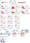
RD histone biosynthesis relative to S phase entry. (A) Schematic of geminin sensor and live-cell tracking (52). (B) Scatter of EdU, IF, or RNA FISH signal in MCF10A cells aligned to the rise of geminin following timelapse imaging (Left column). Average protein or mRNA signal as well as 95% CIs as shaded bands; EdU incorporation was used to estimate the start and end of late S phase (dashed lines; Right column). Axes were determined by maximum and minimum signal for each protein and mRNA. (C) Overlay of average and 95% CI for H1.1 (red), H3.1 (orange), and H4.2 (pink) histone mRNA with either EdU (dark blue), HLB intensity (light blue), or SLBP (green) aligned to geminin rise. (D) An updated model based on data herein, in which RD histone biosynthesis is coupled to both cell-cycle entry and DNA replication. Cell counts can be found in SI Appendix.

References

    1. Hu Z., et al. ., Nucleosome loss leads to global transcriptional up-regulation and genomic instability during yeast aging. Genes Dev. 28, 396–408 (2014). - PMC - PubMed
    1. Oberdoerffer P., An age of fewer histones. Nat. Cell Biol. 12, 1029–1031 (2010). - PubMed
    1. Mendiratta S., Gatto A., Almouzni G., Histone supply: Multitiered regulation ensures chromatin dynamics throughout the cell cycle. J. Cell Biol. 218, 39–54 (2019). - PMC - PubMed
    1. Duronio R. J., Marzluff W. F., Coordinating cell cycle-regulated histone gene expression through assembly and function of the Histone Locus Body. RNA Biol. 14, 726–738 (2017). - PMC - PubMed
    1. Luger K., Mäder A. W., Richmond R. K., Sargent D. F., Richmond T. J., Crystal structure of the nucleosome core particle at 2.8 A resolution. Nature 389, 251–260 (1997). - PubMed

Publication types

LinkOut - more resources