. 2021 Jul 29;13(14):19013-19027.
doi: 10.18632/aging.203351. Epub 2021 Jul 29.

Glucagon-like peptide-1 attenuated carboxymethyl lysine induced neuronal apoptosis via peroxisome proliferation activated receptor-γ

Affiliations

Glucagon-like peptide-1 attenuated carboxymethyl lysine induced neuronal apoptosis via peroxisome proliferation activated receptor-γ

Haoqiang Zhang et al. Aging (Albany NY). .

Abstract

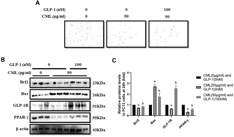
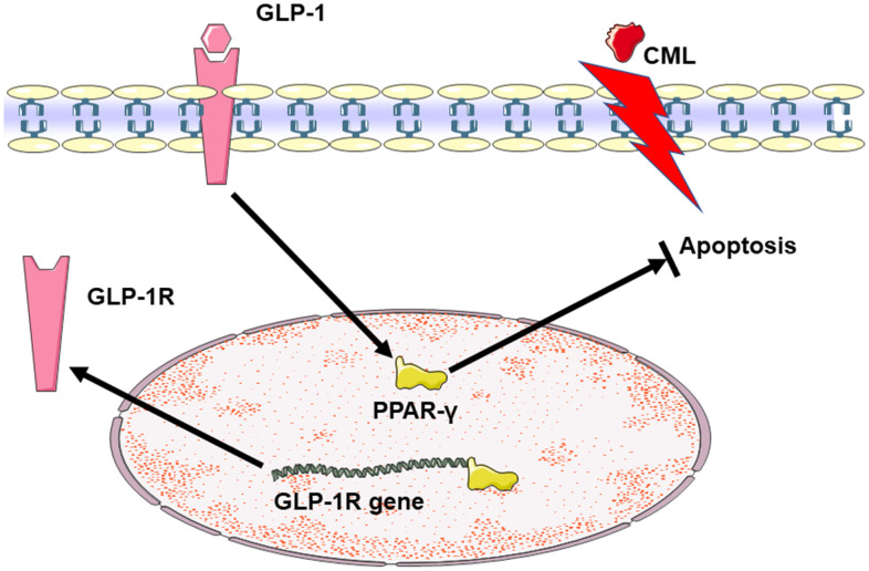
Backgrounds and aims: The role of peroxisome proliferator activated receptor-γ (PPAR-γ) in neuronal apoptosis remains unclear. We aim to investigate the role of PPAR-γ in glucagon-like peptide-1 (GLP-1) alleviated neuronal apoptosis induced by carboxymethyl-lysine (CML).

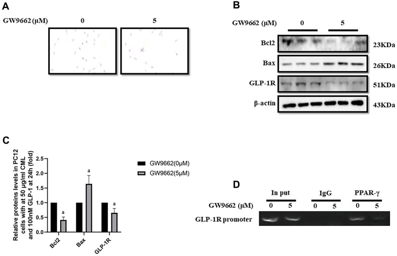
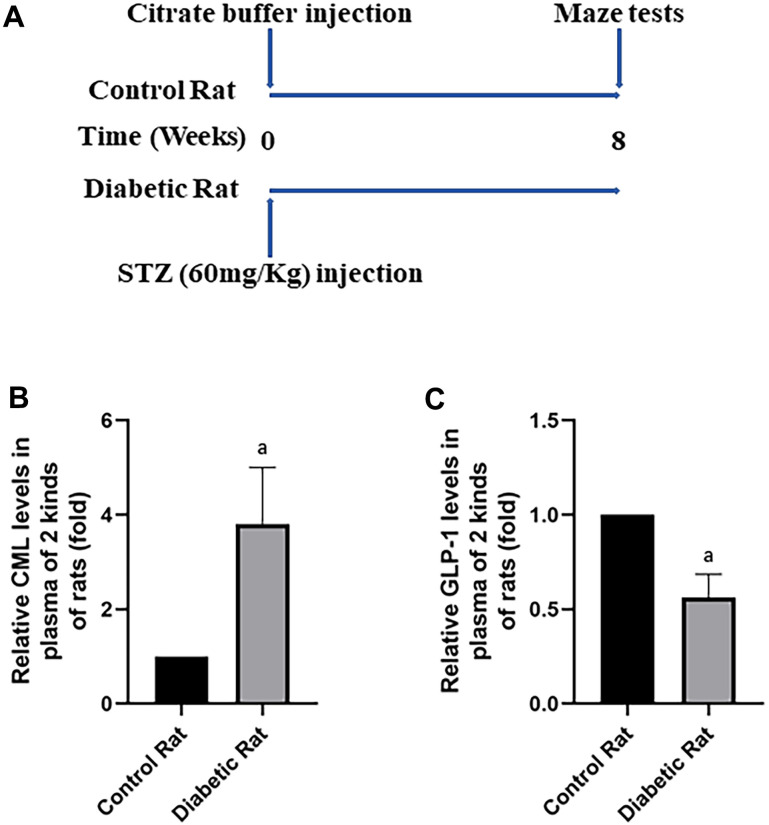
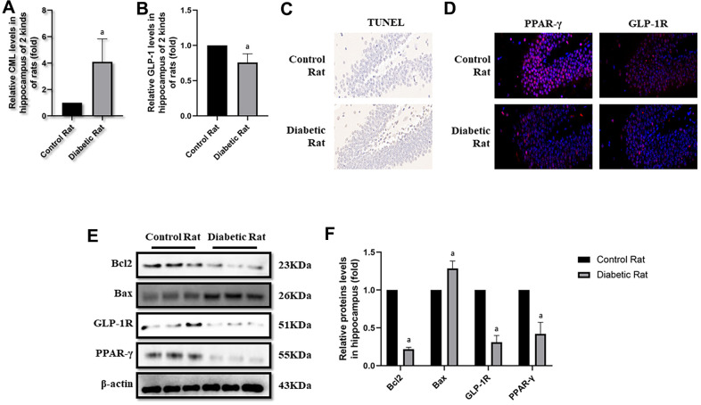
Materials and methods: In vitro, PC12 cells were treated by CML/GLP-1. Moreover. the function of PPAR-γ was blocked by GW9662. In vivo, streptozotocin (STZ) was used to induce diabetic rats with neuronal apoptosis. The cognitive function of rats was observed by Morris water maze. Apoptosis was detected by TUNEL assay. Bcl2, Bax, PPAR-γ and receptor of GLP-1 (GLP-1R) were measured by western blotting or immunofluorescence.

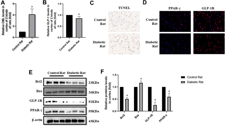
Results: In vitro experiment, CML triggered apoptosis, down-regulated GLP-1R and PPAR-γ. Moreover, GLP-1 not only alleviated the apoptosis, but also increased levels of PPAR-γ. GW9662 abolished the neuroprotective effect of GLP-1 on PC12 cells from apoptosis. Furthermore, GLP-1R promoter sequences were detected in the PPAR-γ antibody pulled mixture. GPL-1 levels decreased, while CML levels increased in diabetic rats, compared with control rats. Additionally, we observed elevated bax, decreased bcl2, GLP-1R and PPAR-γ in diabetic rats.

Conclusions: GLP-1 could attenuate neuronal apoptosis induced by CML. Additionally, PPAR-γ involves in this process.

Keywords: apoptosis; carboxymethyl-lysine; glucagon-like peptide-1; peroxisome proliferator activated receptor-γ.


Conflict of interest statement

CONFLICTS OF INTEREST: The authors declare that they have no conflicts of interest.

Figures

Figure 1
Figure 1
GLP-1 attenuated apoptosis of PC12 cells induced by CML. Results in (A) showed that GLP-1 restored the PC12 cells apoptosis induced by CML. (B) Showed the western blotting results of bcl2, bax, GLP-1R and PPAR-γ. “a” in (C) showed the down-regulated bcl2, PPAR-γ and GLP-1R as well as up-regulated bax in PC12 cells with 50 μg/ml CML, compared with those without CML (all P<0.05). “b” in (C) showed up-regulated bcl2, PPAR-γ and GLP-1R as well as down-regulated bax in PC12 cells with 50 μg/ml CML and 100 nM/ml GLP-1, compared with those with 50 μg/ml CML but without GLP-1 (all P<0.05). Data are represented as mean ± SD; n = 3 per group for results of western blotting.
Figure 2
Figure 2
PPAR-γ inhibition abolished the protective effect of GLP-1 on PC12 cells from apoptosis induced by CML. Results in (A) showed GW9662 abolished the protective effect of GLP-1 on PC12 cells from apoptosis induced by CML. (B) Showed the western blotting results of bcl2, bax, and GLP-1R. “a” in (C) showed the down-regulated bcl2 and GLP-1R as well as up-regulated bax levels between PC12 cells (treated by 50ug/ml CML and 100 nM GLP-1) with and without 5uM GW9662. Results in (D) showed the direct interactivity between PPAR-γ and GLP-1R promoter sequence. Data are represented as mean ± SD; n = 3 per group for results of western blotting.
Figure 3
Figure 3
Diabetic rats showed increased CML and decreased GLP-1 levels in plasma. In (A), we showed treatments of Wistar rats. Each rat was given a large doze of STZ or citrate buffer (PH 4.5) to get diabetic rat or control rat. After 8 weeks, cognitive functions of all rats were tested by water maze. “a” in (B) showed increased CML levels, while in (C) showed decreased GLP-1 levels in the plasma of diabetic rats, compared with control rats. Data are represented as mean ± SD; n=4 per group for results of ELISA.
Figure 4
Figure 4
Levels of apoptosis were elevated in hippocampus of diabetic rats. “a” in (A) showed elevated CML levels in hippocampus of diabetic rats, compared with control rats (P<0.05). “a” in (B) showed decreased GLP-1 levels in hippocampus of diabetic rats, compared with control rats (P<0.05). Results in (C) showed increased apoptosis in hippocampus of diabetic rats, compared with control rats. Results in (D) showed down-regulated PPAR-γ and GLP-1R in hippocampus of diabetic rats, compared with control rats. (E) Showed the western blotting results of bcl2, bax, GLP-1R and PPAR-γ. “a” of (F) showed down-regulated bcl2, GLP-1R and PPAR-γ levels and up-regulated bax levels in hippocampus of diabetic rats, compared with control rats (all P<0.05). Data are represented as mean ± SD; n=4 per group for results of ELISA. n=3 per group for results of western blotting.
Figure 5
Figure 5
Levels of cells apoptosis were elevated in cortex of diabetic rats. “a” in (A) showed elevated CML levels in cortex of diabetic rats, compared with control rats (P<0.05). “a” in (B) showed decreased GLP-1 levels in cortex of diabetic rats, compared with control rats (P<0.05). Results in (C) showed increased apoptosis in cortex of diabetic rats, compared with control rats. Results in (D) showed down-regulated PPAR-γ and GLP-1R in cortex of diabetic rats, compared with control rats. (E) Showed the western blotting results of bcl2, bax, GLP-1R and PPAR-γ. “a” of (F) showed down-regulated bcl2, GLP-1R and PPAR-γ levels and up-regulated bax levels in cortex of diabetic rats, compared with control rats (all P<0.05). Data are represented as mean ± SD; n=4 per group for results of ELISA. n=3 per group for results of western blotting.
Figure 6
Figure 6
Summary diagram of the relationship among GLP-1, CML and PPAR in cells apoptosis. CML could induce the apoptosis of PC12 cells. GLP-1 could induce the expression of PPAR-γ by binding to GLP-1R. And then, PPAR-γ could attenuate the neuronal apoptosis. Additionally, PPAR-γ may promote the expression of GLP-1R by the interaction between PPAR-γ and the promoter sequence of GLP-1R.

References

    1. Chan JC, Zhang Y, Ning G. Diabetes in China: a societal solution for a personal challenge. Lancet Diabetes Endocrinol. 2014; 2:969–79. 10.1016/S2213-8587(14)70144-5 - DOI - PubMed
    1. Wang L, Gao P, Zhang M, Huang Z, Zhang D, Deng Q, Li Y, Zhao Z, Qin X, Jin D, Zhou M, Tang X, Hu Y, Wang L. Prevalence and Ethnic Pattern of Diabetes and Prediabetes in China in 2013. JAMA. 2017; 317:2515–23. 10.1001/jama.2017.7596 - DOI - PMC - PubMed
    1. Xu Y, Wang L, He J, Bi Y, Li M, Wang T, Wang L, Jiang Y, Dai M, Lu J, Xu M, Li Y, Hu N, et al., and 2010 China Noncommunicable Disease Surveillance Group. Prevalence and control of diabetes in Chinese adults. JAMA. 2013; 310:948–59. 10.1001/jama.2013.168118 - DOI - PubMed
    1. Cho NH, Shaw JE, Karuranga S, Huang Y, da Rocha Fernandes JD, Ohlrogge AW, Malanda B. IDF Diabetes Atlas: Global estimates of diabetes prevalence for 2017 and projections for 2045. Diabetes Res Clin Pract. 2018; 138:271–81. 10.1016/j.diabres.2018.02.023 - DOI - PubMed
    1. Alzheimer’s Association. 2016 Alzheimer’s disease facts and figures. Alzheimers Dement. 2016; 12:459–509. 10.1016/j.jalz.2016.03.001 - DOI - PubMed

Publication types

MeSH terms