. 2021 Jul 29;11(1):15467.
doi: 10.1038/s41598-021-95055-x.

Sensitive assay design for detection of anti-drug antibodies to biotherapeutics that lack an immunoglobulin Fc domain

Affiliations

Sensitive assay design for detection of anti-drug antibodies to biotherapeutics that lack an immunoglobulin Fc domain

Derrick Johnson et al. Sci Rep. .

Abstract

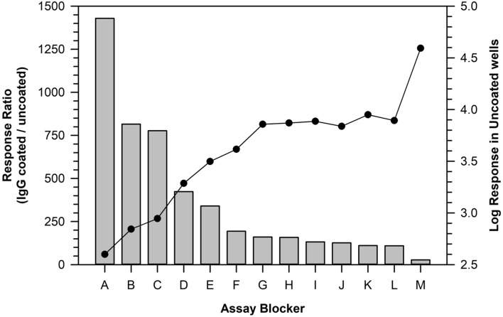
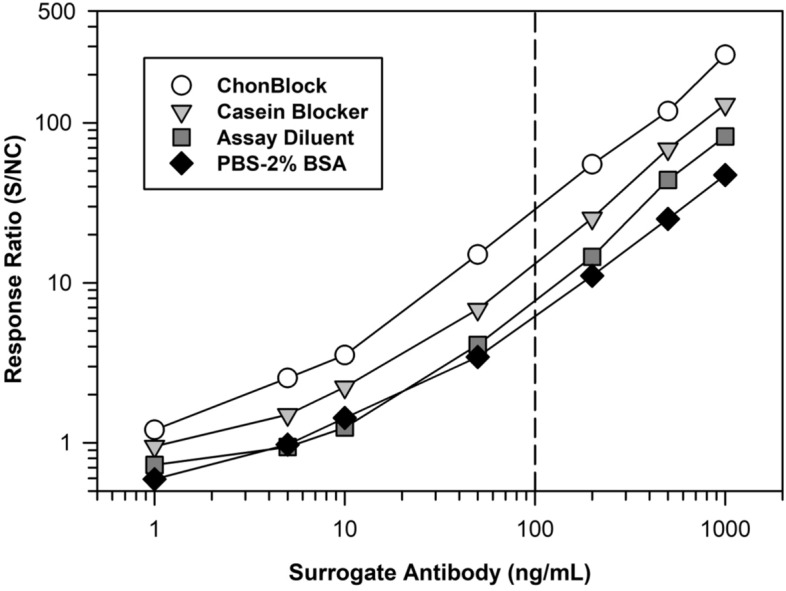
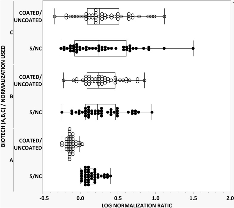
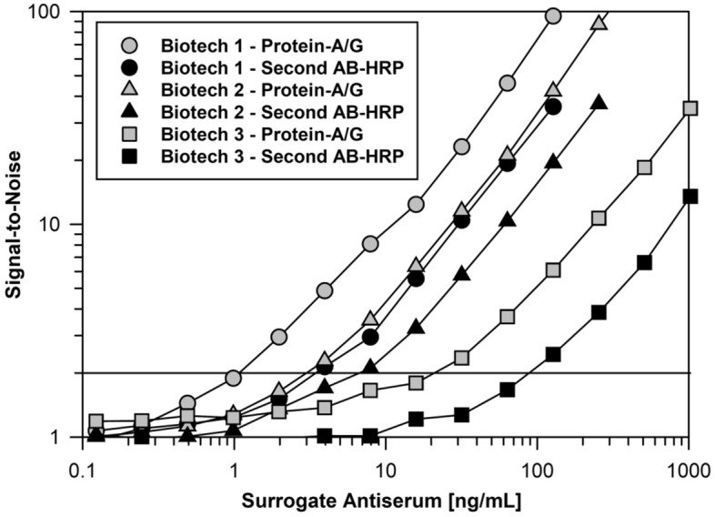
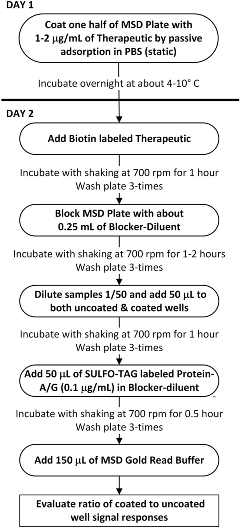
Today the evaluation of unwanted immunogenicity is a key component in the clinical safety evaluation of new biotherapeutic drugs and macromolecular delivery strategies. However, the evolving structural complexity in contemporary biotherapeutics creates a need for on-going innovation in assay designs for reliable detection of anti-drug antibodies, especially for biotherapeutics that may not be well-suited for testing by a bridging assay. We, therefore, initiated systematic optimization of the direct binding assay to adapt it for routine use in regulatory-compliant assays of serum anti-drug antibodies. Accordingly, we first prepared a SULFO-TAG labeled conjugate of recombinant Protein-A/G to create a sensitive electrochemiluminescent secondary detection reagent with broad reactivity to antibodies across many species. Secondly, we evaluated candidate blocker-diluents to identify ones producing the highest signal-to-noise response ratios. Lastly, we introduced use of the ratio of signal responses in biotherapeutic-coated and uncoated wells as a data transformation strategy to identify biological outliers. This alternative data normalization approach improved normality, reduced skewness, and facilitated application of a parametric screening cut point. We believe the optimized direct binding assay design employing SULFO-TAG labeled Protein-A/G represents a useful analytical design for detecting serum ADA to biotherapeutics that lack an immunoglobulin Fc domain.


Conflict of interest statement

All co-authors are employees of B2S Life Sciences (Franklin, IN) and have no conflicts of interest to declare. No writing assistance was used to prepare this manuscript.

Figures

Figure 1
Figure 1
Detection of different human immunoglobulin classes by SULFO-TAG Protein-A/G. A MSD plate was coated with varying concentrations of human IgG, IgM, or IgA diluted in PBS and incubated it overnight at 4 °C. On the following day, the plate was washed and blocked with PBS-2% BSA. After another wash, the coated immunoglobulins were detected using 0.2 µg/mL SULFO-TAG labeled Protein-A/G diluted in PBS-2% BSA. The plot shows the ratio of the relative luminescence (RLU) signal responses for coated wells relative to uncoated wells.
Figure 2
Figure 2
Effectiveness of different plate blocker and sample diluents for use in detection of serum immunoglobulins by SULFO-TAG labeled Protein-A/G. MSD plate wells were coated overnight with either 2 µg/mL human IgG or PBS (uncoated). On the following day, the wells were block using the different blocker-diluent buffers (A through M). A lot of pooled human serum was diluted 1/50 in the various blocker-diluents, added to the plate wells in duplicate and incubated at ambient temperature. After 1 h the plate was washed, and the IgG was detected by incubation with 0.2 µg/mL of SULFO-TAG labeled Protein-A/G diluted in PBS-2% BSA. The response ratio (bar graph) is RLU responses from the IgG coated wells relative to uncoated wells. Assay Blocker A is ChonBlock, B is Assay Diluent, C is Blocker casein in PBS, D is Low Cross buffer, and E is Monster Block. Blockers F through L, which produced similar response ratios, consisted of Starting Block, Super Block, Neptune Block, SynBlock and various combinations of BSA/HSA with PBS and TBS. The least effective blocker, M, was PBS alone. Closed circles depict the Log RLU from the uncoated wells and shows the wide range in non-specific binding responses seen among the different diluent blockers.
Figure 3
Figure 3
Sensitivity comparison of the optimal blocker-diluents for detection of serum ADA. On day 1 the MSD plate was coated overnight at 4 °C with 2 µg/mL of a representative biotherapeutic. On day 2, the plate was washed and blocked with the various candidate blocker-diluents. A standard curve of a surrogate specific rabbit antibody was prepared in human serum and then diluted 1/100 in the various buffers. The assay was performed, as described in the “Materials and methods” section, using detection with 0.1 µg/mL of SULFO-TAG labeled Protein-A/G diluted in LowCross Buffer. The reported response ratio is the RLU of the surrogate antibody serum calibrators/RLU from the NC (i.e., human serum pool not supplemented with surrogate antibody).
Figure 4
Figure 4
Comparison of different data transformation strategies for normalization of ADA signal responses. Three candidate biotherapeutic drugs were evaluated for determination of an ADA screening cut point. Individual signal responses were normalized by both the conventional approach involving S/NC (closed circle) and the modified approach using coated/uncoated wells (open circle) and then subjected to one round of outlier removal using box plot analysis (JMP, SAS institute, ver. 15.2.1). Results are reported in Table 1. As shown above the data transformation involving coated/uncoated wells yielded superior distributional normality with decreased skewness.
Figure 5
Figure 5
Comparison of serum immunoglobulin detection by optimized assay using SULFO-TAG labeled Protein-A/G versus conventional ELISA with horseradish peroxidase. MSD plates were coated overnight at 4 °C with 2 µg/mL of 3 different biotherapeutics. On the following day, the plates were washed and blocked with ChonBlock. Standard curves of surrogate MAbs specific to the various biotherapeutics were prepared in human serum and then diluted 1/100 in ChonBlock. The remainder of the assay was performed as described in the “Materials and methods” section using detection with 0.1 µg/mL of SULFO-TAG labeled Protein-A/G or a 10,000-fold dilution of HRP labeled Protein-A/G diluted in LowCross Buffer. For all three surrogate antibodies, the response ratios were appreciably greater when SULFO-TAG labeled Protein-A/G was used as the detection reagent with 2-times background responses occurring at about 1, 3, and 20 ng/mL, respectively.
Figure 6
Figure 6
Workflow for Tier 1 screening ADA assay using SULFO-TAG labeled Protein-A/G.
Figure 7
Figure 7
Generic workflow of the harmonized multi-tiered ADA sample testing scheme used to detect and characterize serum samples for the presence of reactive antibodies.

References

    1. Wang YM, et al. Evaluating and reporting the immunogenicity impact for biological products: a clinical pharmacology perspective. AAPS J. 2016 doi: 10.1208/s12248-015-9857-y. - DOI - PMC - PubMed
    1. Shankar G, et al. The quintessence of immunogenicity reporting for biotherapeutics. Nat. Biotechnol. 2015 doi: 10.1038/nbt.3181/33(4):334-6. - DOI - PubMed
    1. Chirmule N, Jawa V, Meibohm B. Immunogenicity to therapeutic protein: impact on PK/PD and efficacy. AAPS J. 2012 doi: 10.1208/s12248-012-9340-y. - DOI - PMC - PubMed
    1. Dingman R, Balu-Iyer SV. Immunogenicity of Protein Pharmaceuticals. J. Pharm. Sci. 2019;108(5):1637–1654. doi: 10.1016/j.xphs.2018.12.014. - DOI - PMC - PubMed
    1. Pineda C, Hernandez GC, Jacobs IA, Alverez DF, Carini C. Assessing the immunogenicity of biopharmaceuticals. BioDrugs. 2016 doi: 10.1007/s40259-016-0174-5/2016. - DOI - PMC - PubMed

Publication types