

Mitochondrial regulation of ferroptosis
- PMID: 34328510
- PMCID: PMC8329737
- DOI: 10.1083/jcb.202105043
Mitochondrial regulation of ferroptosis
Abstract
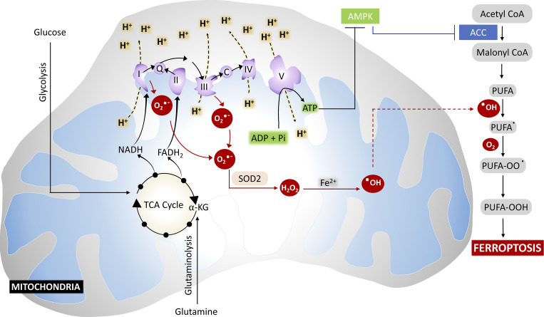
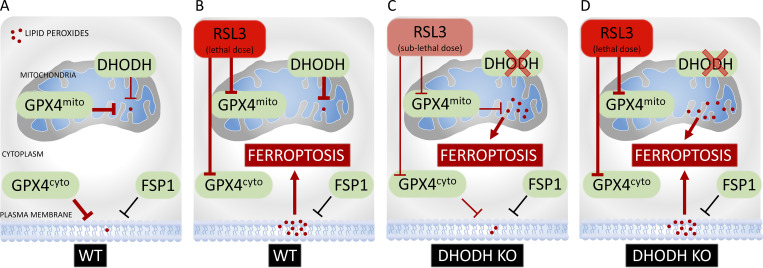
Ferroptosis is a form of iron-dependent regulated cell death driven by uncontrolled lipid peroxidation. Mitochondria are double-membrane organelles that have essential roles in energy production, cellular metabolism, and cell death regulation. However, their role in ferroptosis has been unclear and somewhat controversial. In this Perspective, I summarize the diverse metabolic processes in mitochondria that actively drive ferroptosis, discuss recently discovered mitochondria-localized defense systems that detoxify mitochondrial lipid peroxides and protect against ferroptosis, present new evidence for the roles of mitochondria in regulating ferroptosis, and outline outstanding questions on this fascinating topic for future investigations. An in-depth understanding of mitochondria functions in ferroptosis will have important implications for both fundamental cell biology and disease treatment.
© 2021 Gan.
Figures

Similar articles
-
The Chemistry and Biology of Ferroptosis.Cell Chem Biol. 2020 Apr 16;27(4):365-375. doi: 10.1016/j.chembiol.2020.03.013. Cell Chem Biol. 2020. PMID: 32294465 Free PMC article. Review.
-
Ferroptosis is involved in alcohol-induced cell death in vivo and in vitro.Biosci Biotechnol Biochem. 2020 Aug;84(8):1621-1628. doi: 10.1080/09168451.2020.1763155. Epub 2020 May 18. Biosci Biotechnol Biochem. 2020. PMID: 32419644
-
DHODH tangoing with GPX4 on the ferroptotic stage.Signal Transduct Target Ther. 2021 Jun 18;6(1):244. doi: 10.1038/s41392-021-00656-7. Signal Transduct Target Ther. 2021. PMID: 34145214 Free PMC article. No abstract available.
-
Bavachin Induces Ferroptosis through the STAT3/P53/SLC7A11 Axis in Osteosarcoma Cells.Oxid Med Cell Longev. 2021 Oct 18;2021:1783485. doi: 10.1155/2021/1783485. eCollection 2021. Oxid Med Cell Longev. 2021. PMID: 34707773 Free PMC article.
-
Investigating Nonapoptotic Cell Death Using Chemical Biology Approaches.Cell Chem Biol. 2020 Apr 16;27(4):376-386. doi: 10.1016/j.chembiol.2020.03.005. Epub 2020 Mar 26. Cell Chem Biol. 2020. PMID: 32220334 Free PMC article. Review.
Cited by
-
Epigenetic modification of ferroptosis by non-coding RNAs in cancer drug resistance.Mol Cancer. 2024 Aug 27;23(1):177. doi: 10.1186/s12943-024-02088-7. Mol Cancer. 2024. PMID: 39192329 Free PMC article. Review.
-
Age-Related Macular Degeneration and Mitochondria-Associated Autoantibodies: A Review of the Specific Pathogenesis and Therapeutic Strategies.Int J Mol Sci. 2024 Jan 28;25(3):1624. doi: 10.3390/ijms25031624. Int J Mol Sci. 2024. PMID: 38338904 Free PMC article. Review.
-
Investigation of ferroptosis-associated molecular subtypes and immunological characteristics in lupus nephritis based on artificial neural network learning.Arthritis Res Ther. 2024 Jul 3;26(1):126. doi: 10.1186/s13075-024-03356-z. Arthritis Res Ther. 2024. PMID: 38961491 Free PMC article.
-
Lipid Peroxidation and Iron Metabolism: Two Corner Stones in the Homeostasis Control of Ferroptosis.Int J Mol Sci. 2022 Dec 27;24(1):449. doi: 10.3390/ijms24010449. Int J Mol Sci. 2022. PMID: 36613888 Free PMC article. Review.
-
Uncovering the IL-1β-PCAF-NNT axis: A new player in ferroptosis and tumor immune evasion.Cancer Commun (Lond). 2023 Sep;43(9):1048-1050. doi: 10.1002/cac2.12473. Epub 2023 Jul 24. Cancer Commun (Lond). 2023. PMID: 37488943 Free PMC article. No abstract available.
References
-
- Basit, F., van Oppen L.M., Schöckel L., Bossenbroek H.M., van Emst-de Vries S.E., Hermeling J.C., Grefte S., Kopitz C., Heroult M., Hgm Willems P., and Koopman W.J.. 2017. Mitochondrial complex I inhibition triggers a mitophagy-dependent ROS increase leading to necroptosis and ferroptosis in melanoma cells. Cell Death Dis. 8:e2716. 10.1038/cddis.2017.133 - DOI - PMC - PubMed
Publication types
MeSH terms
Substances
Grants and funding
LinkOut - more resources
Full Text Sources
Medical
Research Materials
