. 2021 Jul 30;40(1):244.
doi: 10.1186/s13046-021-02037-y.

Molecular features and vulnerabilities of recurrent chordomas

Affiliations

Molecular features and vulnerabilities of recurrent chordomas

Carolin Seeling et al. J Exp Clin Cancer Res. .

Abstract

Background: Tumor recurrence is one of the major challenges in clinical management of chordoma. Despite R0-resection, approximately 50% of chordomas recur within ten years after initial surgery. The underlying molecular processes are poorly understood resulting in the lack of associated therapeutic options. This is not least due to the absence of appropriate cell culture models of this orphan disease.

Methods: The intra-personal progression model cell lines U-CH11 and U-CH11R were compared using array comparative genomic hybridization, expression arrays, RNA-seq, and immunocytochemistry. Cell line origin was confirmed by short tandem repeat analysis. Inter-personal cell culture models (n = 6) were examined to validate whether the new model is representative. Cell viability after HOX/PBX complex inhibition with small peptides was determined by MTS assays.

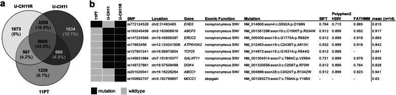
Results: Using whole genome microarray analyses, striking differences in gene expression between primary and recurrent chordomas were identified. These expression differences were confirmed in the world's first intra-personal model of chordoma relapse consisting of cell lines established from a primary (U-CH11) and the corresponding recurrent tumor (U-CH11R). Array comparative genomic hybridization and RNA-sequencing analyses revealed profound genetic similarities between both cell lines pointing to transcriptomic reprogramming as a key mechanism of chordoma progression. Network analysis of the recurrence specific genes highlighted HOX/PBX signaling as a common dysregulated event. Hence, HOX/PBX complexes were used as so far unknown therapeutic targets in recurrent chordomas. Treating chordoma cell lines with the complex formation inhibiting peptide HXR9 induced cFOS mediated apoptosis in all chordoma cell lines tested. This effect was significantly stronger in cell lines established from chordoma relapses.

Conclusion: Clearly differing gene expression patterns and vulnerabilities to HOX/PBX complex inhibition in highly therapy resistant chordoma relapses were identified using the first intra-personal loco-regional and further inter-personal chordoma progression models. For the first time, HOX/PBX interference was used to induce cell death in chordoma and might serve as the basic concept of an upcoming targeted therapy for chordomas of all progression stages.

Keywords: Apoptosis; Cell lines; Chordoma; HOX; PBX; Progression model.


Conflict of interest statement

The authors declare that they have no competing interests.

Figures

Fig. 1
Fig. 1
U-CH11 and U-CH11R cells are clonally related. a Hematoxylin/eosin staining of the primary and the recurrent chordoma tissues and in vitro light microscopy of U-CH11 and U-CH11R. The physaliferous cell morphology is conserved. b Immunocytochemical staining of the chordoma markers brachyury, S100-protein, vimentin, pan-cytokeratin, epithelial membrane antigen (EMA), and Ki-67. c Mean in vitro population doubling times (±SD; n = 3) of U-CH11 and U-CH11R at different cell densities (**p < 0.01) d Overlay of the array comparative genomic hybridization ratio plots of U-CH11 (green) and U-CH11R (blue) showing a high concordance of alterations. e Hierarchical cluster analysis of U-CH11 and U-CH11R in comparison to 12 additional chordoma cell lines. Clustering was performed in a Euclidean distance measure and single linkage rule
Fig. 2
Fig. 2
RNA-Sequencing. a Venn diagram illustrating the mutational concordance of exonic variants in the primary chordoma tissue (11PT), the derived primary cell line U-CH11 and the recurrent chordoma cell line U-CH11R. b Deleterious mutations observed in the samples (colour coded, refer to legend). SIFT, Polyphen2-HDIV and FATHMM represent three examples of all implemented SNP analysis tools (n = 14)
Fig. 3
Fig. 3
U-CH11 and U-CH11R present distinct transcriptomic profiles representative for sacral primary and recurrent chordoma lines. a Volcano plot showing statistical significance (−log10p-value) versus fold change (log2FC) of microarray gene expression data from U-CH11R (n = 3) versus U-CH11 (n = 2). The top five diminished (blue) and enhanced (red) genes in U-CH11R are annotated. Cnetplots illustrating the top three enriched GO-terms and the associated genes of the enhanced (b) and diminished (c) genes in U-CH11R. The fold change of gene expression is indicated by the color, the size of each node represents the number of linked genes to each term. d Rank-rank hypergeometric overlap (RRHO) heatmap for matched and unmatched dataset comparison used to identify significantly concordant transcriptional profiles from two independently conducted microarray gene expression analyses. RRHO was applied to the matched and unmatched data of primary and recurrent chordoma cell lines revealing high overlap between both datasets. The -log10 transformed hypergeometric p-value indicates the strength of enrichment (refer to legend). Concordantly up- and downregulated genes are located in A and D, respectively, disconcordantly expressed genes are in B and C. e Gene set enrichment analysis of the concordantly upregulated genes in recurrent chordoma cell lines. The top six enriched GO-terms and associated genes are given
Fig. 4
Fig. 4
The HOX/PBX network is dysregulated by diminished miR-196a-5p levels in recurrent chordoma cell lines. a Scheme of the HOX clusters and miRNAs of the miR-196 family located within the cluster. Micro-RNA-196a-5p expression levels of matched (b) and unmatched (c) primary and recurrent chordoma cell lines. Relative fold change levels are given on the y-axis. (d) miR-196a-5p target sites (underlined nucleotides) in the 3’UTRs of HOXA7 and HOXB7 predicted by TargetScan 7.2. (e) Response to miR-196a-5p in U-CH1 cells. Firefly luciferase reporters containing either the complementary site of HOXA7 and HOXB8 or the perfect antisense sequence of miR-196a were co-transfected with a miR-196a mimic or a scrambled control, respectively. Firefly luciferase activity was normalized to Renilla luciferase activity. (f) Overexpression of miR-196a-5p in U-CH1 cells confirmed by qRT-PCR. (g) The effect of miR-196a-5p on HOXA7 levels in U-CH1 cells assessed by Western blot analysis. Experiments were performed in biological triplicates. T-tests were performed to determine statistical significance (*p < 0.05, **p < 0.01, ***p < 0.001). (h) GO-term analysis of the predicted target genes (n = 50) illustrated as cnetplot. (i) STRING analysis of the predicted miR-196a-5p target genes (n = 50) revealed an interaction network between HOX transcription factors and PBX co-factors
Fig. 5
Fig. 5
Recurrent chordoma cell lines are susceptible towards HOX/PBX inhibition. (a) Mean IC50 ± SD of HXR9 in U-CH11 (70.26 μM ±17.2) compared to U-CH11R (29.59 μM ±5.5) and (b) unmatched primary (n = 3, 43.91 μM ±12.3) versus recurrent (n = 3, 31.65 μM ±10.4) chordoma derived cell lines. Representative inhibition curves of one representative replicate of the cell lines treated with HXR9 (solid curves) and CXR9 (dotted curves) are depicted. (c) Overall response to HXR9 in chordoma (n = 8; 40.82 μM ±17.2) in comparison to fibroblasts (n = 3; 108.3 μM ±19.9). The inhibition curves of one representative example of fibroblasts are given. (d) Colony formation assay. Values are normalized to the mean amount of colonies after CXR9 treatment (n = 3). Statistical differences were determined by Student’s t-tests (*p < 0.05, **p < 0.01, ***p < 0.001)
Fig. 6
Fig. 6
HXR9-induced apoptosis is mediated via cFOS. Activation of cleaved Caspase-3/7 (488 nm; green channel) in U-CH11 (a) and U-CH11R (b) treated for 24 h with 30 μM CXR9 or HXR9. The percentage of cleaved Caspase 3/7 positive cells following each treatment was determined by cell counts of representative image sections (n = 12; c). Immunoblots showing cleaved PARP, Procaspase 3, cleaved Caspase-3 and cleaved Caspase-7 in chordoma cell lines treated with 30 μM of HXR9 or CXR9 for 24 h (d). CFOS expression levels in response to treatment with HXR9 or two control compounds (CXR9 and DMSO) in U-CH1 versus U-CH19 (e) and U-CH11 compared to U-CH11R (f) cell lines quantified by qRT-PCR and normalized against GAPDH. Gene expression experiments were performed in technical and biological triplicates. Statistical differences were determined by Student’s t-tests (*p < 0.05, **p < 0.01, ***p < 0.001)

References

    1. McMaster ML, Goldstein AM, Bromley CM, Ishibe N, Parry DM. Chordoma: incidence and survival patterns in the United States, 1973-1995. Cancer Causes Control. 2001;12(1):1–11. doi: 10.1023/A:1008947301735. - DOI - PubMed
    1. Miettinen M, Wang Z, Lasota J, Heery C, Schlom J, Palena C. Nuclear Brachyury expression is consistent in Chordoma, common in germ cell tumors and small cell carcinomas, and rare in other carcinomas and sarcomas: an Immunohistochemical study of 5229 cases. Am J Surg Pathol. 2015;39(10):1305–1312. doi: 10.1097/PAS.0000000000000462. - DOI - PMC - PubMed
    1. Sharifnia T, Wawer MJ, Chen T, Huang QY, Weir BA, Sizemore A, Lawlor MA, Goodale A, Cowley GS, Vazquez F, Ott CJ, Francis JM, Sassi S, Cogswell P, Sheppard HE, Zhang T, Gray NS, Clarke PA, Blagg J, Workman P, Sommer J, Hornicek F, Root DE, Hahn WC, Bradner JE, Wong KK, Clemons PA, Lin CY, Kotz JD, Schreiber SL. Small-molecule targeting of brachyury transcription factor addiction in chordoma. Nat Med. 2019;25(2):292–300. doi: 10.1038/s41591-018-0312-3. - DOI - PMC - PubMed
    1. Hsu W, Mohyeldin A, Shah SR, Rhys CM a, Johnson LF, Sedora-Roman NI, Kosztowski TA, Awad OA, McCarthy EF, Loeb DM, et al. Generation of chordoma cell line JHC7 and the identification of Brachyury as a novel molecular target. J Neurosurg. 2011;115(4):760–769. doi: 10.3171/2011.5.JNS11185. - DOI - PMC - PubMed
    1. Yang XR, Ng D, Alcorta DA, Liebsch NJ, Sheridan E, Li S, Goldstein AM, Parry DM, Kelley MJ. T (brachyury) gene duplication confers major susceptibility to familial chordoma. Nat Genet. 2009;41(11):1176–1178. doi: 10.1038/ng.454. - DOI - PMC - PubMed

LinkOut - more resources