. 2021 Jul 30;7(31):eabf7906.
doi: 10.1126/sciadv.abf7906. Print 2021 Jul.

FAN1-MLH1 interaction affects repair of DNA interstrand cross-links and slipped-CAG/CTG repeats

Affiliations

FAN1-MLH1 interaction affects repair of DNA interstrand cross-links and slipped-CAG/CTG repeats

Antonio Porro et al. Sci Adv. .

Abstract

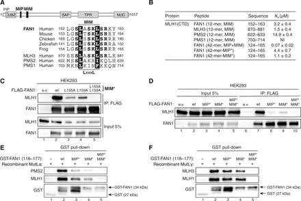
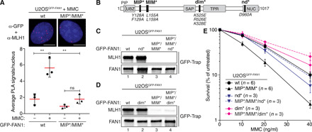
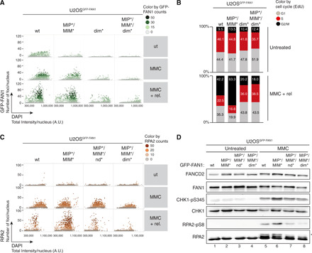
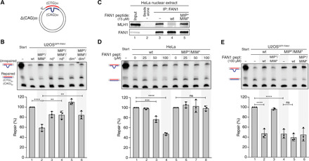
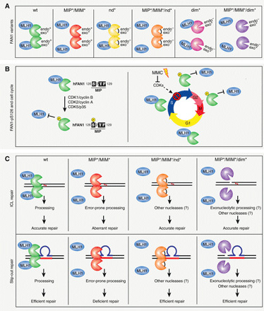
FAN1, a DNA structure-specific nuclease, interacts with MLH1, but the repair pathways in which this complex acts are unknown. FAN1 processes DNA interstrand crosslinks (ICLs) and FAN1 variants are modifiers of the neurodegenerative Huntington's disease (HD), presumably by regulating HD-causing CAG repeat expansions. Here, we identify specific amino acid residues in two adjacent FAN1 motifs that are critical for MLH1 binding. Disruption of the FAN1-MLH1 interaction confers cellular hypersensitivity to ICL damage and defective repair of CAG/CTG slip-outs, intermediates of repeat expansion mutations. FAN1-S126 phosphorylation, which hinders FAN1-MLH1 association, is cell cycle-regulated by cyclin-dependent kinase activity and attenuated upon ICL induction. Our data highlight the FAN1-MLH1 complex as a phosphorylation-regulated determinant of ICL response and repeat stability, opening novel paths to modify cancer and neurodegeneration.


Figures

Fig. 1
Fig. 1. FAN1 contains a MIP box that is negatively regulated by CDK-mediated phosphorylation at S126.
(A) Top: Protein domain architecture of human FAN1 indicating the location of the MIP box. PIP, proliferating cell nuclear antigen–interacting peptide box; UBZ, ubiquitin-binding zinc finger; SAP, SAF-A/B, Acinus, and PIAS; TPR, tetratricopeptide repeat; NUC, nuclease. Bottom: Sequence alignment of the FAN1 orthologs with the MIP box of human EXO1 and BLM. (B) Dissociation constants (Kd) indicating the binding affinity of MLH1 CTD for the indicated peptides from FAN1, EXO1, and BLM as determined by ITC. NI, no interaction (weak and constant signal under the condition used). (C) Immunoblots of FLAG-M2 affinity resin immunoprecipitations (IPs) of extracts from HEK293 cells transfected either with empty vector (e.v.), FLAG-FAN1 wild type (wt), or the variant constructs. The Y128A/F129A mutation is denoted as MIP*. (D) Immunoblots of FLAG-M2 affinity resin IPs of extracts from HEK293 cells transfected with the indicated FLAG-FAN1 constructs. (E) Immunoblots of FLAG-M2 affinity resin IPs of extracts from HEK293 cells cotransfected with FLAG-FAN1 and vectors expressing hemagglutinin (HA)–tagged dominant-negative (dn) forms of CDK1 or CDK2. (F) Immunoblots of green fluorescent protein (GFP)–Trap IPs of extracts from HeLaGFP-FAN1 cells synchronized in mitosis by thymidine-nocodazole block. Mitotic cells were replated in fresh medium for the indicated time periods. The relative ratios of pS126 to FAN1 and MLH1 to FAN1 in GFP-Trap samples were quantified by densitometry using ImageJ. (C to F) The antibodies used are shown on the left.
Fig. 2
Fig. 2. An adjacent MLH1-interacting motif (MIM) in FAN1 synergizes with the MIP box to promote stable MLH1 binding.
(A) Top: Protein domain architecture of human FAN1 indicating the location of the MIP box and MIM. Bottom: Sequence alignment of FAN1 orthologs with the putative MIM of human MLH3, PMS2, and PMS1. (B) ITC was used to determine the Kd indicating the binding affinity of MLH1-CTD for the indicated peptides. (C) Immunoblots of FLAG-M2 affinity resin IPs of extracts from HEK293 cells transfected either with e.v. or indicated FLAG-FAN1 expression constructs. The L155A/L159A mutation is denoted as MIM*. (D) Immunoblots of FLAG-M2 affinity resin IPs of extracts from HEK293 cells transfected either with e.v. or indicated FLAG-FAN1 expression constructs. (E and F) Recombinant MutLα (E) or MutLγ (F) (200 nM) was subjected to pull-down reactions using the indicated recombinant GST-FAN1 (amino acids 118 to 177) variants. (C to F) The antibodies used are shown on the left.
Fig. 3
Fig. 3. FAN1-MLH1 interaction promotes resistance to cross-link damage.
(A) PLA was used to evaluate FAN1-MLH1 association in U2OSGFP-FAN1 wt and U2OSGFP-FAN1 MIP*/MIM* cells mock-treated or treated with MMC (150 ng/ml) for 24 hours. Representative images are shown. Scale bars, 10 μm. Scatterplot displays quantification of the PLA signals per nucleus from at least 100 cells. Data display the means ± SD from three independent experiments. Statistical significance was calculated by unpaired t test. **P < 0.01; ns, not significant. (B) Schematic representation of human FAN1, highlighting positions of mutations used, including MIP*, MIM*, dimerization-defective (dim*), and nuclease-defective (nd*) FAN1 variants. (C and D) Immunoblots of GFP-Trap IPs of extracts from indicated U2OSGFP-FAN1 cells. The antibodies used are shown on the left. (E) Clonogenic survival assay of the indicated U2OSGFP-FAN1 cells exposed to increasing doses of MMC. Viability of untreated cells was defined as 100%. Data are presented as the means ± SEM.
Fig. 4
Fig. 4. FAN1-MLH1 interaction prevents extensive ssDNA formation following cross-link damage.
(A) QIBC analysis of GFP-FAN1 foci in U2OSGFP-FAN1 cells mock-treated, treated with MMC (20 ng/ml) for 24 hours, or MMC-treated and then released for 72 hours. Color-coded scatterplots indicate the number of GFP-FAN1 foci per nucleus. ut, untreated. (B) Same cells as in (A) were pulse-labeled with ethynyl deoxyuridine (EdU) during the last 30 min before harvesting and subjected to the Click-IT reaction. Cell cycle distribution was evaluated by QIBC using the 4′,6-diamidino-2-phenylindole (DAPI) and EdU signals (fig. S5C). (C) QIBC of RPA2 foci in U2OSGFP-FAN1 cells mock-treated, treated with MMC (20 ng/ml) for 24 hours, or MMC-treated and then released for 72 hours. Color-coded scatterplots indicate the number of RPA2 foci per nucleus. A.U., arbitrary units. (D) Same cells as in (C) were treated with MMC (300 ng/ml) for 24 hours, and lysates were analyzed by immunoblotting using the indicated antibodies. Asterisk indicates hyperphosphorylated form of RPA2.
Fig. 5
Fig. 5. FAN1-MLH1 interaction promotes repair of CAG/CTG DNA slip-outs.
(A) Scheme of the slipped (CTG)30/(CAG)50 substrate. (B) The (CAG)20 slip-out substrate was incubated with extracts from U2OSGFP-FAN1 cells expressing indicated FAN1 variants. Repair efficiency was quantified by densitometric analysis of Southern blots. Values represent the mean of three independent experiments. Error bars represent ±SD. Statistical significance was calculated by ordinary one-way analysis of variance (ANOVA) test followed by Tukey’s multiple comparisons. **P < 0.005, ***P < 0.0005, ****P < 0.00005. (C) HeLa nuclear extract was incubated with FAN1-derived 60-mer peptides (amino acids 118 to 177) containing wt or mutant MIP-MIM, immunoprecipitated using anti-FAN1 antibody, and immunoblotted with the indicated antibodies. (D) Extracts from HeLa cells were supplemented with increasing concentrations of FAN1 peptides and incubated with the (CAG)20 slip-out substrate. Repair efficiency was calculated as described in (B). (E) Extracts of U2OSGFP-FAN1 wt and U2OSGFP-FAN1 MIP*/MIM* cells were supplemented with indicated FAN1 peptides and incubated with the (CAG)20 slip-out substrate. Repair efficiency was calculated as described in (B).
Fig. 6
Fig. 6. FAN1-MLH1 interaction affects the repair of ICLs and slipped-DNA repeats.
Summary of the FAN1-MLH1 complex interaction and a plausible model for its involvement in ICL and slip-out repair. (A) Scheme of the FAN1-MLH1 interaction in different states of wt or mutated FAN1. Protein orientation is unknown and arbitrarily presented for ease. (B) Left: FAN1 is subjected to CDK-mediated phosphorylation at S126 located within the MIP box. Right: Regulation of FAN1-MLH1 interaction through the cell cycle and in response to ICL damage. (C) Top: The FAN1-MLH1 complex localizes to ICL damaged chromatin to preserve genome stability and ensure cell viability. In cells, devoid of the FAN1-MLH1 interaction aberrant ICL repair and cell death ensues. Inactivation of FAN1’s endo- and exonuclease activities by mutating D960A or inhibition of FAN1 dimerization, affecting FAN1’s endo- but not exonucleolytic activity, would alleviate the toxicity of the FAN1 MIP/MIM-mutated variant and promote cell proliferation and survival. Bottom: Inhibition of the FAN1-MLH1 interaction (MIP*/MIM*) blocks repair of slipped-DNAs. Repair defects were rescued when the MLH1-interacting–defective FAN1 was also defective either in both its endonuclease activities or in dimer formation (endo- but not exonuclease defective). Therefore, the regulatory aspects of the FAN1 binding to MLH1 aligns the pathway of ICL repair with that of slipped-DNA processing. Mutations: MIP*, Y128A/F129A; MIM*, L155A/L159A; nd*, D960A; dim*, K525E/R526E/K528E.

Comment in

References

    1. Liu T., Ghosal G., Yuan J., Chen J., Huang J., FAN1 acts with FANCI-FANCD2 to promote DNA interstrand cross-link repair. Science 329, 693–696 (2010). - PubMed
    1. Smogorzewska A., Desetty R., Saito T. T., Schlabach M., Lach F. P., Sowa M. E., Clark A. B., Kunkel T. A., Harper J. W., Colaiácovo M. P., Elledge S. P., A genetic screen identifies FAN1, a Fanconi anemia-associated nuclease necessary for DNA interstrand crosslink repair. Mol. Cell 39, 36–47 (2010). - PMC - PubMed
    1. Kratz K., Schöpf B., Kaden S., Sendoel A., Eberhard R., Lademann C., Cannavo E., Sartori A. A., Hengartner M. O., Jiricny J., Deficiency of FANCD2-associated nuclease KIAA1018/FAN1 sensitizes cells to interstrand crosslinking agents. Cell 142, 77–88 (2010). - PubMed
    1. MacKay C., Déclais A.-C., Lundin C., Agostinho A., Deans A. J., MacArtney T. J., Hofmann K., Gartner A., West S. C., Helleday T., Lilley D. M. J., Rouse J., Identification of KIAA1018/FAN1, a DNA repair nuclease recruited to DNA damage by monoubiquitinated FANCD2. Cell 142, 65–76 (2010). - PMC - PubMed
    1. Cannavo E., Gerrits B., Marra G., Schlapbach R., Jiricny J., Characterization of the interactome of the human MutL homologues MLH1, PMS1, and PMS2. J. Biol. Chem. 282, 2976–2986 (2007). - PubMed

Publication types

MeSH terms