. 2021 Aug 3;118(31):e2108079118.
doi: 10.1073/pnas.2108079118.

GHB analogs confer neuroprotection through specific interaction with the CaMKIIα hub domain

Affiliations

GHB analogs confer neuroprotection through specific interaction with the CaMKIIα hub domain

Ulrike Leurs et al. Proc Natl Acad Sci U S A. .

Abstract

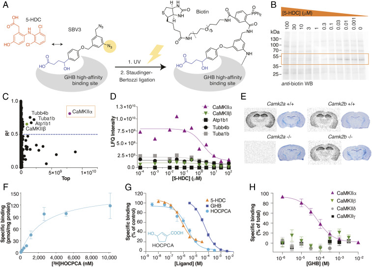
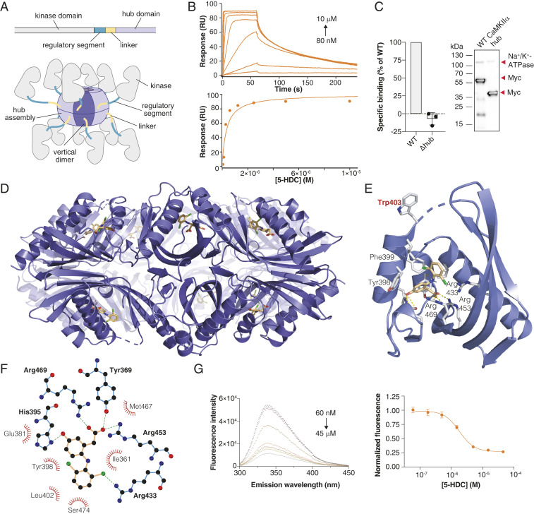
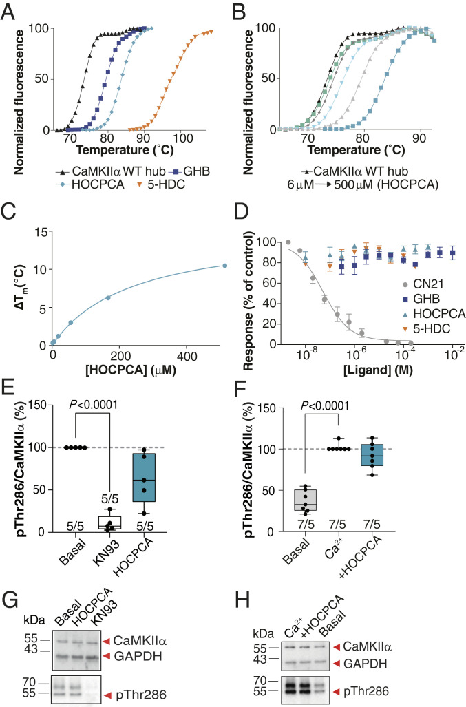
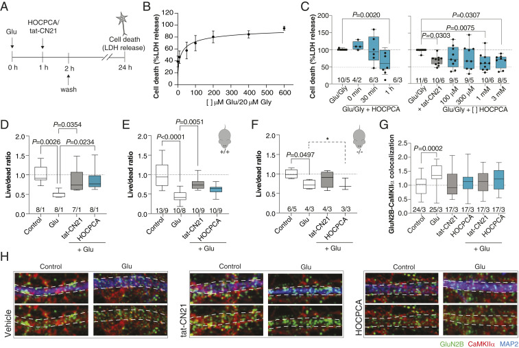
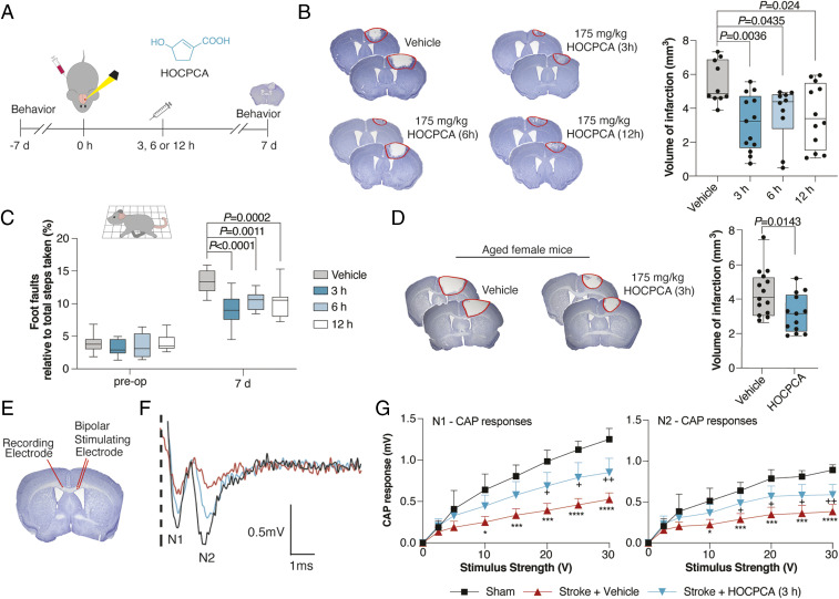
Ca2+/calmodulin-dependent protein kinase II alpha subunit (CaMKIIα) is a key neuronal signaling protein and an emerging drug target. The central hub domain regulates the activity of CaMKIIα by organizing the holoenzyme complex into functional oligomers, yet pharmacological modulation of the hub domain has never been demonstrated. Here, using a combination of photoaffinity labeling and chemical proteomics, we show that compounds related to the natural substance γ-hydroxybutyrate (GHB) bind selectively to CaMKIIα. By means of a 2.2-Å x-ray crystal structure of ligand-bound CaMKIIα hub, we reveal the molecular details of the binding site deep within the hub. Furthermore, we show that binding of GHB and related analogs to this site promotes concentration-dependent increases in hub thermal stability believed to alter holoenzyme functionality. Selectively under states of pathological CaMKIIα activation, hub ligands provide a significant and sustained neuroprotection, which is both time and dose dependent. This is demonstrated in neurons exposed to excitotoxicity and in a mouse model of cerebral ischemia with the selective GHB analog, HOCPCA (3-hydroxycyclopent-1-enecarboxylic acid). Together, our results indicate a hitherto unknown mechanism for neuroprotection by a highly specific and unforeseen interaction between the CaMKIIα hub domain and small molecule brain-penetrant GHB analogs. This establishes GHB analogs as powerful tools for investigating CaMKII neuropharmacology in general and as potential therapeutic compounds for cerebral ischemia in particular.

Keywords: HOCPCA; excitotoxicity; photoaffinity labeling; photothrombotic stroke; x-ray crystallography.


Conflict of interest statement

Competing interest statement: The University of Copenhagen and Otago Innovation Ltd. have licensed the patent rights for GHB derivatives and their uses (WO/2019/149329) to Ceremedy Ltd., of which B.F., B.R.K., and P.W. are cofounders.

Figures

Fig. 1.
Fig. 1.
Identification of CaMKIIα as the specific GHB high-affinity target. (AD) Target identification using a combination of photoaffinity labeling (PAL), affinity purification, and chemical quantitative proteomics. (A) Bioorthogonal approach using the photolabile diazide–labeled GHB analog SBV3 (GHB moiety in blue), in competition with 5-HDC (orange) for PAL followed by biotin-ligation. (B) Representative anti-biotin Western blot after PAL and concentration-dependent competition with 5-HDC (see also SI Appendix, Fig. S2). (C) Identification of CaMKIIα from LC-MS/MS data as the best hit from nonlinear regression analysis for all proteins and (D) concentration-dependent competition of the best hits (proteins were quantified using the label-free quantification [LFQ] algorithm) (see also Dataset S1). (E) Target validation by [3H]HOCPCA autoradiography using brain slices from Camk2a and Camk2b+/+ and -/- mice (cresyl violet staining for tissue visualization) (see also SI Appendix, Fig. S3). (FH) Target validation by [3H]HOCPCA binding to whole-cell homogenate from HEK293T cells transfected with CaMKIIα. (F) [3H]HOCPCA saturation binding to CaMKIIα (n = 5); shown is one representative curve (means ± SD). (G) [3H]HOCPCA competition binding to CaMKIIα in the presence of GHB (n = 3), HOCPCA (n = 5), and 5-HDC (n = 3), pooled data (means ± SEM) (see also SI Appendix, Table S1). (H) Subtype-selective specific [3H]HOCPCA binding for CaMKIIα cf CaMKIIβ. Data are pooled (n = 3) for each subtype and depicted as specific binding (% of total) (see also SI Appendix, Fig. S4A).
Fig. 2.
Fig. 2.
GHB analogs bind the CaMKIIα hub domain. (A) Schematic of a single CaMKIIα subunit composed of a kinase domain (gray), regulatory segment (green), linker (yellow), and hub domain (lilac). Twelve to 14 hub domains oligomerize into the holoenzyme, shown here in an activated form. (B) Concentration-dependent binding of 5-HDC to immobilized CaMKIIα 6x Hub measured by surface plasmon resonance (Top); Langmuir-binding isotherm (Bottom), representative data (see also SI Appendix, Fig. S4 BF). (C) Absence of [3H]HOCPCA binding to the CaMKIIα mutant lacking the hub domain (Δhub) with representative Western blot showing expected sizes. Na+/K+-ATPase was used as a loading control; red arrows indicate the relevant bands. (D) X-ray crystal structure of 5-HDC bound to the CaMKIIα 6x Hub (14-mer). (E) Close-up view of a single hub subunit showing the key molecular interactions with displacement (flip) of Trp403 with ligand bound highlighted. (F) Ball and stick model of key binding residues (bold), nearby residues, and hydrogen bonds in green-dashed lines (for electron densities, reference SI Appendix, Fig. S5). (G) Quenching of intrinsic tryptophan fluorescence caused by Trp403 flip (6× Hub) with increasing concentrations of 5-HDC (Left) and resulting inhibition curve (Right) (n = 8), pooled data (means ± SEM) (see also SI Appendix, Fig. S6 AC).
Fig. 3.
Fig. 3.
GHB analogs stabilize the hub but fail to affect CaMKII enzymatic activity under basal, nonpathological conditions. (A) Right-shifted thermal shift assay melting curves of CaMKIIα WT hub upon binding of GHB, HOCPCA and 5-HDC, (B) HOCPCA concentration dependence, and (C) saturation isotherm; representative data (see also SI Appendix, Fig. S5 D and E). (D) GHB analogs do not affect syntide-II phosphorylation by CaMKIIα, CN21 as positive control, pooled data (n = 3, means ± SEM). (E and F) CaMKIIα Thr286 autophoshorylation quantified by Western blot in cultured cortical neurons (days in vitro [DIV] 18 to 20). No effect of HOCPCA under (E) basal and (F) Ca2+-stimulated conditions. Shown is quantification of mean band intensities of Ca2+-stimulated pThr286 levels normalized to total CaMKIIα expression with 50 to 100 μM Ca2+ alone or together with HOCPCA (3 mM) for 1 h. (G and H) Respective representative Western blots. GAPDH was used as loading control. (E and F) Number in bar diagrams indicates number of experiments/individual cultures. Box plots (boxes, 25 to 75%; whiskers, minimum and maximum; lines, median) (one-way ANOVA, post hoc Dunnett’s test).
Fig. 4.
Fig. 4.
Neuroprotective effects of HOCPCA in cultured neurons. (A and C) Excitotoxicity experiments in cultured cortical neurons (DIV 16 to 18). (A and B) Timeline and Glu–Gly response curve for testing neuroprotective effects of compounds by lactate dehydrogenase (LDH) release (see also SI Appendix, Fig. S7). Maximum cell death was obtained with 100 μM Glu/20 μM Gly and above (data normalized to maximum cell death), pooled data from five different cultures (means ± SEM). (C) Time- (Left) and concentration-dependent effects of HOCPCA (0.1 to 3 mM) (Right) on cell survival at 24 h in cultured cortical neurons stimulated with 100 to 200/20 µM Glu/Gly for 1 h (tat-CN21 as control). Cell death was normalized to maximum cell death as measured by LDH release (one-way ANOVA, post hoc Dunnett’s test). (D and H) Excitotoxicity experiments in cultured hippocampal neurons isolated from either FvB or C57/B6 mice. (D) HOCPCA (1 mM) improves cell survival when applied 1 h after a Glu-excitotoxic insult (400 μM Glu) in pooled FvB neurons (DIV 16 to 17) (tat-CN21 as control). Live–dead cell ratio was measured by calcein AM and ethidium homodimer-1 staining and normalized to control condition (one-way ANOVA, post hoc Dunnett’s test). (E and F) Cell survival effects in neurons (DIV 16 to 17) isolated from Camk2a+/+ and -/- littermates (C57/B6), showing consistent Glu-induced cell death. The effect of HOCPCA observed in +/+ neurons is partial, yet completely absent in -/- neurons (95% CI, test for equality; dotted line and marked by *). (G) Quantification of GluN2B–CaMKIIα colocalization in hippocampal neurons (DIV 14 to 19) exposed to Glu (400 μM) for 2 min and immediately fixed. (H) Representative immunostained images. The dotted lines indicate the outlines of the dendrite. Colocalization of CaMKIIα (red) with the GluN2B-positive dots (green) was measured within the dotted lines. Note the punctuated CaMKIIα pattern in the dendrite upon stimulation in the vehicle condition, which is absent with HOCPCA (2 mM) and tat-CN21. (C and G) Number in bar diagrams indicates number of experiments/individual cultures. Box plots (boxes, 25 to 75%; whiskers, minimum and maximum; lines, median) (one-way ANOVA, post hoc Dunnett’s test). Number in bar diagrams indicates number of experiments/individual cultures. Box plots (boxes, 25 to 75%; whiskers, minimum and maximum; lines, median) (one-way ANOVA, post hoc Dunnett’s test).
Fig. 5.
Fig. 5.
Neuroprotective effects of HOCPCA in vivo. (A) Timeline for testing of HOCPCA by systemic administration in mice. (B) Effect on infarct size after treatment with a single dose of HOCPCA at 3, 6, or 12 h postphotothrombosis in young male mice (one-way ANOVA, post hoc Dunnett’s test), representative cresyl violet staining with doses and n’s indicated (Left) and quantification of stroke volumes (Right). (C) Functional recovery of young male mice assessed in grid-walking task (preoperative, preoperation; two-way ANOVA (time, treatment), post hoc Dunnett’s test) (see also SI Appendix, Fig. S10). (D) Treatment of aged female mice (20 to 24 mo) with HOCPCA (175 mg/kg) (two-tailed Student’s t test), representation as for B. (EG) Axonal function assessed by electrophysiological recording of compound action potentials (CAPs) 14 d poststroke. Young male mice were treated with a single dose of HOCPCA (175 mg/kg) at 3 h poststroke (blue) cf vehicle (red) and sham (black). (F) Representative recording of CAPs showing negative peak for myelinated (N1) and unmyelinated axons (N2). (G) Amplitudes of CAP peaks for N1 (Right) and N2 (Left). Means ± SD, two-way ANOVA (stimulus strength, treatment), post hoc Tukey’s test, +P < 0.05, ++P < 0.01, sham compared with stroke + HOCPCA, *P < 0.05, ***P < 0.001, ****P < 0.0001 sham compared with stroke + vehicle. Box plots (boxes, 25 to 75%; whiskers, minimum and maximum; lines, median).

References

    1. De Koninck P., Schulman H., Sensitivity of CaM kinase II to the frequency of Ca2+ oscillations. Science 279, 227–230 (1998). - PubMed
    1. McSpadden E. D., et al. ., Variation in assembly stoichiometry in non-metazoan homologs of the hub domain of Ca2+ /calmodulin-dependent protein kinase II. Protein Sci. 28, 1071–1082 (2019). - PMC - PubMed
    1. Rosenberg O. S., et al. ., Oligomerization states of the association domain and the holoenyzme of Ca2+/CaM kinase II. FEBS J. 273, 682–694 (2006). - PubMed
    1. Hoelz A., Nairn A. C., Kuriyan J., Crystal structure of a tetradecameric assembly of the association domain of Ca2+/calmodulin-dependent kinase II. Mol. Cell 11, 1241–1251 (2003). - PubMed
    1. Rosenberg O. S., Deindl S., Sung R. J., Nairn A. C., Kuriyan J., Structure of the autoinhibited kinase domain of CaMKII and SAXS analysis of the holoenzyme. Cell 123, 849–860 (2005). - PubMed

Publication types

MeSH terms

Substances