. 2021 Jul 30;12(1):4625.
doi: 10.1038/s41467-021-24930-y.

Toxin import through the antibiotic efflux channel TolC

Affiliations

Toxin import through the antibiotic efflux channel TolC

Nicholas G Housden et al. Nat Commun. .

Abstract

Bacteria often secrete diffusible protein toxins (bacteriocins) to kill bystander cells during interbacterial competition. Here, we use biochemical, biophysical and structural analyses to show how a bacteriocin exploits TolC, a major outer-membrane antibiotic efflux channel in Gram-negative bacteria, to transport itself across the outer membrane of target cells. Klebicin C (KlebC), a rRNase toxin produced by Klebsiella pneumoniae, binds TolC of a related species (K. quasipneumoniae) with high affinity through an N-terminal, elongated helical hairpin domain common amongst bacteriocins. The KlebC helical hairpin opens like a switchblade to bind TolC. A cryo-EM structure of this partially translocated state, at 3.1 Å resolution, reveals that KlebC associates along the length of the TolC channel. Thereafter, the unstructured N-terminus of KlebC protrudes beyond the TolC iris, presenting a TonB-box sequence to the periplasm. Association with proton-motive force-linked TonB in the inner membrane drives toxin import through the channel. Finally, we demonstrate that KlebC binding to TolC blocks drug efflux from bacteria. Our results indicate that TolC, in addition to its known role in antibiotic export, can function as a protein import channel for bacteriocins.


Conflict of interest statement

The authors declare no competing interests.

Figures

Fig. 1
Fig. 1. Structure of the KlebC–TolC-binding domain and its binding to KqTolC.
a Cartoon representation of the KlebC51-254 TolC-binding domain crystal structure. Leu86, Ala107, Tyr177 and Leu198, mutated to form disulphide bonds are shown as yellow spheres. Side chains of FRET pair Trp81 (green sticks) and Gln204 (red sticks), which was mutated to Cys, are also shown. b ITC titration of 84 µM KlebC1-254 into 7.6 µM KqTolC (filled squares) and 89 µM KlebC51-254 into 5.9 µM KqTolC (offset by 0.06 µcal s−1 in top panel and open circles in bottom panel). Data were fitted to a one set of sites fit to give Kd = 35 ± 7 nM, N = 0.90 ± 0.03 binding sites per TolC trimer and ΔH 30.6 ± 2.6 kcal mol−1 and Kd = 368 ± 16 nM, N = 1.34 ± 0.12 binding sites per TolC trimer and ΔH 20.8 ± 1.0 kcal mol−1 for KlebC1-254 and KlebC51-254, respectively. Typical traces are shown, values are averages of duplicate experiments. c Pre-equilibrium fluorescence increase in tryptophan emission upon complex formation between 0.5 µM KqTolC and 7.5 µM KlebC1-254. Single exponential fit to the data to determine kapp is shown in red. Inset, Dependence of kapp on KlebC1-254 concentration. Data are averages of duplicate experiments fitted to a straight line, the gradient of which gives the association rate constant, 1.9 ± 0.1 × 103 M−1 s−1. d Liquid growth curves of K. quasipneumoniae Qmp M1-977, with the addition of KlebC-E9 DNase (black), A107C, Y177C KlebC-E9 DNase (reduced, solid red; oxidised, dashed red), L86C, L198C KlebC-E9 DNase (reduced, solid blue; oxidised, dashed blue), or no KlebC-E9 DNase (green), added after 60 min. Data shown are from one representative experiment out of three biological repeats. e In total, 200 µl KqTolC (green), KqTolC + L86C, L198C KlebC1-254 (reduced, solid blue; oxidised, dashed blue), and KqTolC + A107C, Y177C KlebC1-254 (reduced, solid red; oxidised, dashed red) were loaded onto a 10/300 S200 column. f Fluorescence emission of 1 µM Q204CAEDANS KlebC1-254 in the absence (green) and presence (red) of 1 µM KqTolC. λEx = 280 nm, slit widths = 2 nm.
Fig. 2
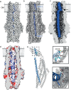
Fig. 2. KlebC1-254 binds within the lumen of the KqTolC trimer.
a Single-particle cryo-EM map of the KqTolC-KlebC1-254 complex with KlebC1-254 density (blue) resolved within KqTolC (grey). The map resolution extends from 3 to 4 Å. b Cut through of KqTolC-KlebC1-254 complex map in panel a with the fitted model, shows KlebC1-254 density (blue) within the KqTolC channel (grey). c Surface representation of the KqTolC-KlebC1-254 complex highlighting fit of KlebC1-254 in the lumen of KqTolC. The shape of the KqTolC channel combined with interactions between the helices of KlebC1-254 and KqTolC, result in KlebC1-254 forming a kink (denoted by the arrow) in order to pass through the pore unobstructed. d Model of the electrostatic surface for the KqTolC-KlebC1-254 complex (the protein sequence for helix 1 is depicted in place of polyalanine used to solve the structure), highlighting the charge matching between the two proteins. Electrostatics generated in chimera. e KqTolC chain C shown as a cartoon (grey) interacts with KlebC1-254 (blue cartoon) helix 2. Insets show zoomed view of key interactions between KqTolC chain C (grey sticks) and KlebC1-254 (cyan sticks), which are localised predominantly to the N-terminal end of helix 2. Cryo-EM map density for modelled residues is shown as inset bottom right.
Fig. 3
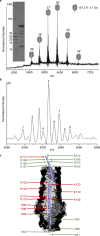
Fig. 3. Mapping the regions of KlebC1-254 protected from trypsin by KqTolC.
a Native-state ESI-MS spectrum of the trypsin-digested KqTolC-KlebC1-254 complex. Representative spectra shown from n = 3 technical replicates. Inset, 10–20% SDS-PAGE analysis of trypsin-digested KqTolC-KlebC1-254 complex after purification by gel-filtration chromatography. b Detailed view of the 26+ charge state showing heterogeneity of the sample. Numbered peaks are assigned in Supplementary Table 3. c Cartoon representation of KlebC67-163 bound within the lumen of KqTolC. The figure was constructed by superposing residues 63–81 and residues 145–172 of the KlebC51-254 crystal structure onto the KlebC77-151 seen within the TolC lumen of the cryo-EM structure. Cleaved trypsin sites are highlighted in green whilst uncut sites are shown in red.
Fig. 4
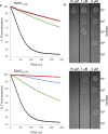
Fig. 4. KlebC fragment-based inhibition of TolC efflux activity resulting in decreased viability of K. quasipneumoniae.
a Inhibition of Nile Red Efflux through TolC in the presence of 100 µM (red), 10 µM (blue), 1 µM (green) or 0 µM (black) KlebC1-254 or KlebC51-254. Data shown are one representative experiment of two biological repeats. b Growth of serial dilution of K. quasipneumoniae prepared in the presence of 10, 1 or 0 µM KlebC1-254 or KlebC51-254 when plated onto LB-Agar in the presence of 1 mg ml−1 Rhodamine 6G.
Fig. 5
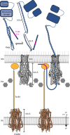
Fig. 5. Model depicting how the KlebC toxin crosses the OM through TolC.
The N-terminal domain of KlebC (blue) exists as a folded hairpin in solution, with a disordered N-terminal domain containing a TonB-box sequence (magenta). The helical hairpin is likely in equilibrium with an unfurled counterpart, which is the binding-competent form of KlebC. We speculate that electrostatic attraction brings the positively charged N-terminus of KlebC into the negatively charged TolC channel. Thereafter, KlebC snakes through the TolC channel, resulting in ultra-slow association kinetics for the complex. The disordered N-terminal region of KlebC then passes through the periplasmic entrance of TolC, which likely requires flexing of the iris helices to accommodate a disordered polypeptide. Around 70 residues at the N-terminus of KlebC escape the iris leaving the helical regions of KlebC docked within the TolC channel. The disordered nature of these residues means that a wide search radius is possible. Toxin translocation is driven by TonB, which associates with the KlebC TonB box (residues 16–20), in conjunction with the PMF-linked inner membrane stator complex ExbB-ExbD. Previous work on TonB-dependent bacteriocins has shown that this protein–protein interaction is sufficient to drive entry into the cell. How the nuclease then translocates to the cytoplasm is not known but may involve the inner membrane AAA+ATPase/protease FtsH, as has been shown for E. coli-specific colicins, .

References

    1. Nikaido H. Molecular basis of bacterial outer membrane permeability revisited. Microbiol. Mol. Biol. Rev. 2003;67:593–656. doi: 10.1128/MMBR.67.4.593-656.2003. - DOI - PMC - PubMed
    1. Pagès JM, James CE, Winterhalter M. The porin and the permeating antibiotic: a selective diffusion barrier in Gram-negative bacteria. Nat. Rev. Microbiol. 2008;6:893–903. doi: 10.1038/nrmicro1994. - DOI - PubMed
    1. Koronakis V, Eswaran J, Hughes C. Structure and function of TolC: the bacterial exit duct for proteins and drugs. Annu. Rev. Biochem. 2004;73:467–489. doi: 10.1146/annurev.biochem.73.011303.074104. - DOI - PubMed
    1. Wandersman C, Delepelaire P. TolC, an Escherichia coli outer membrane protein required for hemolysin secretion. Proc. Natl Acad. Sci. USA. 1990;87:4776–4780. doi: 10.1073/pnas.87.12.4776. - DOI - PMC - PubMed
    1. Cascales E, et al. Colicin biology. Microbiol. Mol. Biol. Rev. 2007;71:158–229. doi: 10.1128/MMBR.00036-06. - DOI - PMC - PubMed

Publication types

MeSH terms

Supplementary concepts