. 2021 Dec;26(12):7746-7759.
doi: 10.1038/s41380-021-01238-3. Epub 2021 Jul 30.

Autoantibodies against NMDA receptor 1 modify rather than cause encephalitis

Affiliations

Autoantibodies against NMDA receptor 1 modify rather than cause encephalitis

Justus B H Wilke et al. Mol Psychiatry. 2021 Dec.

Abstract

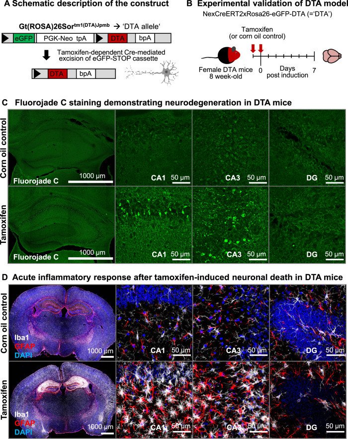
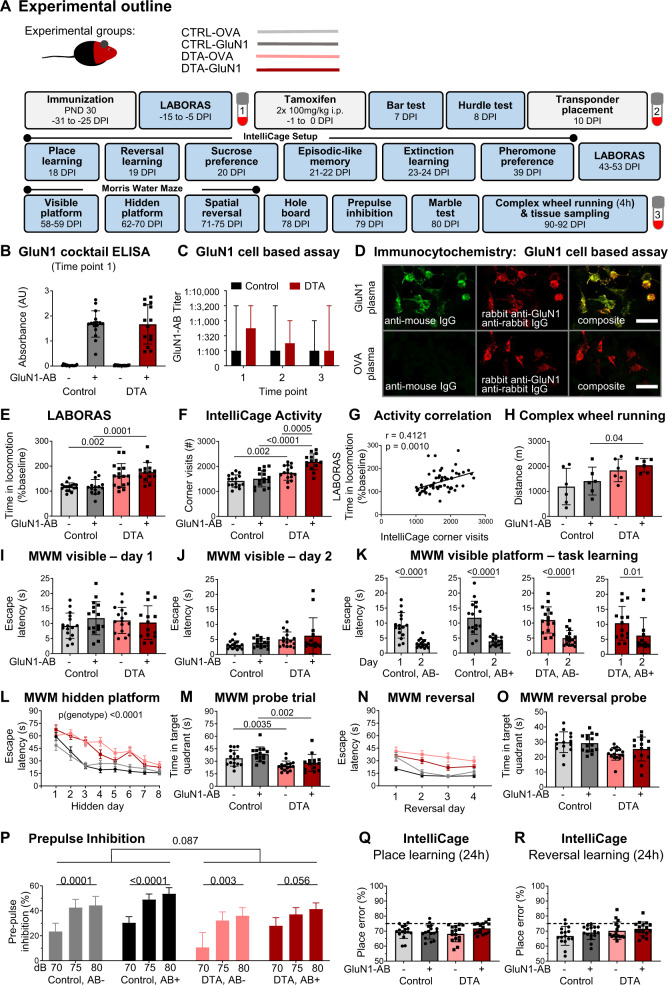
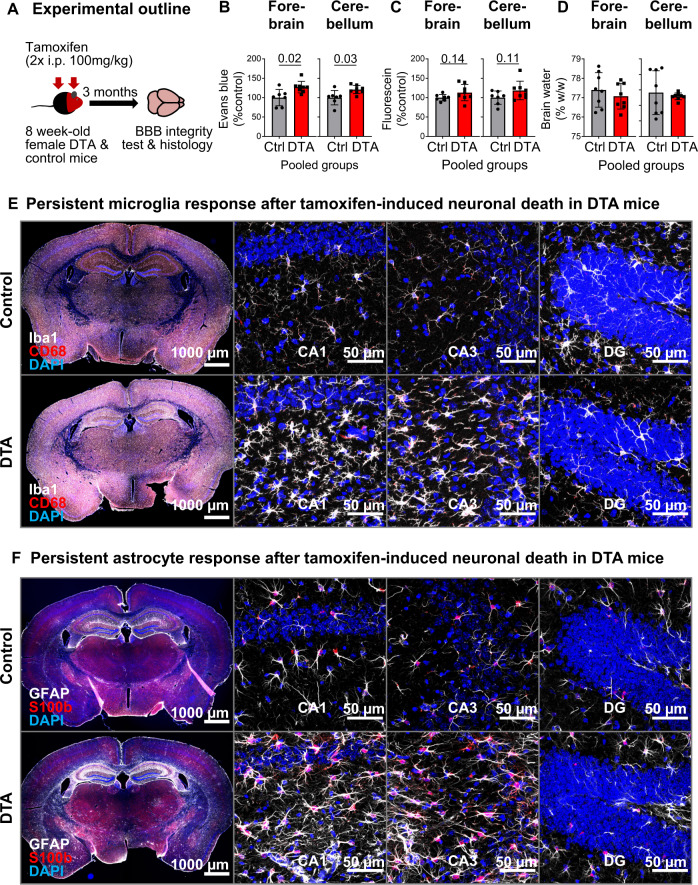
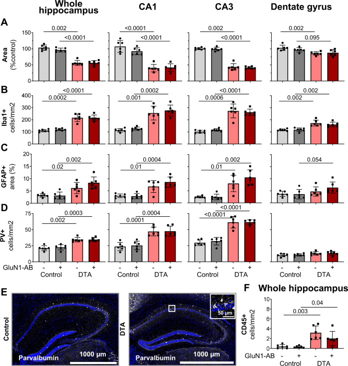
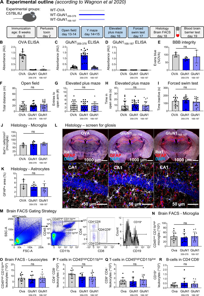
The etiology and pathogenesis of "anti-N-methyl-D-aspartate-receptor (NMDAR) encephalitis" and the role of autoantibodies (AB) in this condition are still obscure. While NMDAR1-AB exert NMDAR-antagonistic properties by receptor internalization, no firm evidence exists to date that NMDAR1-AB by themselves induce brain inflammation/encephalitis. NMDAR1-AB of all immunoglobulin classes are highly frequent across mammals with multiple possible inducers and boosters. We hypothesized that "NMDAR encephalitis" results from any primary brain inflammation coinciding with the presence of NMDAR1-AB, which may shape the encephalitis phenotype. Thus, we tested whether following immunization with a "cocktail" of 4 NMDAR1 peptides, induction of a spatially and temporally defined sterile encephalitis by diphtheria toxin-mediated ablation of pyramidal neurons ("DTA" mice) would modify/aggravate the ensuing phenotype. In addition, we tried to replicate a recent report claiming that immunizing just against the NMDAR1-N368/G369 region induced brain inflammation. Mice after DTA induction revealed a syndrome comprising hyperactivity, hippocampal learning/memory deficits, prefrontal cortical network dysfunction, lasting blood brain-barrier impairment, brain inflammation, mainly in hippocampal and cortical regions with pyramidal neuronal death, microgliosis, astrogliosis, modest immune cell infiltration, regional atrophy, and relative increases in parvalbumin-positive interneurons. The presence of NMDAR1-AB enhanced the hyperactivity (psychosis-like) phenotype, whereas all other readouts were identical to control-immunized DTA mice. Non-DTA mice with or without NMDAR1-AB were free of any encephalitic signs. Replication of the reported NMDAR1-N368/G369-immunizing protocol in two large independent cohorts of wild-type mice completely failed. To conclude, while NMDAR1-AB can contribute to the behavioral phenotype of an underlying encephalitis, induction of an encephalitis by NMDAR1-AB themselves remains to be proven.


Conflict of interest statement

WS is a member of the board and holds stocks in Euroimmun AG. All other authors declare no competing financial or other interests.

Figures

Fig. 1
Fig. 1. Acute inflammatory response upon tamoxifen-induced Cre recombination in NexCreERT2xRosa26-eGFP-DTA (DTA) mice.
A Schematic description of the Rosa26-eGFP-DTA allele. Upon tamoxifen-dependent Cre translocation, a loxP- (triangles) flanked eGFP-STOP cassette is excised, resulting in expression of diphtheria toxin chain A and ultimately cell death. B Experimental validation of the acute inflammatory response in DTA mice. Female DTA mice, 8-week old, received 2 intraperitoneal injections of either tamoxifen or solvent control (corn oil). C, D Brains were collected after 7 days and stained for neurodegeneration/cell death with Fluorojade C, as well as microglia (Iba1) and astrocytes (GFAP) as indicators of reactive gliosis. High-resolution images of CA1, CA3, and dentate gyrus (DG) regions were acquired as 10 µm Z-stacks and are displayed as maximum-intensity projections.
Fig. 2
Fig. 2. Pathophysiological relevance of NMDAR1-AB (=GluN1-AB) in the context of gray-matter inflammation.
A Experimental outline indicating experimental groups, as well as order and time of behavioral tests (DPI = days post induction, i.e., after the last tamoxifen injection). B NMDAR1-AB validation by ELISA. C Cell-based (HEK293T) clinical standard assay for NMDAR1-AB (Euroimmun). D Immunocytochemical colocalization (CBA, Euroimmun) with a commercial rabbit GluN1-AB directed against the C-terminal domain. E Intra individual change of locomotor activity assessed in LABORAS at baseline and after tamoxifen induction. For each mouse, time in locomotion after tamoxifen induction was normalized to time in locomotion prior to tamoxifen application (baseline). F Activity (number of corner visits) over a 7-day IntelliCage session. G Pearson correlation between IntelliCage activity and intra individual changes of LABORAS locomotor activity. H Locomotor activity assessed by 4 h voluntary complex wheel running. I–O Cognitive testing in Morris water maze (MWM). I–K Visible platform task comprising 2 training days, demonstrating the ability for fast escape and simple task learning using within-maze cues. L Training of hidden platform task using extra-maze cues. DTA mice performed significantly worse than control mice (repeated-measures ANOVA, p < 0.0001), whereas no effect of NMDAR1-AB was observed in either DTA (p = 0.3273) or control mice (p = 0.5972). M Evaluation of spatial memory in the probe trial. N Spatial reversal of the hidden platform. O Evaluation of cognitive flexibility and reversal learning in a second probe trial after spatial reversal training. P Prepulse inhibition of acoustic startle. Intra group comparisons performed using repeated-measure one-way ANOVA; inter group comparison between genotypes for 75 and 80 dB prepulses performed using mixed ANOVA. Q IntelliCage-based evaluation of place learning and R reversal learning within 24 h sessions shows similar performance across groups. Dashed lines indicate performance at chance level (75%). Experiments were performed with 14–16 mice/group, except for CRW (H, n = 6 mice/group). Data presented as mean  ±  SD, except for repeated measure data (L, N, P; mean  ±  SEM) and AB titers (C, median, range).
Fig. 3
Fig. 3. Long-lasting inflammatory response at 3 months after tamoxifen induction.
A Abbreviated experimental outline (detailed schematic in Fig. 2A). Blood–brain barrier (BBB) permeability, irrespective of NMDAR1-AB status, assessed by Evans blue (B) and fluorescein (C) extravasation, as well as brain-water content (D) in the forebrain and cerebellum of DTA and control mice. Data presented as mean  ± SD. E, F Representative images demonstrating persistent inflammatory changes in the hippocampus of DTA mice, including increased microglia density and apparent changes in morphology (E, quantifications in Fig. 4B) and increased expression of glial fibrillary acidic protein, GFAP (F), quantified in Fig. 4C). High-resolution images of CA1, CA3, and dentate gyrus (DG) regions were acquired as 10 µm Z-stacks and displayed as maximum-intensity projections.
Fig. 4
Fig. 4. Histological quantification of typical encephalitis readouts in the hippocampus.
A For assessment of atrophy, the respective hippocampal areas were normalized to the mean of control mice. Normalized area shows the average of 6 sections per mouse (within Bregma −1.34 mm and −1.64 mm). B Number of Iba1+ cells (microglia). C GFAP+ area determined densitometrically upon uniform thresholding. D Density of parvalbumin (PV)-positive interneurons. E Representative images of parvalbumin stainings in control and DTA mice. F Leukocyte (CD45+ and Iba1− cells/mm2) infiltration into the hippocampal parenchyma. Data from 6 mice/group displayed as mean  ± SD.
Fig. 5
Fig. 5. Summary of the results from the replication study.
A Experimental outline, following the protocol of Wagnon et al. [17]. B–D Experimental validation of immunization success using OVA-ELISA, GluN1359–378-ELISA, and GluN1168-187-ELISA. E Blood–brain-barrier (BBB) integrity assessed through Evans blue extravasation. F–I Results of behavioral phenotyping, showing locomotor activity in the open field, anxiety-related behavior in elevated plus maze, and depression-like behavior in the forced-swim test. J–L Histological quantification using 8 mice/immunization with focus on reactive gliosis, showing microglia numbers, GFAP+ area (densitometry), and representative images of quantified stainings. High-resolution images of CA1 were acquired as 10 µm Z-stacks and displayed as maximum-intensity projections. M–R Characterization of the brains’ immune cell compartment by flow cytometry of 11–12 mice/group. M Gating strategy. Quantification of CD11bhighCD45mid cells (microglia). Quantification of CD11blowCD45high leukocytes, CD4+ T cells, CD8+ T cells, and CD19+ B cells. Data displayed as mean ±SD.

References

    1. Dalmau J, Gleichman AJ, Hughes EG, Rossi JE, Peng X, Lai M, et al. Anti-NMDA-receptor encephalitis: case series and analysis of the effects of antibodies. Lancet Neurol. 2008;7:1091–8. - PMC - PubMed
    1. Dalmau J, Tüzün E, Wu HY, Masjuan J, Rossi JE, Voloschin A, et al. Paraneoplastic anti-N-methyl-D-aspartate receptor encephalitis associated with ovarian teratoma. Ann Neurol. 2007;61:25–36. - PMC - PubMed
    1. Hughes EG, Peng X, Gleichman AJ, Lai M, Zhou L, Tsou R, et al. Cellular and synaptic mechanisms of anti-NMDA receptor encephalitis. J Neurosci. 2010;30:5866–75. - PMC - PubMed
    1. Manto M, Dalmau J, Didelot A, Rogemond V, Honnorat J. In vivo effects of antibodies from patients with anti-NMDA receptor encephalitis: further evidence of synaptic glutamatergic dysfunction. Orphanet J Rare Dis. 2010;5:1–12. - PMC - PubMed
    1. Mikasova L, De Rossi P, Bouchet D, Georges F, Rogemond V, Didelot A, et al. Disrupted surface cross-talk between NMDA and Ephrin-B2 receptors in anti-NMDA encephalitis. Brain. 2012;135:1606–21. - PubMed

Publication types

Substances