

doi: 10.1007/978-1-0716-1593-5_6.
Multiplexed Tissue Tomography
Affiliations
- PMID: 34331280
- PMCID: PMC8559895
- DOI: 10.1007/978-1-0716-1593-5_6
Item in Clipboard
Multiplexed Tissue Tomography
Methods Mol Biol.
2021.
Abstract
Multiplexed tissue tomography enables comprehensive spatial analysis of markers within a whole tissue or thick tissue section. Clearing agents are often used to make tissue transparent and facilitate deep tissue imaging. Many methods of clearing and tissue tomography are currently used in a variety of tissue types. Here we detail a workflow known as transparent tissue tomography (T3), which builds upon previous methods and can be applied to difficult to clear tissues such as tumors.
Keywords: Antibody; Immunofluorescence; Microscopy; Multiplex imaging; Tissue clearing; Tomography.
© 2021. Springer Science+Business Media, LLC, part of Springer Nature.
Figures
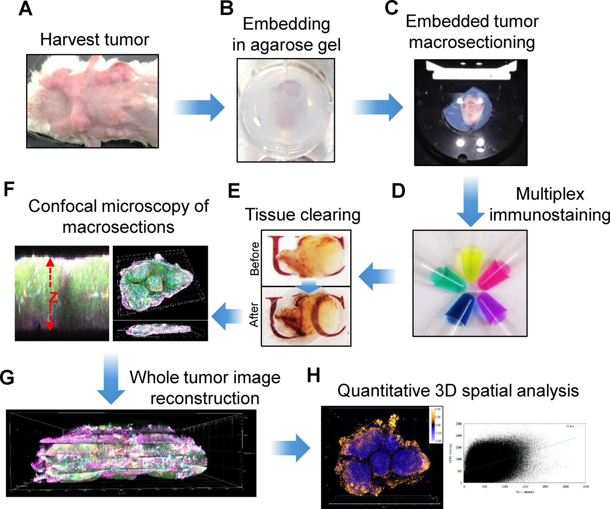
A) Harvesting whole tumors followed by light tissue fixation; B) Tissue embedding in 2% agarose gel; C) Collecting thick tissue sections (macrosections) from a vibrating microtome; D) Staining with a cocktail of fluorescent primary antibodies; E) Optical clearing of the macrosections using D-fructose solutions; F) Three-dimensional confocal imaging of multiple fluorophores; G) Image reconstruction of whole tumors by concatenating image data; H) 3D analysis of markers throughout whole tumor. Reproduced from Lee et al., 2017 with permission from Nature (reference [6])

A) Collection of a tissue cylinder (core) followed by light fixation. B) Placement of core in a pre-cast agarose well. C) Staining with a cocktail of fluorescent primary antibodies followed by washing and fixation. D) Optical clearing using D-fructose. E) Confocal imaging of both sides of the core. F) Fusion of half-cylinder images and reconstruction of the whole core; G) 3D spatial analysis of multiple markers. H) Removal of D-fructose and tissue fixation; I) 2D chromogenic immunohistochemistry (IHC) of each marker; J) Correlation between 2D and 3D image data. Reproduced from Lee et al., 2018 with permission from Nature (reference [7])

Top left: 3D rendering of a reconstructed mouse tumor revealing Her2+ cancer cells, CD45+ immune cells, proliferating cells (Ki-67), CD31+ endothelial cells, and PD-L1+ cells. Top left inset shows the appearance of the tumor mass after excision. Top right inset shows a representative 2D image with Her2+ cells. Top right: Lateral view of the reconstructed tumor shows the depth of the whole tumor. Bottom: 3D (left) and 2D (right) individual channel images for each of the five markers. Adapted from Lee et al., 2017 with permission from Nature (reference [6])
References
-
- Chung K, Wallace J, Kim S-Y, Kalyanasundaram S, Andalman AS, Davidson TJ, Mirzabekov JJ, Zalocusky KA, Mattis J, Denisin AK, Pak S, Bernstein H, Ramakrishnan C, Grosenick L, Gradinaru V, Deisseroth K (2013) Structural and molecular interrogation of intact biological systems. Nature 497:332–337. doi: 10.1038/nature12107 - DOI - PMC - PubMed
Publication types
MeSH terms
Substances
Grants and funding
LinkOut - more resources
Full Text Sources
