Review
. 2021 Sep;303(1):83-102.
doi: 10.1111/imr.13014. Epub 2021 Jul 30.

Know your enemy or find your friend?-Induction of IgA at mucosal surfaces

Affiliations
Review

Know your enemy or find your friend?-Induction of IgA at mucosal surfaces

Mats Bemark et al. Immunol Rev. 2021 Sep.

Abstract

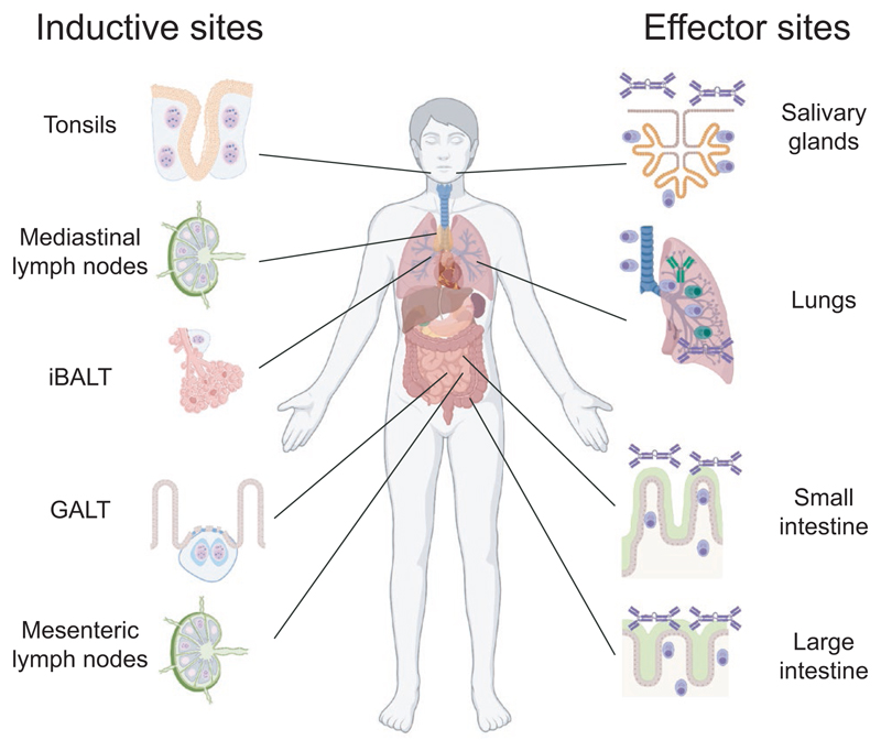
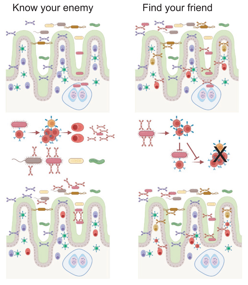
Most antibodies produced in the body are of the IgA class. The dominant cell population producing them are plasma cells within the lamina propria of the gastrointestinal tract, but many IgA-producing cells are also found in the airways, within mammary tissues, the urogenital tract and inside the bone marrow. Most IgA antibodies are transported into the lumen by epithelial cells as part of the mucosal secretions, but they are also present in serum and other body fluids. A large part of the commensal microbiota in the gut is covered with IgA antibodies, and it has been demonstrated that this plays a role in maintaining a healthy balance between the host and the bacteria. However, IgA antibodies also play important roles in neutralizing pathogens in the gastrointestinal tract and the upper airways. The distinction between the two roles of IgA - protective and balance-maintaining - not only has implications on function but also on how the production is regulated. Here, we discuss these issues with a special focus on gut and airways.

Keywords: IgA; commensal microbiota; infection; mucosa.


Conflict of interest statement

Conflict of Interest

The authors do not declare any conflicts of interest.

Figures

Figure 1
Figure 1
Inductive and effector sites for gut and airway antibody mucosal responses. The mucosal systems for immune responses in the airways and the intestinal tract contain several sites involved in activation of adaptive immune response (inductive sites) and production of antibodies (effector sites). Inductive sites for airways include tonsils (considered the nasal-associated lymphoid tissue (NALT) equivalent in humans) in the upper airways, inducible bronchus-associated lymphoid tissues (iBALT) in the lower airways and mediastinal lymph nodes (medLN) that drain the lungs. Gut-associated lymphoid tissues (GALT), primarily made up of Peyer’s patches (PP) and isolated lymphoid follicles (ILF), function as inductive sites in the gut while mesenteric lymph nodes (MLN) drain the gut. Plasma cells producing IgA antibodies are found both in the upper airways, exemplified here by a salivary gland, and lower airways where both IgA and IgG are produced, and represent airway effector sites. In the gut, the lamina propria of the large and small intestine function as an effector site and is filled with plasma cells that produce IgA antibodies
Figure 2
Figure 2. Antigen-specific B cell responses in Peyer´s patches.
(A) Peyer´s patches (PP) are supported by blood and lymph vessels that enables B and T cells to enter via high endothelial venules in areas between B cell follicles. These, as well as plasmablasts generated during the response, leave the organ via the lymph that subsequently pass the mesenteric lymph nodes before returning to blood when the thoracic ducts enters into the left subclavian vein. (B) Naive and memory B cells will enter the PP and migrate toward the subepithelial dome (SED) where they encounter antigens transported into the structure through M cells. When an antigen-specific naive or memory B cell encounters antigen, it will be activated in a process that is not dependent on antigen interactions but do not require high affinity. (C) Following activation, antigen-specific B cell will proliferate within the SED. The process is expedited if they interact with antigen-specific T cells and it may also benefit from non-cognate interactions between B and T cells. (D) Some activated B cells will enter into the preformed germinal centers (GC) where they will continue to proliferate, but SED proliferation will still be maintained. Entry and further proliferation within the GC will be dependent on antigen affinity as well as cognate interactions with T cells. B cells migrating from the SED may carry specific antigens using their B cell receptors that are loaded onto follicular dendritic cells in the GC. B cells showing signs of already having been in the GC are also present in the SED. It is not known if these migrate directly from the GC or leave as activated memory cells that subsequently reenter to the SED from blood. (E) Antigen-specific B cells and plasmablasts can leave the PP through the lymph. During the response, many of the B cells leaving appear to still have an activated phenotype
Figure 3
Figure 3
Commensal binding of IgA antibodies and consequences of the induction pathways. Two mutually non-exclusive models for IgA binding to commensal microbiota has been proposed, and these have implications on the induction of antibodies. According to a "Know your enemy" model, bacterial strains able to trigger inflammation will activate antigen-specific responses in GALT, likely through T cell-dependent mechanisms, which ensures that this bacterial strain cannot invade into tissues again. IgA antibodies will cover other bacterial strains due to cross-reactivity between epitopes. According to the "Find your friend" model, broadly reactive IgA antibodies are produced in a T independent manner before the bacterial encounter, possibly even in the absence of antigen-specific interactions due to triggering of pattern recognition receptors on B cells. Bacteria covered with non-inflammatory IgA will trigger regulatory T cell responses if the bacteria manage to invade into tissue, which in turn ensures that specific responses or inflammation are not triggered
Figure 4
Figure 4
Plasmablast responses during COVID-19 disease. Plasmablasts responses were studied in hospitalized COVID-19 patients during acute disease and three months after recovery. Plasmablast made up 10% of all CD19+ B cells in blood during active disease, but less than 1% 3 months after recovery or in healthy controls. Plasmablasts encountered in blood during active disease expressed higher levels of integrin β1, CCR10, and CD138 than those after recovery, and produced antibodies of IgA1, IgG1, and IgM class, which suggested that they many generated in a mucosal immune response. Antibodies of all classes produced by the plasmablasts during disease reacted with SARS-CoV-2, and both IgA and IgG antibodies showed cross-reactivity to endemic coronaviruses. After recovery, the plasmablasts in blood produced low levels of IgG antibodies against SARS-CoV-2 while antibodies of other classes could not be detected, and they did not show cross-reactivity

References

    1. Pabst R, Russell MW, Brandtzaeg P. Tissue distribution of lymphocytes and plasma cells and the role of the gut. Trends Immunol. 2008;29:206–208. - PubMed
    1. Corthésy B. Multi-faceted functions of secretory IgA at mucosal surfaces. Front Immunol. 2013;4:185. - PMC - PubMed
    1. Brandtzaeg P. Secretory IgA: Designed for anti-microbial defense. Front Immunol. 2013;4:222. - PMC - PubMed
    1. Xu Z, Takizawa F, Casadei E, et al. Specialization of mucosal immunoglobulins in pathogen control and microbiota homeostasis occurred early in vertebrate evolution. Sci Immunol. 2020;5:eaay3254. - PMC - PubMed
    1. Yu Y, Wang Q, Huang Z, Ding L, Xu Z. Immunoglobulins, mucosal immunity and vaccination in teleost fish. Front Immunol. 2020;11:567941. - PMC - PubMed

Publication types

Substances