

Effects of different intensities of continuous training on vascular inflammation and oxidative stress in spontaneously hypertensive rats
- PMID: 34331512
- PMCID: PMC8419160
- DOI: 10.1111/jcmm.16813
Effects of different intensities of continuous training on vascular inflammation and oxidative stress in spontaneously hypertensive rats
Abstract
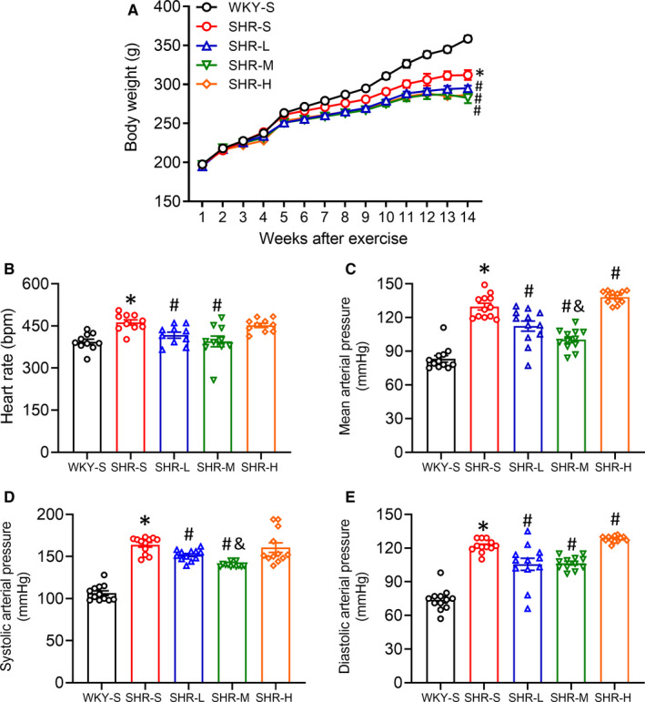
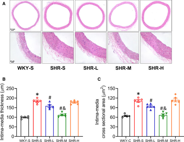
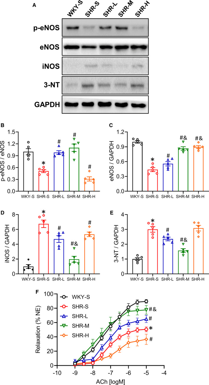
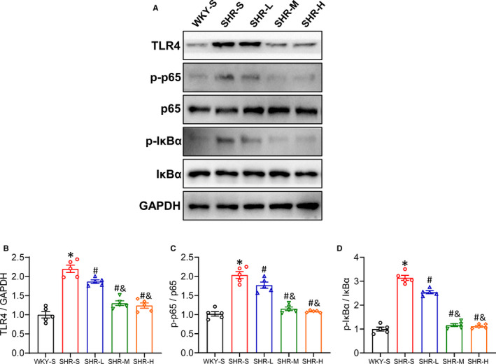
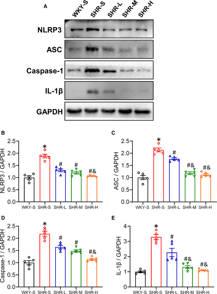
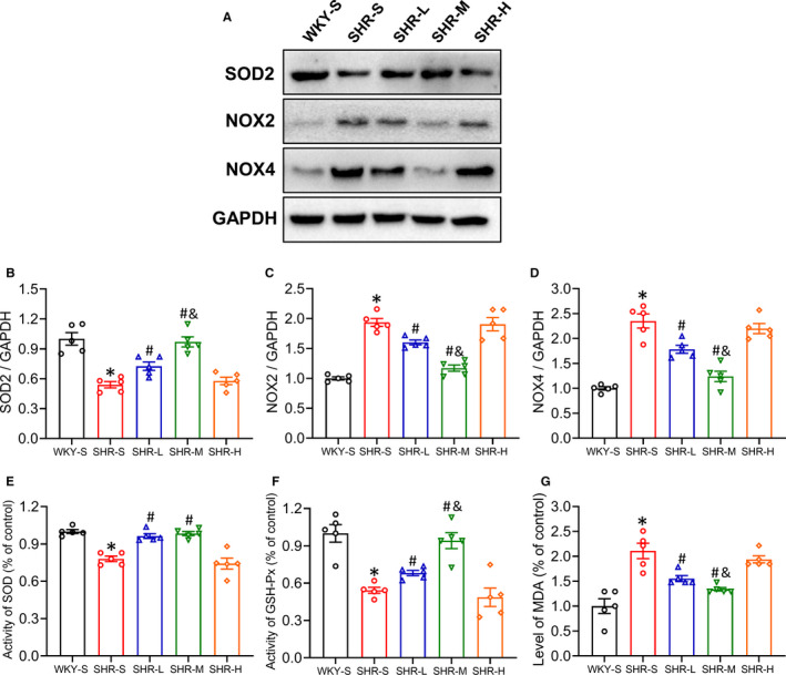
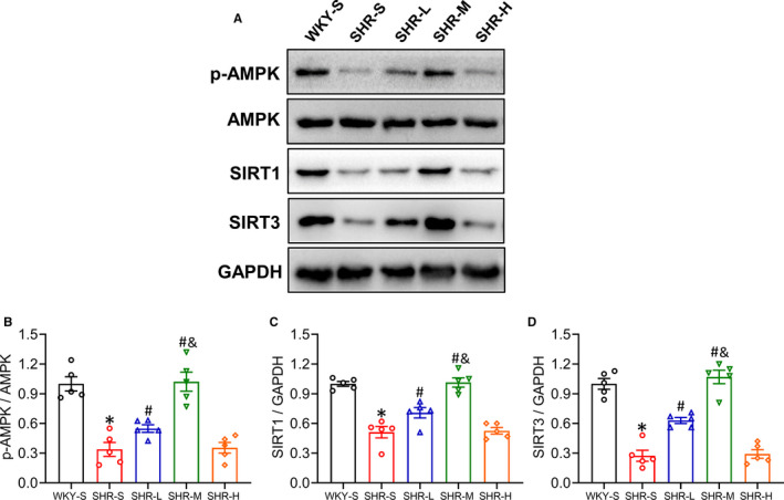
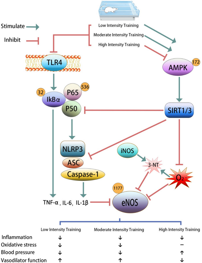
We aimed to study the effects and underlying mechanism of different intensities of continuous training (CT) on vascular inflammation and oxidative stress in spontaneously hypertensive rats (SHR). Rats were divided into five groups (n = 12): Wistar-Kyoto rats sedentary group (WKY-S), sedentary group (SHR-S), low-intensity CT group (SHR-L), medium-intensity CT group (SHR-M) and high-intensity CT group (SHR-H). Changes in body mass, heart rate and blood pressure were recorded. The rats were euthanized after 14 weeks, and blood and vascular tissue samples were collected. Haematoxylin and Eosin staining was used to observe the aortic morphology, and Western blot was used to detect the expression of mesenteric artery proteins. After CT, the mean arterial pressures improved in SHR-L and SHR-M and increased in SHR-H compared with those in SHR-S. Vascular inflammation and oxidative stress levels significantly subsided in SHR-L and SHR-M (p < 0.05), whereas in SHR-H, only vascular inflammation significantly subsided (p < 0.05), and oxidative stress remained unchanged (p > 0.05). AMPK and SIRT1/3 expressions in SHR-L and SHR-M were significantly up-regulated than those in SHR-S (p < 0.05). These results indicated that low- and medium-intensity CT can effectively reduce the inflammatory response and oxidative stress of SHR vascular tissue, and high-intensity CT can improve vascular tissue inflammation but not oxidative stress.
Keywords: blood vessel; oxidative stress; spontaneously hypertensive rats; training intensity; vascular inflammation.
© 2021 The Authors. Journal of Cellular and Molecular Medicine published by Foundation for Cellular and Molecular Medicine and John Wiley & Sons Ltd.
Conflict of interest statement
The authors declare no conflict of interest.
Figures
Similar articles
-
Coconut oil supplementation and physical exercise improves baroreflex sensitivity and oxidative stress in hypertensive rats.Appl Physiol Nutr Metab. 2015 Apr;40(4):393-400. doi: 10.1139/apnm-2014-0351. Epub 2015 Feb 9. Appl Physiol Nutr Metab. 2015. PMID: 25659569
-
Impact of moderate- and high-intensity exercise on the endothelial ultrastructure and function in mesenteric arteries from hypertensive rats.Life Sci. 2019 Apr 1;222:36-45. doi: 10.1016/j.lfs.2019.01.058. Epub 2019 Feb 27. Life Sci. 2019. PMID: 30825543 Free PMC article.
-
Time-dependent effects of training on cardiovascular control in spontaneously hypertensive rats: role for brain oxidative stress and inflammation and baroreflex sensitivity.PLoS One. 2014 May 1;9(5):e94927. doi: 10.1371/journal.pone.0094927. eCollection 2014. PLoS One. 2014. PMID: 24788542 Free PMC article.
-
[Effects and mechanisms of negative air ions on blood pressure, oxidative stress and inflammation levels in SHR rats].Wei Sheng Yan Jiu. 2024 Mar;53(2):300-309. doi: 10.19813/j.cnki.weishengyanjiu.2024.02.019. Wei Sheng Yan Jiu. 2024. PMID: 38604968 Chinese.
-
Cardiac changes in spontaneously hypertensive rats: Modulation by aerobic exercise.Prog Biophys Mol Biol. 2023 Jan;177:109-124. doi: 10.1016/j.pbiomolbio.2022.11.001. Epub 2022 Nov 5. Prog Biophys Mol Biol. 2023. PMID: 36347337 Review.
Cited by
-
MICT ameliorates hypertensive nephropathy by inhibiting TLR4/NF-κB pathway and down-regulating NLRC4 inflammasome.PLoS One. 2024 Jul 25;19(7):e0306137. doi: 10.1371/journal.pone.0306137. eCollection 2024. PLoS One. 2024. PMID: 39052650 Free PMC article.
-
Measurement of blood pressure in rats: Invasive or noninvasive methods?Physiol Rep. 2024 Sep;12(17):e70041. doi: 10.14814/phy2.70041. Physiol Rep. 2024. PMID: 39266877 Free PMC article. Review.
-
Recurrent Hypoglycemia Impaired Vascular Function in Advanced T2DM Rats by Inducing Pyroptosis.Oxid Med Cell Longev. 2022 Jul 23;2022:7812407. doi: 10.1155/2022/7812407. eCollection 2022. Oxid Med Cell Longev. 2022. PMID: 35915611 Free PMC article.
-
HIIT and MICT mitigate endothelial dysfunction in early atherosclerotic mice via PCSK9 inhibition.Sci Rep. 2025 Aug 19;15(1):30411. doi: 10.1038/s41598-025-05206-7. Sci Rep. 2025. PMID: 40830352 Free PMC article.
-
Aerobic exercise inhibits renal EMT by promoting irisin expression in SHR.iScience. 2023 Jan 14;26(2):105990. doi: 10.1016/j.isci.2023.105990. eCollection 2023 Feb 17. iScience. 2023. PMID: 36798442 Free PMC article.
References
-
- Tanito M, Nakamura H, Kwon YW, et al. Enhanced oxidative stress and impaired thioredoxin expression in spontaneously hypertensive rats. Antioxid Redox Signal. 2004;6:89‐97. - PubMed
-
- Siti HN, Kamisah Y, Kamsiah J. The role of oxidative stress, antioxidants and vascular inflammation in cardiovascular disease (a review). Vascul Pharmacol. 2015;71:40‐56. - PubMed
-
- Hayden MS, Ghosh S. Shared principles in NF‐kappaB signaling. Cell. 2008;132(3):344‐362. - PubMed
Publication types
MeSH terms
LinkOut - more resources
Full Text Sources
Medical
