. 2022 Jan;22(1):46-57.
doi: 10.1111/ajt.16780. Epub 2021 Aug 31.

Kidney transplantation from triple-knockout pigs expressing multiple human proteins in cynomolgus macaques

Affiliations

Kidney transplantation from triple-knockout pigs expressing multiple human proteins in cynomolgus macaques

David Ma et al. Am J Transplant. 2022 Jan.

Abstract

Porcine cells devoid of three major carbohydrate xenoantigens, αGal, Neu5GC, and SDa (TKO) exhibit markedly reduced binding of human natural antibodies. Therefore, it is anticipated that TKO pigs will be better donors for human xenotransplantation. However, previous studies on TKO pigs using old world monkeys (OWMs) have been disappointing because of higher anti-TKO pig antibodies in OWMs than humans. Here, we show that long-term survival of renal xenografts from TKO pigs that express additional human transgenes (hTGs) can be achieved in cynomolgus monkeys. Kidney xenografts from TKO-hTG pigs were transplanted into eight cynomolgus recipients without pre-screening for low anti-pig antibody titers. Two recipients of TKO-hTG xenografts with low expression of human complement regulatory proteins (CRPs) (TKO-A) survived for 2 and 61 days, whereas six recipients of TKO-hTG xenografts with high CRP expression (TKO-B) survived for 15, 20, 71, 135, 265, and 316 days. Prolonged CD4+ T cell depletion and low anti-pig antibody titers, which were previously reported important for long-term survival of αGal knock-out (GTKO) xenografts, were not always required for long-term survival of TKO-hTG renal xenografts. This study indicates that OWMs such as cynomolgus monkeys can be used as a relevant model for clinical application of xenotransplantation using TKO pigs.

Keywords: immunosuppression/immune modulation; translational research/science; xenoantibody; xenoantigen; xenotransplantation.


Conflict of interest statement

The authors of this manuscript have conflicts of interest to disclose as described by the American Journal of Transplantation. Research funding was provided by eGenesis Inc. eGenesis has filed patent applications on the transgenic pig technology described in this paper. Wenning Qin, Yinan Kan, Jacob V. Layer, Violette B. Paragas, Kathryn Stiede, Katherine C. Hall, Michele E. Youd, Luis M. Queiroz, William F. Westlin, Michael Curtis are employees of eGenesis Bio. and Luhan Yang is a former employee of eGenesis with current affiliation to Qihan Bio. James F. Markmann and Robert B. Colvin are consultants of eGenesis. Other authors have no conflicts of interest to disclose.

Figures

FIGURE 1
FIGURE 1
Immunosuppressive regimen. After induction with ATG and anti‐CD20 (aCD20) mAb, recipients received renal xenograft from either GTKO/CD55 or TKO/hTG pigs. After transplantation, either anti‐CD40 (aCD40) or anti‐CD154 (aCD154) mAb on days 0, 2, 5, 7,12 and weekly (20 mg/kg), daily mycophenolate mofetil (MMF) and a 2‐month course of daily tacrolimus (Tac) or rapamycin (Rapa) were administered. Methylprednisolone was also administered intramuscularly for 1 month. One to two additional doses of anti‐CD20 mAb were administered to maintain CD20+ B cell depletion at least until day 100
FIGURE 2
FIGURE 2
Serum creatinine in recipients transplanted with GTKO/CD55 xenografts. Two recipients treated with the anti‐CD154 mAb‐based regimen survived for 76 and 93 days, while both recipients treated with anti‐CD40 mAb quickly lost their xenografts due to either rejection or TMA on day 11 and 15
FIGURE 3
FIGURE 3
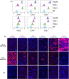
(A) Expression of human proteins in TKO‐A (EGEN‐2528) and TKO‐B (EGEN‐2536) pig donors. Human protein expression was determined by flow cytometry using ear punch–derived cells. Histograms representing the expression of indicated transgenes. Expression of complement regulatory genes (CRPs) was low in TKO‐A, while their expression was high in TKO‐B cells. On the other hand, expression of HLA‐E/B2M and CD47 was high in TKO‐A, but lower in TKO‐B. PDL‐1 was expressed only in TKO‐A. HUVEC, human umbilical vein endothelial cells; WT, ear punch–derived cells from wild type pigs; HLAE, HLA‐E; B2M, beta‐2‐microglobulin. (B) Expression of human proteins in kidneys from TKO‐A and TKO‐B. Immunofluorescence staining of the human transgenic proteins in TKO‐A (EGEN‐2528), TKO‐B (EGEN‐2536), and wild‐type kidney cryosections and FFPE sections, as described in Section 2. Nuclear counterstaining was performed with the Hoechst dye. Scale bars, 50 μm. TKO‐B kidneys showed very high expression of hCD46 and moderate expression of hCD55, hCD59 HLA‐E, and hCD47. In contrast, expression of CRPs in TKO‐A kidneys was weak (CD46) or absent (CD55 and CD59), while expression of CD47 and PD‐L1 was high
FIGURE 4
FIGURE 4
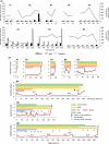
(A) Anti‐donor IgG and IgM antibodies and platelet counts pre‐ and posttransplant. Antibody bindings to donor pig endothelial cells were measured by flow cytometry as described in the method. The each column shows the ratio of mean fluorescent intensity (MFI) of IgG (black) and IgM (gray) antibody binding to the background MFI without serum. Dotted lines indicate platelet counts (×103/mm3). Antibody titers against donor endothelial cells started to elevate after reduction of immunosuppression. Severe TMA was associated with thrombocytopenia in B3 and B6. (B) Clinical courses of TKO‐A and TKO‐B recipients. Clinical courses and immunosuppressive medications of eight TKO‐A and B recipients were depicted. Red lines indicated serum creatinine levels (mg/dl). Recipients were treated with weekly anti‐CD154 mAb (green), daily MMF (yellow), daily rapamycin (pink), or tacrolimus (blue) for two months and methylprednisolone (pred, gray) for 1 month. Parvovirus infection (♦) and several episodes of bacteremia (▼) and UTI (U) were observed during the post‐op course of long‐term survivors (B4, B5, and B6). These infectious complications necessitated to reduce the dose of immunosuppressive medications, which resulted in terminal rejections. Bx, kidney xenograft biopsy
FIGURE 5
FIGURE 5
Histopathological findings in two long‐term survivors. (A–D) (B4): Biopsy on day 203 showed no rejection (H&E, ×10) (A) and no TMA (Pas, 20×) (B). However, after reduction of immunosuppression, biopsy on day 237 showed early TCMR and focal glomerular TMA (C). Terminal rejection on d265 (D). (E–H) (B6): Biopsy taken on day 217 showed no rejection (E), no TMA (F), and no C4d deposition (G). Autopsy on day 316 showed pyelonephritis, TCMR, and CAMR (H)
FIGURE 6
FIGURE 6
Lymphocyte subsets and transplant outcome (TKO‐B recipients). Absolute counts (mean ± SE) of various lymphocyte subsets in the long‐term (>100 days) survivors (B4–B6, magenta) were compared with those in the short‐term (<100 days) survivors (B1–B3, blue). Naïve (CD95CD28+), central memory (TCM, CD95+CD28+), and effector memory (TEM, CD95+CD28) T cells (CD3+CD4+ or CD3+CD8+) recovered quickly by day 10, while NK cells (NKG2a+CD16+CD8+CD3) recovered slowly by day 100. B cells (CD3CD20+) were deleted from the peripheral blood up to 260 days by 2–3 doses of rituximab. There was no statistically significant difference in these lymphocyte counts between short‐term and long‐term survivors but effector memory T cells were more depressed in the short‐term survivors [Color figure can be viewed at wileyonlinelibrary.com]

References

    1. UNOS Transplant trends. 2020. https://unos.org/data/transplant‐trends/. Accessed February 20, 2021.
    1. Cooper DKC, Gaston R, Eckhoff D, et al. Xenotransplantation‐the current status and prospects. Br Med Bull. 2018;125(1):5‐14. - PMC - PubMed
    1. Nunes dos Santos RM, Carneiro D'Albuquerque LA, Reyes LM, et al. CRISPR/Cas and recombinase‐based human‐to‐pig orthotopic gene exchange for xenotransplantation. J Surg Res. 2018;229:28‐40. - PubMed
    1. Estrada JL, Martens G, Li P, et al. Evaluation of human and non‐human primate antibody binding to pig cells lacking GGTA1/CMAH/beta4GalNT2 genes. Xenotransplantation. 2015;22(3):194‐202. - PMC - PubMed
    1. Yamamoto T, Iwase H, Patel D, et al. Old World Monkeys are less than ideal transplantation models for testing pig organs lacking three carbohydrate antigens (Triple‐Knockout). Sci Rep. 2020;10(1):9771. - PMC - PubMed

Publication types