. 2021 Jul 31;19(1):228.
doi: 10.1186/s12951-021-00969-3.

Directional homing of glycosylation-modified bone marrow mesenchymal stem cells for bone defect repair

Affiliations

Directional homing of glycosylation-modified bone marrow mesenchymal stem cells for bone defect repair

Long Chen et al. J Nanobiotechnology. .

Abstract

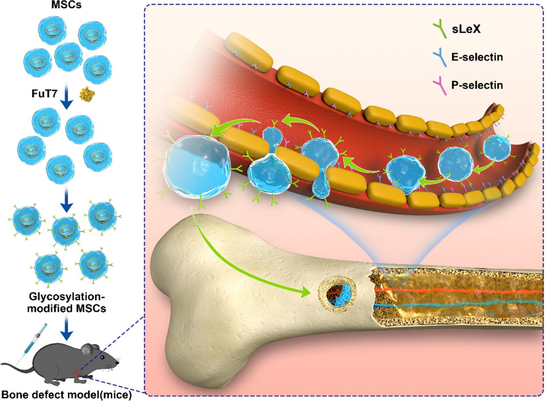
Background: One of the greatest challenges for tissue-engineered bone is the low survival rate of locally grafted cells. The cell homing technology can effectively increase the number of these grafted cells, therefore, enhancing the repair of bone defects. Here we explore the effect of fucosylation modification on the directional homing of bone marrow mesenchymal stem cells (BMSCs) and their ability to repair bone defects.

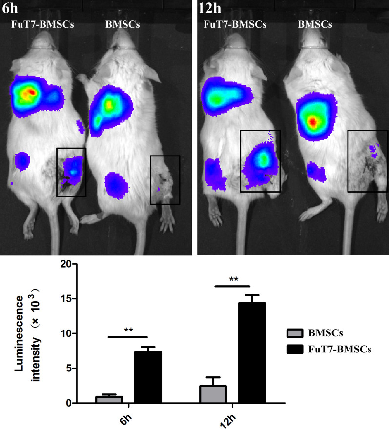
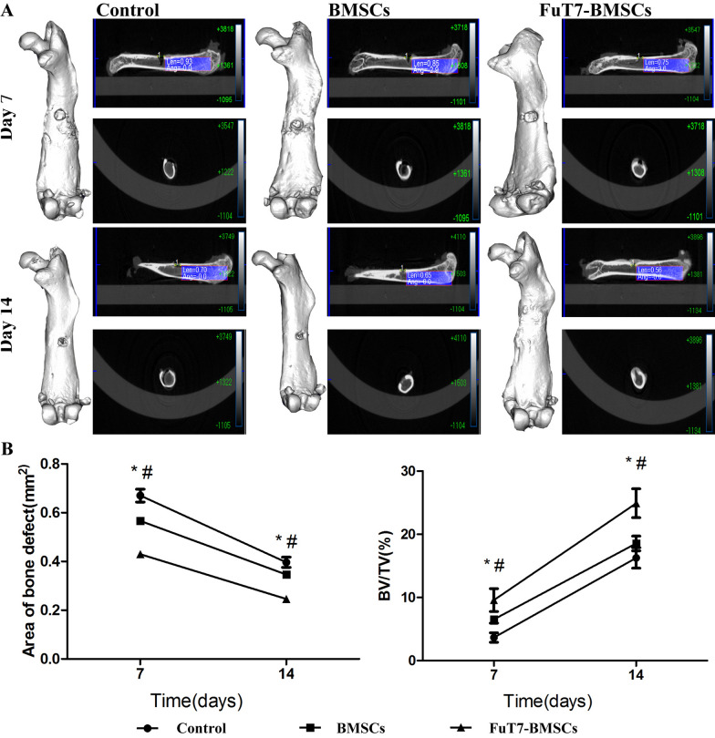
Results: Glycosylated BMSCs expressed high levels of the Sialyl Lewis-X (sLeX) antigen, which enabled the cells to efficiently bind to E- and P-selectins and to home to bone defect sites in vivo. Micro-CT and histological staining results confirmed that mice injected with FuT7-BMSCs showed an improved repair of bone defects compared to unmodified BMSCs.

Conclusions: The glycosylation modification of BMSCs has significantly enhanced their directional homing ability to bone defect sites, therefore, promoting bone repair. Our results suggest that glycosylation-modified BMSCs can be used as the source of the cells for the tissue-engineered bone and provide a new approach for the treatment of bone defects.

Keywords: Bone defect repair; Bone marrow mesenchymal stem cells; Directional homing; Glycosylation.


Conflict of interest statement

The authors declare that they have no competing interests.

Figures

Fig. 1
Fig. 1
Identification of BMSCs. Representative images showing A morphology of BMSCs, scale bar = 100 μm; B Expression of CD44, CD105 and CD34 using flow cytometry; C alizarin red staining (osteogenesis) at day 21, scale bar = 100 μm; D Oil Red O staining (adipogenesis) at day 14, scale bar = 100 μm
Fig. 2
Fig. 2
Surface glycosylation modification of BMSCs. Representative images presenting A FuT7 and empty vector expression in BMSCs, scale bar = 100 μm; B FuT7 and sLeX (CD15S) for surface glycosylation modification of BMSCs by western blotting; C relative gene expression of FuT7 for surface glycosylation modification of BMSCs, **p < 0.01; D cell proliferation analysis as shown by CCK-8 for FuT7-BMSCs and BMSCs, #p > 0.05; E alizarin red staining (osteogenesis) at day 21 for FuT7-BMSCs and BMSCs, scale bar = 200 μm; F relative gene expression of Col I and Runx2 for FuT7-BMSCs and BMSCs, **p < 0.01, #p > 0.05
Fig. 3
Fig. 3
The migration and static cell adhesion abilities of glycosylated BMSCs. Representative images presenting A Transwell cell migration assay for glycosylated BMSCs, scale bar = 200 μm, **p < 0.05; B The static adhesion abilities of glycosylated BMSCs, scale bar = 100 μm, **p < 0.05
Fig. 4
Fig. 4
The binding capacities of glycosylated BMSCs to E- and P- selectins detected by flow cytometry
Fig. 5
Fig. 5
The bone marrow flushing method for detection of the directional homing ability of glycosylated BMSCs, scale bar = 200 μm, **p < 0.05
Fig. 6
Fig. 6
The in vivo imaging method for detection of the directional homing ability of glycosylated BMSCs, **p < 0.05
Fig. 7
Fig. 7
The result of Micro-CT for regenerated bone in femoral bone defect mouse model at 7 and 14 days. Representative images presenting A Post-injection micro-CT images for regenerated bone in femoral bone defect mouse model at 7 and 14 days; B The area of bone defect for femoral bone defect mouse model at 7 and 14 days after injection, *FuT7-BMSCs versus BMSCs p < 0.05, #FuT7-BMSCs vs Control p < 0.05; (C) Bone volume (BV)/Tissue volume (TV) ratio for femoral bone defect mouse model at 7 and 14 days after injection, *FuT7-BMSCs versus BMSCs p < 0.05, #FuT7-BMSCs vs Control p < 0.05
Fig. 8
Fig. 8
The H&E and Masson staining for regenerated bone in femoral bone defect mouse model at 10 days, scale bar = 200 μm
None

Similar articles

Cited by

References

    1. Yagi H, Soto-Gutierrez A, Parekkadan B, Kitagawa Y, Tompkins RG, Kobayashi N, Yarmush ML. Mesenchymal stem cells: mechanisms of immunomodulation and homing. Cell Transplant. 2010;19(6):667–679. doi: 10.3727/096368910X508762. - DOI - PMC - PubMed
    1. Karp JM, Leng Teo GS. Mesenchymal stem cell homing: the devil is in the details. Cell Stem Cell. 2009;4(3):206–216. doi: 10.1016/j.stem.2009.02.001. - DOI - PubMed
    1. Chen L, Wu J, Wu C, Xing F, Li L, He Z, Peng K, Xiang Z. Three-dimensional co-culture of peripheral blood-derived mesenchymal stem cells and endothelial progenitor cells for bone regeneration. J Biomed Nanotechnol. 2019;15(2):248–260. doi: 10.1166/jbn.2019.2680. - DOI - PubMed
    1. Peng J, Chen L, Peng K, Chen X, Wu J, He Z, Xiang Z. Bone marrow mesenchymal stem cells and endothelial progenitor cells co-culture enhances large segment bone defect repair. J Biomed Nanotechnol. 2019;15(4):742–755. doi: 10.1166/jbn.2019.2735. - DOI - PubMed
    1. Fong EL, Chan CK, Goodman SB. Stem cell homing in musculoskeletal injury. Biomaterials. 2011;32(2):395–409. doi: 10.1016/j.biomaterials.2010.08.101. - DOI - PMC - PubMed

LinkOut - more resources