Review
. 2021 Oct:211:108707.
doi: 10.1016/j.exer.2021.108707. Epub 2021 Jul 29.

Redox chemistry of lens crystallins: A system of cysteines

Affiliations
Review

Redox chemistry of lens crystallins: A system of cysteines

Eugene Serebryany et al. Exp Eye Res. 2021 Oct.

Abstract

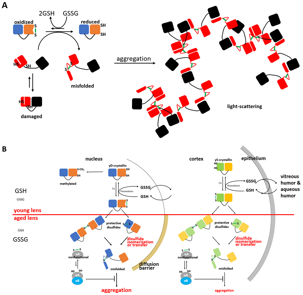
The nuclear region of the lens is metabolically quiescent, but it is far from inert chemically. Without cellular renewal and with decades of environmental exposures, the lens proteome, lipidome, and metabolome change. The lens crystallins have evolved exquisite mechanisms for resisting, slowing, adapting to, and perhaps even harnessing the effects of these cumulative chemical modifications to minimize the amount of light-scattering aggregation in the lens over a lifetime. Redox chemistry is a major factor in these damages and mitigating adaptations, and as such, it is likely to be a key component of any successful therapeutic strategy for preserving or rescuing lens transparency, and perhaps flexibility, during aging. Protein redox chemistry is typically mediated by Cys residues. This review will therefore focus primarily on the Cys-rich γ-crystallins of the human lens, taking care to extend these findings to the β- and α-crystallins where pertinent.

Keywords: Cataract; Crystallins; Disulfide exchange; Lens biochemistry; Protein aggregation.


Figures

Figure 1:
Figure 1:. Molecular and tissue-level consequences of disulfide flow in the aging lens.
(A) The redox “hot potato” mechanism driven by aggregation. Oxidizing conditions in the lens fiber cell cytoplasm generate an excess of oxidized glutathione (GSSG) over the reduced form. A monomeric γ-crystallin (blue/orange) takes on the role of redox buffer, becoming oxidized at an easily accessible site. The resulting disulfide generates conformational frustration and is therefore reactive (prone to isomerization). A covalently damaged crystallin molecule (red/black), whose partial unfolding exposes a normally buried pair of Cys residues, receives the reactive disulfide. The resulting trapped misfolded conformer self-assembles via specific non-native interactions into aggregates large enough to scatter visible light, leading to lens opacification (adapted from (Serebryany et al., 2020; Serebryany et al., 2018)). (B) A simplified schematic of changes in redox flow and the consequent driving forces for aggregation in young vs. aged lens tissues. Young lens cortices are well supplied with reducing equivalents due to glutathione uptake from the vitreous and aqueous humors and recycling in the lens epithelium, and young lens nuclei uptake reduced glutathione from the lens cortex. Aged lenses are characterized by formation of a diffusion barrier preventing glutathione uptake by the nucleus from the cortex, which creates a highly oxidized environment, particularly in the nucleus. Disulfide formation is minimized by methylation of susceptible cysteines. Despite the evolutionary adaptation of increased thermodynamic and kinetic stability of γC/D-crystallins, which are abundant in the nucleus, relative to γS-crystallins, which are abundant in the cortex, most cases of age-related cataract are nuclear, as expected from this schematic.

Similar articles

Cited by

References

    1. Abkevich VI, Shakhnovich EI, 2000. What can disulfide bonds tell us about protein energetics, function and folding: simulations and bioninformatics analysis. J. Mol. Biol 300, 975–985. - PubMed
    1. Anderson DM, Nye-Wood MG, Rose KL, Donaldson PJ, Grey AC, Schey KL, 2020. MALDI imaging mass spectrometry of β- and γ-crystallins in the ocular lens. J. Mass Spectrom 55, 11. - PMC - PubMed
    1. Barber-Zucker S, Shaanan B, Zarivach R, 2017. Transition metal binding selectivity in proteins and its correlation with the phylogenomic classification of the cation diffusion facilitator protein family. Sci. Rep 7, 16381. - PMC - PubMed
    1. Basak A, Bateman O, Slingsby C, Pande A, Asherie N, Ogun O, Benedek GB, Pande J, 2003. High-resolution X-ray crystal structures of human γD crystallin (1.25 Å) and the R58H mutant (1.15 Å) associated with aculeiform cataract. J. Mol. Biol 328, 1137–1147. - PubMed
    1. Benedek G, 1983. Protein microstructure: Why the eye lens is transparent. Nature 302, 383–384. - PubMed

Publication types