. 2021 Aug 1;8(1):168.
doi: 10.1038/s41438-021-00602-2.

Cuticle and skin cell walls have common and unique roles in grape berry splitting

Affiliations

Cuticle and skin cell walls have common and unique roles in grape berry splitting

Ben-Min Chang et al. Hortic Res. .

Abstract

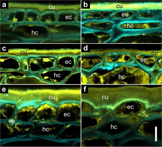
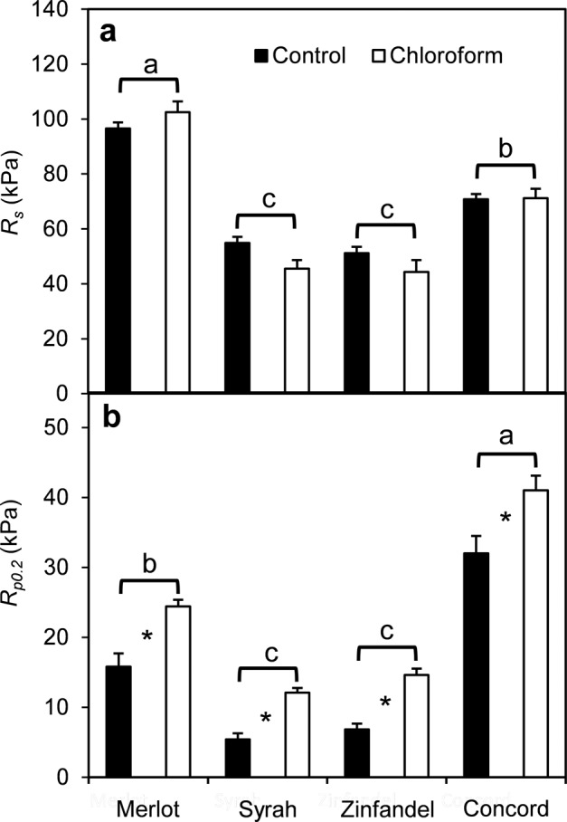
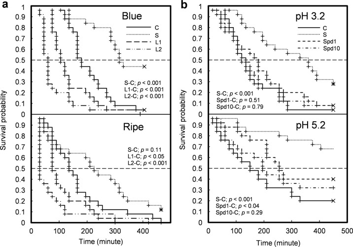
The skin protects a fruit from environmental stresses and supports the fruit's structure. Failure of the skin leads to fruit splitting and may compromise commercial production for fruit growers. The mechanical properties of the cuticle and skin cell walls might influence the splitting susceptibility of fleshy fruits. Thin shell theory and fracture mechanics were utilized in this study to target the potential factors contributing to splitting susceptibility. The study analyzed the structure of the cuticle and epidermis in ripening grape berries and examined the temporal dynamics of berry splitting. Cuticular waxes were partially removed, and skin cell walls were manipulated using wall stiffening and loosening solutions that altered reactions involving hydrogen peroxide. A more than twofold difference in cuticle thickness among grape cultivars did not account for their differences in splitting resistance. However, while removing predominantly epicuticular wax did not alter the berries' splitting resistance, their surface appearance and increasing yield strength following partial wax removal support the notion that cuticular waxes contribute to berry mechanical properties. Immersing berries in H2O2-based cell wall loosening solutions increased the splitting probability and accelerated berry splitting, whereas cell wall stiffening solutions decreased the splitting probability and delayed berry splitting. These results showed that both cuticle and skin cell walls contribute to the mechanical properties of grape berries and to their splitting resistance. The results also suggest that the two current explanations for fruit splitting, the critical turgor model and the zipper model, should be viewed as complementary rather than incompatible.


Conflict of interest statement

The authors declare no competing interests.

Figures

Fig. 1
Fig. 1. Confocal laser scanning micrographs of grape berry skin tissues.
a ‘Merlot’ green hard berry. b ‘Merlot’ overripe berry. c ‘Zinfandel’ green hard berry. d ‘Zinfandel’ overripe berry. e ‘Concord’ green hard berry. f ‘Concord’ overripe berry. The autofluorescence (501–549 nm) from phenolic compounds was visualized by yellow false color. The fluorescence (422–464 nm) from cell walls due to the Calcofluor white staining of cellulose was visualized by blue false color. Overlapping blue and yellow signals are responsible for the greenish appearance of some structures. The letters indicate cuticle (cu), epidermal cells (ec), hypodermal cells (hc), and anticlinal pegs (ap). The vertical scale bar represents 10 µm
Fig. 2
Fig. 2. Effect of partial cuticular wax removal on mechanical properties of grape berries.
a Splitting resistance (Rs). b Offset yield strength (Rp0.2). ‘Merlot’, ‘Syrah’, ‘Zinfandel’, and ‘Concord’ grape berries were immersed in chloroform for 20 s. The error bars indicate standard errors of the mean (n = 10 for ‘Merlot’, ‘Syrah’, ‘Zinfandel’; n = 5 for ‘Concord’). Asterisks indicate significant differences (p < 0.05) between control and chloroform treatment by Student’s t-test. Letters above brackets indicate significant differences among cultivars by Fisher’s least significant differences test
Fig. 3
Fig. 3. Grape berry surface observations.
a Appearance of dehydrated ‘Merlot’ berries before and after immersion in chloroform for 20 s. b Temporal progression of splitting on ‘Concord’ berry immersed in water. The time after the start of berry submersion is noted in each panel. c Distribution of pre-existing microcracks on the receptacle area or floral cap scar on ‘Concord’ berry. The left inset and arrow indicate a concentric microcrack. The right inset and arrow indicate a radial microcrack. d A split propagating bilaterally from a concentric microcrack; the radial whitish traces in the exposed berry flesh are vascular bundles
Fig. 4
Fig. 4. Survival probability of ‘Concord’ grape berries in H2O2-based immersion solutions.
a Blue and ripe berries in control (C), stiffening (S), loosening 1 (L1), and loosening 2 (L2) solutions. All solutions were based on 50 mM sodium citrate buffer at pH 3.3. Control: buffer solution. Stiffening: 50 mM H2O2 added. Loosening 1 & 2: 50 mM H2O2 + 50 mM ascorbate added. Loosening 2 received 15 min pre-treatment in 1 mM FeSO4. All other treatments received 15 min pre-treatment in the buffer. b Overripe berries in control (C), stiffening (S), 1 mM spermidine (Spd1), and 10 mM spermidine (Spd10) solutions with 50 mM sodium citrate buffer at pH 3.2 and pH 5.2. The C group was the reference for comparisons using the Gehan–Breslow–Wilcoxon test. Significance is noted for comparison pairs; p-value. Plus signs indicate individual splitting events. Multiplication signs indicate censored events, i.e., intact berries at the end of the experiment

References

    1. Hardie WJ, O’Brien TP, Jaudzems VG. Morphology, anatomy and development of the pericarp after anthesis in grape, Vitis vinifera L. Aust. J. Grape Wine Res. 1996;2:97–142. doi: 10.1111/j.1755-0238.1996.tb00101.x. - DOI
    1. Ginzberg I, Stern RA. Strengthening fruit-skin resistance to growth strain by application of plant growth regulators. Sci. Hortic. 2016;198:150–153. doi: 10.1016/j.scienta.2015.11.016. - DOI
    1. Martin LBB, Rose JKC. There’s more than one way to skin a fruit: formation and functions of fruit cuticles. J. Exp. Bot. 2014;65:4639–4651. doi: 10.1093/jxb/eru301. - DOI - PubMed
    1. Segado P, Domínguez E, Heredia A. Ultrastructure of the epidermal cell wall and cuticle of tomato fruit (Solanum lycopersicum L.) during development. Plant Physiol. 2016;170:935–946. doi: 10.1104/pp.15.01725. - DOI - PMC - PubMed
    1. Matthews MA, Cheng G, Weinbaum SA. Changes in water potential and dermal extensibility during grape berry development. J. Am. Soc. Hortic. Sci. 1987;112:314–319.