Comparative Study
. 2021 Dec 13;51(6):2931-2942.
doi: 10.3906/sag-2012-62.

Diagnostic performance of shear wave elastography and diffusion-weighted magnetic resonance imaging in cervical lymph nodes: a comparative study

Affiliations
Comparative Study

Diagnostic performance of shear wave elastography and diffusion-weighted magnetic resonance imaging in cervical lymph nodes: a comparative study

Veli Süha Öztürk et al. Turk J Med Sci. .

Abstract

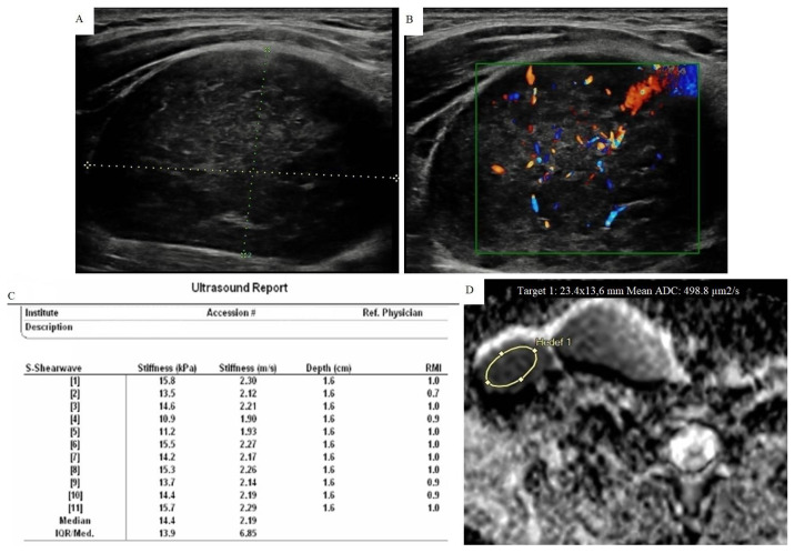
Background/aim: To investigate the potential diagnostic value of point shear wave elastography (pSWE) and the contribution of concurrent diffusion weighted magnetic resonance imaging (DW-MRI) to diagnostic performance in patients with cervical lymphadenopathy.

Materials and methods: This cross-sectional study included 116 cervical lymph nodes of 94 patients. All lymph nodes were evaluated before the treatment or histopathological sampling. Gray scale ultrasonographic features, elastographic stiffness and apparent diffusion coefficient (ADC) values were measured and recorded. Lymph nodes were divided into benign and malignant groups with histopathological findings.

Results: Short axis measurement, axis ratio, hilum morphology, vascularization patterns, pSWE, and ADC values were the most significant parameters in logistic regression tests. The median stiffness of malignant nodes was higher and the mean ADC values were lower than others. Also, lymphoproliferative disorders had the lowest ADC values (p < 0.001). Area under the curve values for pSWE and DW-MRI were 0.852 [95% confidence interval (CI), 0.779–0.925], 0.790 (95% CI, 0.695–0.885), respectively. The accuracy rate increased from 79.3% to 85.3% when the pSWE was combined with the ultrasonography (US).

Conclusion: The use of pSWE combined with conventional US will reduce the number of biopsies and may be sufficient to differentiate the lymph nodes.

Keywords: Lymph nodes; sonoelastography; ultrasound; diffusion weighted MRI.


Conflict of interest statement

All of the authors of this article have reported no disclosures. There are no relevant commercial interests in the study.

Figures

Figure 1
Figure 1
Point shear wave elastography measurement method.
Figure 2
Figure 2
A 48-year-old woman with benign cytology diagnosed with FNAB. A) There was a lymph node that newly developed in follow-up with 13 × 9 mm size, well-circumscribed, smooth border and ovoid-shaped at the level II on the right side of neck, in the case with a history of thyroid papillary cancer on the same side. However, normal fatty hilum could not be identified. B) Doppler US examination showed type II vascularization. C) pSWE parameters are shown in the table. The median stiffness value was found as 27.1 kPa (3.01 m/s) according to the elasticity measurement. D) The ADC map shows the area where the measurement was made. The average ADC value was found to be 0.813 × 10⁻³ mm2/s. It was a remarkable example with low ADC value and showed that MRI could be confusing with unnecessary use.
Figure 3
Figure 3
An 18-year-old woman diagnosed as mixed cellular Hodgkin lymphoma after lymph node excision. A) In the case with multiple lymph nodes in the neck, the largest was at level 5 on the right with a 48 × 31 mm. It showed heterogeneous internal structure and trabecular pattern. Also note that normal echogenic hilum was absent. B) Type III mixed vascularization was seen in CDUS. C) pSWE parameters are shown in a table. The median stiffness value was found as 14.4 kPa (2.19 m/s) according to the elasticity measurement. D) The ADC map shows the area where the measurement was made. The average ADC value is found to be 0.498 × 10⁻³ mm2/s. Nodes in lymphoproliferative diseases may be softer than expected. However, the diagnostic success of ADC measurement in this group was very high.
Figure 4
Figure 4
A 61-year-old woman with squamous cell carcinoma metastasis diagnosed with FNAB. A) In the case with a history of lung cancer, 17 × 12 mm sized lymph node was developed in follow-up in the level V on the right side. Lymph node had smooth contour, round shaped, fairly heterogeneous parenchyma and normal echogenic hilus could not be seen. B) Type III mixed vascularization pattern was observed in CDUS. C) pSWE parameters are shown in a table. The median stiffness value was found as 51.6 kPa (4.15 m/s) according to the elasticity measurement. D) The ADC map shows the area where the measurement was made. The average ADC value was found to be 0.722 × 10⁻³ mm2/s. It was in the malignant group with both elasticity and ADC values.

References

    1. Vassallo P, Wernecke K, Roos N, Peters P. Differentiation of benign from malignant superficial lymphadenopathy: the role of high-resolution US. Radiology. 1992;183(1):215–220. doi: 10.1148/radiology.183.1.1549675. - DOI - PubMed
    1. Ahuja A, Ying M. An overview of neck node sonography. Investigative Radiology. 2002;37(6):333–342. doi: 10.1097/00004424-200206000-00005. - DOI - PubMed
    1. Van den Brekel M, Castelijns JA, Snow GB. Detection of lymph node metastases in the neck: radiologic criteria. Radiology. 1994;192(3):617–618. doi: 10.1148/radiology.192.3.8058923. - DOI - PubMed
    1. Lyshchik A, Higashi T, Asato R, Tanaka S, Ito J, et al. Thyroid gland tumor diagnosis at US elastography. Radiology. 2005;237(1):202–211. doi: 10.1148/radiol.2363041248. - DOI - PubMed
    1. Altay C, Seçil M. Sonoelastografinin Temel İlkeleri. Türk Radyoloji Seminerleri. 2019;7:1–12. doi: 10.5152/trs.2019.749. (in Turkish) - DOI

Publication types

LinkOut - more resources