

Positron emission tomography/computed tomography hypermetabolism of Kikuchi-Fujimoto disease mimicking malignant lymphoma: a case report and literature review
- PMID: 34334002
- PMCID: PMC8326629
- DOI: 10.1177/03000605211032859
Positron emission tomography/computed tomography hypermetabolism of Kikuchi-Fujimoto disease mimicking malignant lymphoma: a case report and literature review
Abstract
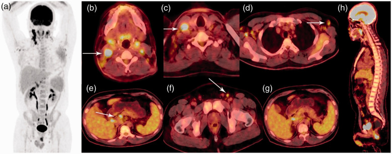
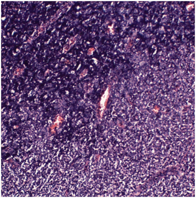
Kikuchi-Fujimoto disease (KFD), also known as histiocytic necrotizing lymphadenitis, is a benign, self-limiting inflammatory disorder of unknown etiology and pathogenesis. This report presents a rare case involving a man with 18F-fluorodeoxyglucose positron emission tomography/computed tomography (FDG PET/CT) hypermetabolism caused by KFD mimicking malignant lymphoma. The PET/CT maximum intensity projection showed multiple hypermetabolic lymphadenopathies and homogeneous FDG uptake in the bone marrow and spleen. Malignant lymphoma was initially suspected. The patient then underwent excision biopsy of one enlarged right cervical lymph node that was selected because it showed the highest FDG uptake in PET/CT, and examination of this biopsy specimen confirmed the diagnosis of KFD. PET/CT is useful for assessing the general condition of patients and can help to select lymph nodes for excision biopsy based on the highest FDG uptake. However, KFD can predispose to localized FDG uptake and limit the specificity of PET/CT by mimicking malignancy. Thus, positive results of PET/CT should be interpreted with caution.
Keywords: Kikuchi–Fujimoto disease; Positron emission tomography/computed tomography; case report; fluorodeoxyglucose; lymphadenopathy; lymphoma.
Conflict of interest statement
Figures
References
-
- Bosch X, Guilabert A, Miquel R, et al.. Enigmatic Kikuchi-Fujimoto disease: a comprehensive review. Am J Clin Pathol 2004; 122: 141–152. - PubMed
-
- Hutchinson CB, Wang E. Kikuchi-Fujimoto disease. Arch Pathol Lab Med 2010; 134: 289–293. - PubMed
-
- Pepe F, Disma S, Teodoro C, et al. . Kikuchi-Fujimoto disease: a clinicopathologic update. Pathologica 2016; 108: 120–129. - PubMed
-
- Kikuchi M. Lymphadenitis showing focal reticulum cell hyperplasia with nuclear debris and phagocytes: a clinicopathological study. Acta Hematol Jpn 1972; 35: 379–380.
-
- Fujimoto Y, Kozima Y, Yamaguchi K. Cervical subacute necrotizing lymphadenitis: a new clinicopathologic entity. Naika 1972; 20: 920–927.
Publication types
MeSH terms
Substances
LinkOut - more resources
Full Text Sources
Medical
