Review
. 2022 Jan;38(1):22-44.
doi: 10.1016/j.tig.2021.07.002. Epub 2021 Jul 29.

Advancing human disease research with fish evolutionary mutant models

Affiliations
Review

Advancing human disease research with fish evolutionary mutant models

Emily A Beck et al. Trends Genet. 2022 Jan.

Abstract

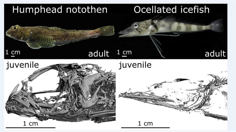
Model organism research is essential to understand disease mechanisms. However, laboratory-induced genetic models can lack genetic variation and often fail to mimic the spectrum of disease severity. Evolutionary mutant models (EMMs) are species with evolved phenotypes that mimic human disease. EMMs complement traditional laboratory models by providing unique avenues to study gene-by-environment interactions, modular mutations in noncoding regions, and their evolved compensations. EMMs have improved our understanding of complex diseases, including cancer, diabetes, and aging, and illuminated mechanisms in many organs. Rapid advancements of sequencing and genome-editing technologies have catapulted the utility of EMMs, particularly in fish. Fish are the most diverse group of vertebrates, exhibiting a kaleidoscope of specialized phenotypes, many that would be pathogenic in humans but are adaptive in the species' specialized habitat. Importantly, evolved compensations can suggest avenues for novel disease therapies. This review summarizes current research using fish EMMs to advance our understanding of human disease.

Keywords: cavefish; electric fish; icefish; killifish; mummichog; notothenioid; platyfish; stickleback; swordtail; teleost.


Conflict of interest statement

Declaration of interests The authors declare no competing interests.

Figures

Figure 1.
Figure 1.. Summary of fish evolutionary mutant models and affected organ systems.
Each circle includes a line drawing of a ray-finned evolutionary mutant model with common name and the diseased organ system primarily modeled in that species. (Mexican cavefish/heart and pancreas; Antarctic Icefish/bones and blood; Turquoise Killifish/ whole body; Platyfish/skin; Electric fish/whole body; threespine stickleback/intestines; Mummichog/brain). Lines connect these groups to their human counterpoints.
Figure 2.
Figure 2.. Summary of fish evolutionary mutant models and affected organ systems.
Phylogenetic relationships among the fish EMMs discussed in this review (in blue), in addition to the traditional model zebrafish (Danio rerio) and the non-teleost outgroup spotted gar (Lepisosteus oculatus). These relationships, published by Rabosky et al. 2018, are time-calibrated, with branch lengths in units of millions of years (MY) and scaled according to the bar below the tree. Also noted is the timing of the teleost genome duplication (TGD) event.
Figure 3.
Figure 3.. Antarctic Icefishes, Models of Anemias.
Compared to their red-blooded relative (e.g., A. Bullhead notothen), the 16 species of white-blooded icefishes lack functional hemoglobin genes (e.g., B. South Georgia icefish), and in six species, also fail to express cardiac myoglobin (e.g., C. Blackfin icefish). Compared to dark-red hearts of red-blooded species (A1), hearts of icefish species expressing myoglobin are pink (B1) and beige in icefish species failing to express myoglobin (C1). The blood of red-blooded species is dark-red and contains a large fraction of erythrocytes (A2; left, fresh blood; right, blood after centrifugation) that are ovoid (A3, Wright-Giemsa stain). In comparison icefish blood appears opalescent white and contains only limited amount of blood cells, majorly white-blood cells and thrombocytes (B2, C2). A few erythropoietic cells are present in icefish blood, however, they display abnormal morphologies and fragility (B3, C3). a, atrium; c, blood cells; e, erythrocytes; o, outflow tract; p, plasma; v, ventricle.
Figure 4.
Figure 4.. Notothenioids, Models of skeletal Dysplasias.
Morphological and molecular analyses of notothenioids revealed alterations in the timing and rates of bone development in several lineages. Compared to bottom dwellers (e.g., Humphead notothen), more buoyant notothenioids (e.g., Ocellated icefish) show delayed mineralization of bony elements, slow rates of osteological development, smaller bones, or even bone loss as seen in CT-scans of craniofacial regions of juveniles of comparable sizes.
Figure 5.
Figure 5.. Threespine Stickleback, A models of Inflammatory Bowel Diseases.
Threespine stickleback from various populations exhibit pronounced differences in neutrophil activity in the gut. Pictured here are two threespine stickleback gut sections stained for neutrophils (black dots). (A) Healthy stickleback gut with neutrophils primarily located outside intestinal villi (arrow). (B) Inflamed stickleback gut with neutrophils congregating inside the villi (box).

References

    1. Ko DC et al. (2009) A Genome-wide In Vitro Bacterial-Infection Screen Reveals Human Variation in the Host Response Associated with Inflammatory Disease. Am. J. of Hum. Genet 85, 214–227 - PMC - PubMed
    1. Torkamani A et al. (2012) Clinical implications of human population differences in genome-wide rates of functional genotypes. Front. Genet 3, 211. - PMC - PubMed
    1. Lu YF et al. (2014) Personalized medicine and human genetic diversity. Cold Spring Harb. Perspect. Med 4, a008581. - PMC - PubMed
    1. Steele FR (2009) Personalized medicine: Something old, something new. Per. Med 6, 1–5 - PubMed
    1. Wangler MF et al. (2017) Model organisms facilitate rare disease diagnosis and therapeutic research. Genetics. 207, 9–27 - PMC - PubMed

Publication types