. 2021 Oct;100(4):406-427.
doi: 10.1124/molpharm.121.000285. Epub 2021 Aug 1.

Membrane-Facilitated Receptor Access and Binding Mechanisms of Long-Acting β 2-Adrenergic Receptor Agonists

Affiliations

Membrane-Facilitated Receptor Access and Binding Mechanisms of Long-Acting β 2-Adrenergic Receptor Agonists

Christopher T Szlenk et al. Mol Pharmacol. 2021 Oct.

Abstract

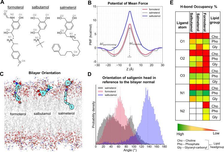
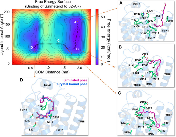
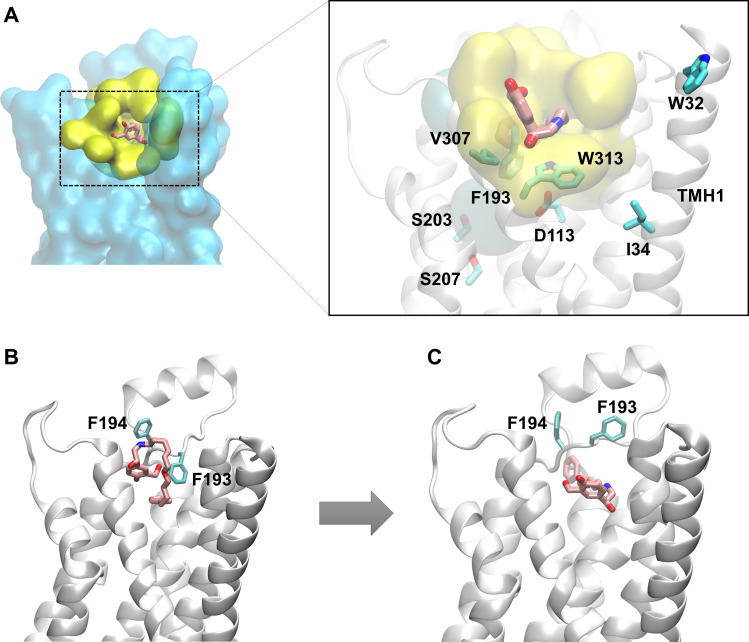
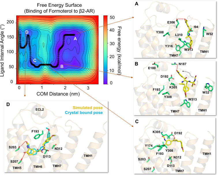
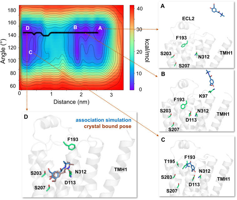
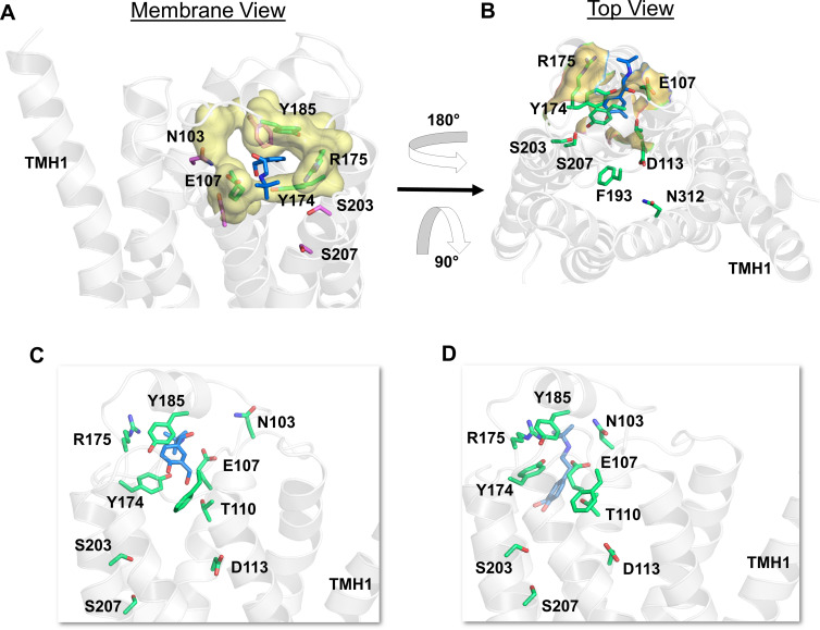
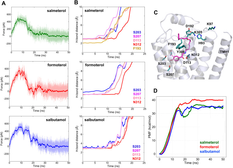
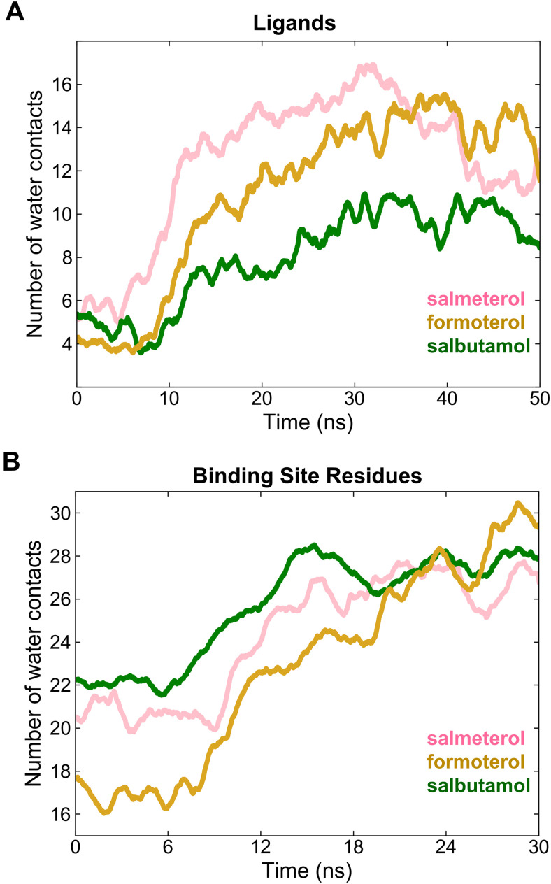
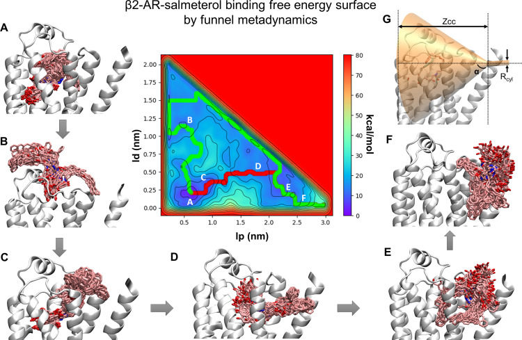
The drugs salmeterol, formoterol, and salbutamol constitute the frontline treatment of asthma and other chronic pulmonary diseases. These drugs activate the β2-adrenergic receptors (β2-AR), a class A G protein-coupled receptor (GPCR), and differ significantly in their clinical onset and duration of actions. According to the microkinetic model, the long duration of action of salmeterol and formoterol compared with salbutamol were attributed, at least in part, to their high lipophilicity and increased local concentrations in the membrane near the receptor. However, the structural and molecular bases of how the lipophilic drugs reach the binding site of the receptor from the surrounding membrane remain unknown. Using a variety of classic and enhanced molecular dynamics simulation techniques, we investigated the membrane partitioning characteristics, binding, and unbinding mechanisms of the ligands. The obtained results offer remarkable insight into the functional role of membrane lipids in the ligand association process. Strikingly, salmeterol entered the binding site from the bilayer through transmembrane helices 1 and 7. The entry was preceded by membrane-facilitated rearrangement and presentation of its phenyl-alkoxy-alkyl tail as a passkey to an access route gated by F193, a residue known to be critical for salmeterol's affinity. Formoterol's access is through the aqueous path shared by other β2-AR agents. We observed a novel secondary path for salbutamol that is distinct from its primary route. Our study offers a mechanistic description for the membrane-facilitated access and binding of ligands to a membrane protein and establishes a groundwork for recognizing membrane lipids as an integral component in the molecular recognition process. SIGNIFICANCE STATEMENT: The cell membrane's functional role behind the duration of action of long-acting β2-adrenergic receptor (β2-AR) agonists such as salmeterol has been a subject of debate for a long time. This study investigated the binding and unbinding mechanisms of the three commonly used β2-AR agonists, salmeterol, formoterol, and salbutamol, using advanced simulation techniques. The obtained results offer unprecedented insights into the active role of membrane lipids in facilitating access and binding of the ligands, affecting the molecular recognition process and thus their pharmacology.


Figures

Fig. 1.
Fig. 1.
Membrane partitioning characteristics of salmeterol, formoterol, and salbutamol in the bilayer, made up of POPC and cholesterol. (A) Two-dimensional structures of the studied β2-AR agonists. (B) The PMF curves show the solvation free energies for membrane partitioning and crossing of the ligands, revealing their energetically favorable bilayer locations (of their COMs). Both salmeterol and formoterol show similar free energy profiles, whereas salbutamol has higher energy barriers for partitioning and crossing of the membrane. (C) The time-average preferred orientations of the ligands within the membrane. The nitrogen atom of the choline, phosphorous atom of the phosphate, and oxygen of the glyceryl carbonyl headgroups are represented as balls in blue, olive green, and red colors, respectively. The lipid alkyl chains are represented as lines in gray color. (D) The distinct bilayer orientation of the saligenin head in each ligand is quantified as a tilt angle between the bilayer normal (z-axis) and a vector, connecting O1 and N1 atoms of the ligands. The vertical dashed lines represent the mean values. (E) The percentage (%) H-bond occupancy of the polar atoms of each ligand with lipid headgroups (choline, phosphate, and glyceryl carbonyls) through the simulation time. H-bonds are counted only if the bond distance and angle are within the cutoff values of 3.5 Å and 40 degrees, respectively.
Fig. 2.
Fig. 2.
The free energy surface for salmeterol’s access and binding to the β2-adrenergic receptor through the TMHs 1 and 7 from its energetically favorable location in the membrane. The two-dimensional free energy surface is characterized by the distance between the COM of the ligand and binding site residues (x-axis) in nm and the internal angle of the salmeterol molecule (y-axis) in degrees. The minimum energy path, as determined by well tempered metadynamics simulations, is given in bold black line, and several representative intermediate states along the path were labeled (A–D). (A) As salmeterol (licorice representation, magenta color) approaches the receptor from within the membrane, its aryl-alkoxy-alkyl tail comes into contact with the hydrophobic residues, including W3137.40, I3097.36, L3107.37, V391.38, W421.41, and I431.42 from TMHs 1 and 7. (B and C) Salmeterol undergoes a significant conformational change from its extended to bent form and engages in H-bond interactions with D192ECL2 through its O1 and O2 groups, which destabilize the salt bridge between D192ECL2 and K3057.32. The breakage of the salt bridge allows salmeterol to move further, disrupting the hydrophobic lock (between F193ECL2 and Y3087.35) while the tail is still anchored by the hydrophobic residues. (D) Outward movement of the side chain indole ring of W3137.40 releases the tail and facilitates salmeterol’s entry into the pocket, which then assumes its final bound pose, mostly similar to that of the crystal structure pose (PDB ID 6MXT) (Masureel et al., 2018). In its final bound pose, salmeterol engages in polar H-bond and salt bridge interactions with S2035.42, S2075.46, D1133.32, and N3127.39. Salmeterol (crystal pose in dark blue) and the binding site residues (green) are illustrated in licorice representation. The receptor is illustrated in secondary structure representation (light blue).
Fig. 3.
Fig. 3.
Salmeterol enters the β2-AR binding site from within the membrane through the transmembrane helices 1 and 7 using its lipophilic tail as a passkey. (A) Hydrophobic residues from TMHs 1 and 7 anchor the lipophilic tail of salmeterol and facilitate salmeterol’s entry into the pocket from within the membrane. (B) However, in several simulations, salmeterol’s entry by its saligenin head is blocked by the side chain phenyl ring of F193ECL2 that acts as a gate. (C) Fascinatingly, salmeterol flips 180° and presents its aryl-alkyl tail as a passkey to the gate, which immediately opens up the channel by flipping F193ECL2 upward to gain entry into the pocket.
Fig. 4.
Fig. 4.
The free energy surface for formoterol’s access and binding to the β2-adrenergic receptor from its energetically favorable location in the membrane. The two-dimensional free energy surface is characterized by the distance between the COM of the ligand and binding site residues (x-axis) and the internal angle of formoterol (y-axis). The minimum energy path, as determined by well tempered metadynamics simulations, is given in bold black line, and several representative low-energy intermediate states along the path were labeled (A–D). (A) As formoterol (licorice representation, yellow color) approaches the receptor from within the membrane, its alkoxy-aryl-alkyl tail comes into contact with TMHs 2 and 7, engaging in hydrophobic contacts with I942.65, L3107.37, and W3137.40. (B and C) Formoterol moves up over the transmembrane helices and remains in a region where it makes extensive H-bond interactions with several polar residues and bulk water molecules. Specifically, the H-bond with D192ECL2 through its O3 appears to destabilize the salt bridge between D192ECL2 and K3057.32. The breakage of the salt bridge allows formoterol to slide down from this region into the binding pocket. As formoterol enters the pocket, the hydrophobic lock (between F193ECL2 and Y3087.35) breaks, resulting in the formation of a π-π interaction between F193’s phenyl ring and the aromatic ring at the tail end of the ligand. (D) Formoterol is seen in its final bound pose, slightly different from its X-ray structure pose (RMSD 2.1Å) bound to turkey β1-AR [PDB ID 6IBL (Lee et al., 2020)]. In its final bound pose, formoterol engages in polar H-bond and salt bridge interactions with S2035.42, S2075.46, D1133.32, and N3127.39. Formoterol (crystal pose in cyan) and the binding site residues (green) are illustrated in licorice representation. The receptor is illustrated in secondary structure representation (light golden yellow).
Fig. 5.
Fig. 5.
The free energy surface and minimum energy path for salbutamol’s access and binding to the β2-adrenergic receptor. The primary binding path for salbutamol is through the common aqueous route, as determined by well tempered metadynamics simulations (minimum energy path, in bold black line). (A) In each simulation, salbutamol first entered the aqueous bulk before it established its first contact with the receptor. (B) Salbutamol first encountered K972.68 (through its saligenin hydroxyl groups). (C) Salbutamol slid along the extracellular vestibule, establishing interactions with F193ECL2 through its t-butyl end and with D1133.32 and N3127.39 with its saligenin end. (D) Salbutamol is seen in its final bound pose, very similar to its crystal X-ray structure pose (RMSD of 2.25Å) bound to turkey β1-AR (PDB ID 2Y04) (Warne et al., 2011). In its final bound pose, salbutamol engages in polar interactions with S2035.42, S2075.46, D1133.32, and N3127.39. Salbutamol (blue) and the binding site residues (green) are illustrated in licorice representation. The receptor is illustrated in secondary structure representation (light pink).
Fig. 6.
Fig. 6.
Salbutamol enters the β2-AR binding site through a novel polar channel. (A) From within the membrane view of the channel entrance (in yellow color and surface representation) displaying critical polar residues (in green color and licorice representation) that make immediate interactions with salbutamol (in blue color and licorice representation) as it enters. (B) The top view of the polar channel illustrating its position that is distal to the common access path lined by F193ECL2. (C and D) The snapshots show the intermediate states of salbutamol before reaching the binding pocket in its final bound pose, as presented in Fig. 6D. The receptor is illustrated in secondary structure representation (white).
Fig. 7.
Fig. 7.
Unbinding paths and critical residue interactions along the dissociation pathways for salmeterol, formoterol, and salbutamol. (A–C) Dissociation paths for salmeterol, formoterol, and salbutamol, respectively. Salmeterol dissociates into the aqueous bulk and spends a transient period there before partitioning into the membrane near the receptor. Formoterol and salbutamol dissociate into the aqueous bulk surrounding the extracellular loop region. The start, intermediate, and final poses of the molecules are represented as density maps in red, gray, and blue colors, respectively. (D–F) Critical residues in contact with the ligands at their bound states and along the dissociation path. (G–I) Pie charts representing residues from various TMHs and ECL2 and the fraction of simulation time (percent occupancy) during which they are within 4 Å of the ligands.
Fig. 8.
Fig. 8.
Force profiles, critical electrostatic/H-bond interactions, and PMF profiles of salmeterol, formoterol, and salbutamol while unbinding from the β2-adrenergic receptor revealed by steered MD simulations. (A) Average force profile for each ligand calculated using three independent SMD simulations. The cumulative force applied increases until critical interactions get disrupted and then decreases to zero as bonds are broken successively. (B) The time evolution of the H-bond distances between the ligands and the binding site residues, indicating the strength and longevity of the interactions contributing to binding affinities of the ligands. (C) Salmeterol bound to the receptor in its crystal structure pose, showing all the critical residues involved in its dissociation path. (D) The PMF profile for each ligand was calculated by Jarzynski equality.
Fig. 9.
Fig. 9.
The number of water contacts as a measure of solvation (wetting or hydration) of the ligands (A) and binding site residues (B) during the dissociation of salmeterol, formoterol, and salbutamol. The water contacts were at a minimum at the start of the simulation (t = 0 ns) when the ligands were bound. The contacts increased gradually as the ligands drifted away from the binding site and the degree of increase was directly proportional to the ligand size in the order of salmeterol > formoterol > salbutamol. Notably, the water contacts decreased for salmeterol, indicating its relocation into the surrounding membrane. Similarly, the number of water contacts with the binding site residues increased gradually, converging to approximately the same number of contacts (∼28).
Fig. 10.
Fig. 10.
The free energy surface of salmeterol binding to β2-AR obtained from the funnel metadynamics simulation using lp, the position of the ligand along the funnel axis, and ld, the distance of the ligand from this axis, as collective variables. Two minimum energy paths, one passing through the aqueous bulk (green) and another through the transmembrane helices (red), reaching the membrane, were determined by the Minimum Energy Path Surface Analysis tool. (A–F) Multiple clusters representing the fully bound state (A) to several intermediate conformational states (B–F) were sampled by several recrossing between bound and unbound states during the entire 900-ns simulation. (G) The funnel placement was based on several association/dissociation simulations during which salmeterol was seen leaving the pocket either by aqueous routes or by a transmembrane route through TMH 1 and 7. The cone region of the funnel was defined by a vertex height Zcc of 3.0 nm from the origin and an α angle of 0.6 rad. The radius of the cylindrical portion of the funnel rcyl was 2 Å.

Similar articles

Cited by

References

    1. (2019)Molecular Operating Environment (MOE), 2013.08, Chemical Computing Group ULC, 1010 Sherbrooke St. West, Suite #910, Montreal, QC, Canada.
    1. Abel R, Young T, Farid R, Berne BJ, Friesner RA (2008) Role of the active-site solvent in the thermodynamics of factor Xa ligand binding. J Am Chem Soc 130:2817–2831. - PMC - PubMed
    1. Abraham MJ, Murtola T, Schulz R, Páll S, Smith JC, Hess B, Lindahl E (2015) GROMACS: high performance molecular simulations through multi-level parallelism from laptops to supercomputers. SoftwareX 1–2:19–25.
    1. Anderson GP, Lindén A, Rabe KF (1994) Why are long-acting beta-adrenoceptor agonists long-acting? Eur Respir J 7:569–578. - PubMed
    1. Baker JG (2010) The selectivity of beta-adrenoceptor agonists at human beta1-, beta2- and beta3-adrenoceptors. Br J Pharmacol 160:1048–1061. - PMC - PubMed

Publication types

MeSH terms