Review
. 2021 Aug 2;6(1):290.
doi: 10.1038/s41392-021-00678-1.

Liquid-liquid phase separation in human health and diseases

Affiliations
Review

Liquid-liquid phase separation in human health and diseases

Bin Wang et al. Signal Transduct Target Ther. .

Abstract

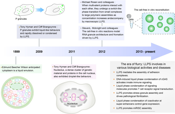
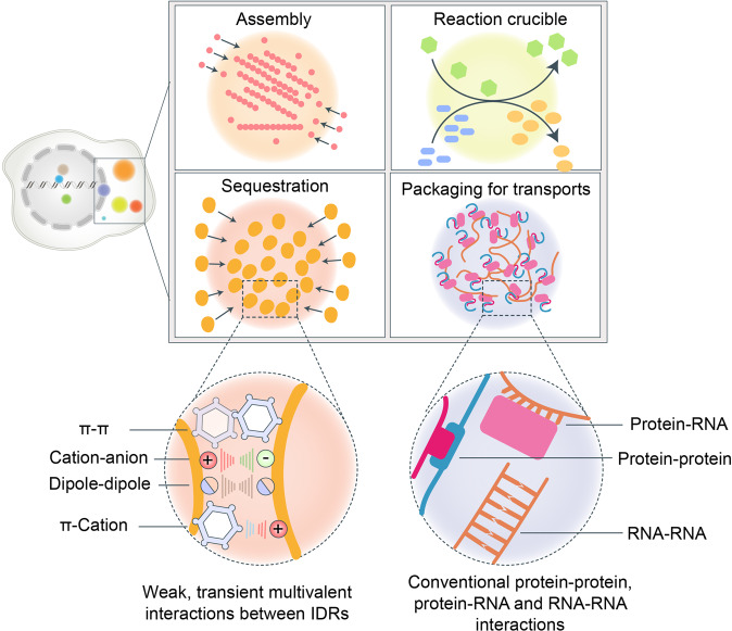
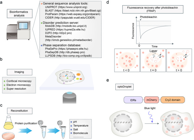
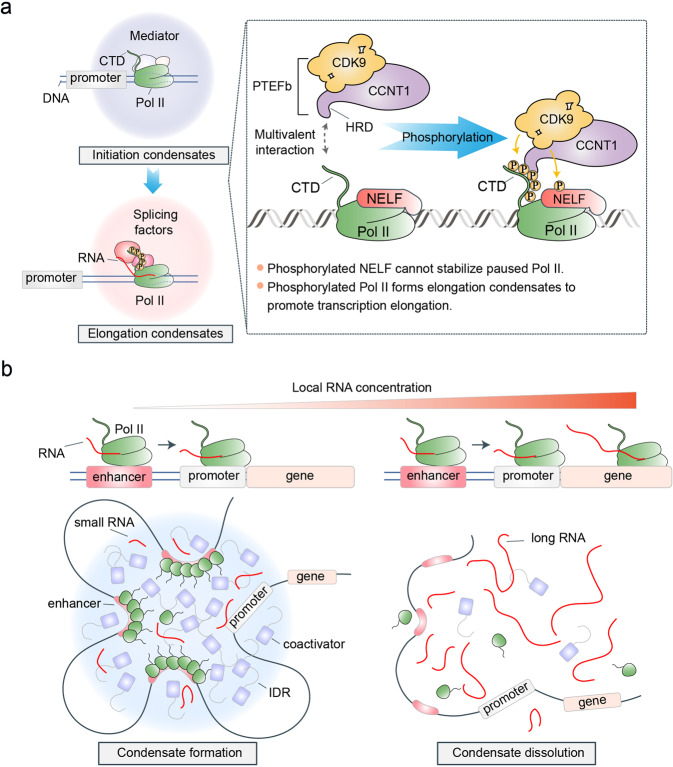
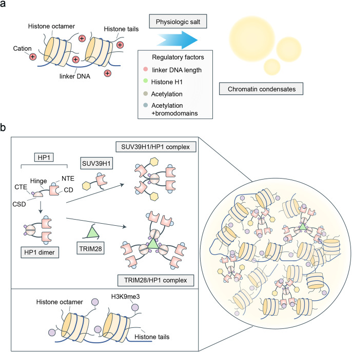
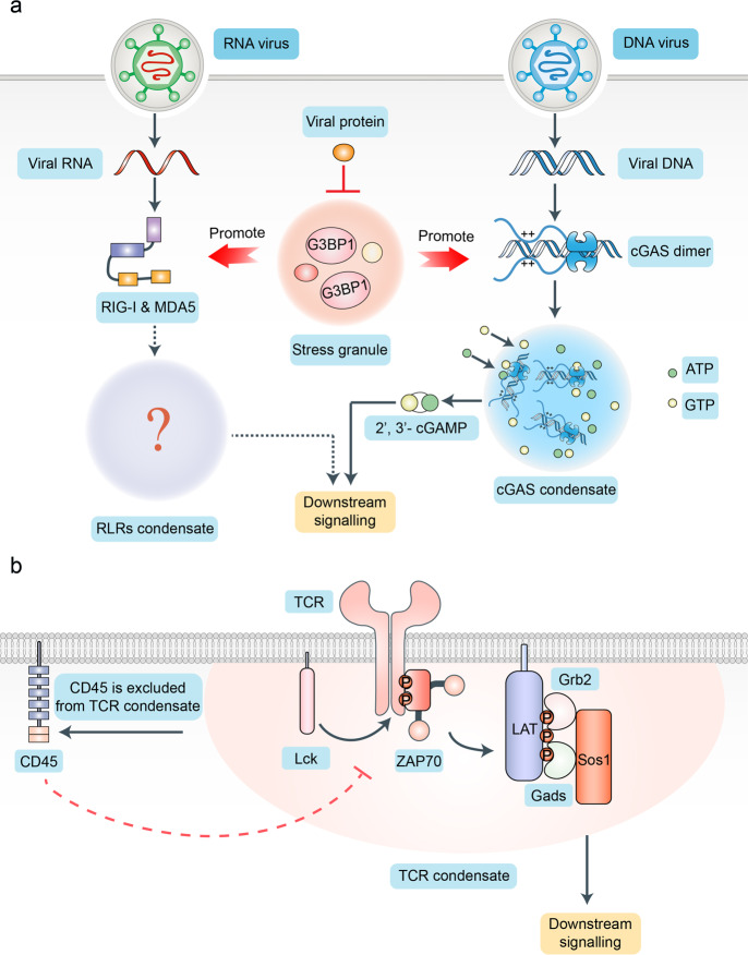
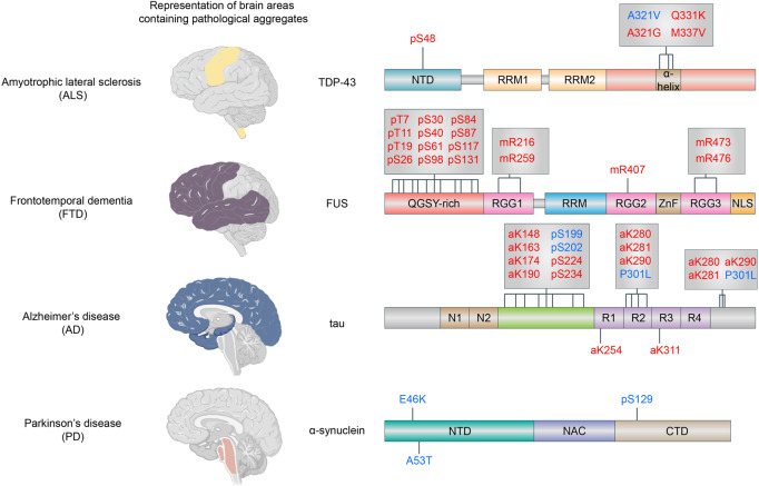
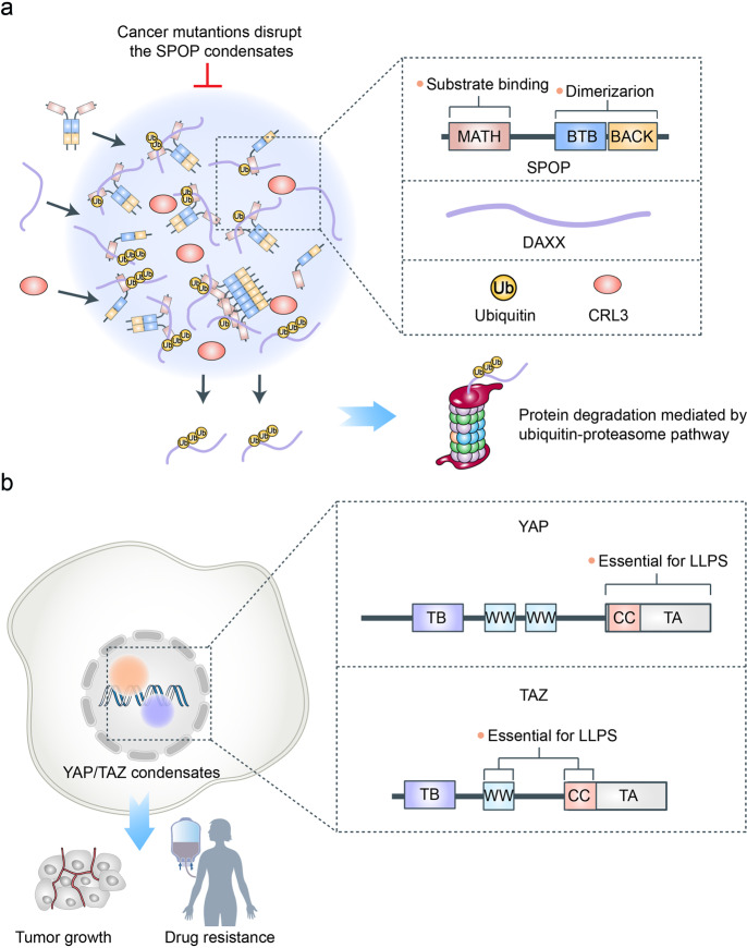
Emerging evidence suggests that liquid-liquid phase separation (LLPS) represents a vital and ubiquitous phenomenon underlying the formation of membraneless organelles in eukaryotic cells (also known as biomolecular condensates or droplets). Recent studies have revealed evidences that indicate that LLPS plays a vital role in human health and diseases. In this review, we describe our current understanding of LLPS and summarize its physiological functions. We further describe the role of LLPS in the development of human diseases. Additionally, we review the recently developed methods for studying LLPS. Although LLPS research is in its infancy-but is fast-growing-it is clear that LLPS plays an essential role in the development of pathophysiological conditions. This highlights the need for an overview of the recent advances in the field to translate our current knowledge regarding LLPS into therapeutic discoveries.


Conflict of interest statement

The authors declare no competing interests.

Figures

Fig. 1
Fig. 1
History of the discovery and development of LLPS. Representative milestone findings promoting the development of LLPS are enumerated in the figure
Fig. 2
Fig. 2
The forces driving LLPS. There are several functions of biomolecular condensates, including the assembly of a large complex, as a reaction crucible promoting biochemical reaction, sequestration of specific proteins to inhibit or promote some reaction, and packaging for transports. Besides, there are two types of multivalent interactions that contribute to LLPS. One is conventional multivalent interactions between protein and protein, protein and RNA, or RNA and RNA. The other is weak, transient, multivalent interactions between intrinsically disordered regions (IDRs), including π–π interactions, cation–anion interactions, dipole–dipole interactions, and π–cation interactions
Fig. 3
Fig. 3
The methods to identify or study LLPS. a Selected bioinformatic tools or databases for studying LLPS. b Electron microscopy, confocal microscopy, and super-resolution imaging techniques can provide detailed information on biomolecular condensates. c The cell-free reconstitution assay can detect the specific phase separation conditions of targeted proteins in vitro. d Fluorescence recovery after photobleaching can detect the material properties and dynamics of biomolecular condensates. After being photobleached by laser, the fluorescence of condensates will recover over time. The less time condensates take to recover, the higher is their fluidity. e OptoDroplet system can regulate multivalency using blue light to promote or reverse the formation of biomolecular condensates in vivo. Cry2, an Arabidopsis thaliana protein domain that forms oligomers following blue-light activation, are fused to the IDRs from targeted proteins and fluorescently tagged proteins
Fig. 4
Fig. 4
The function of LLPS in regulating gene transcription. a Phosphorylation regulates the transformation of initiation condensates to elongation condensates. In transcriptional initiation, RNA polymerase II (Pol II) phase-separates various factors with IDRs, such as transcription factors and coactivators, to form initiation condensates. After the transcription initiation, Pol II does not directly enter the elongation phase but pauses in a region approximately 50 bp downstream of the transcription start site, which is called promoter-proximal pausing. Thereafter, PTEFb can phase-separate into the initiation condensates through the multivalent interactions between the histidine-rich domain and carboxy-terminal domain (CTD) of Pol II. Therefore, CDK9, a subunit domain of PTEFb, can phosphorylate the negative elongation factor (NELF) and CTD. Phosphorylated NELF cannot stabilize paused Pol II and phosphorylated Pol II forms elongation condensates by hyperphosphorylated CTD, thereby achieving transcription elongation. b The transcriptional condensates can also be formed at a super-enhancer. Besides, the local RNA concentration can negatively regulate the formation of super-enhancer condensates. Low levels of RNA at regulatory DNA elements promote the formation of transcriptional condensates, whereas high levels of RNA from gene transcription can dissolve the transcriptional condensates
Fig. 5
Fig. 5
The function of LLPS in regulating genome organization. a The chromatin undergoes LLPS in physiologic salt, which is driven by the positively charged histone tails. Several factors can regulate the formation or properties of chromatin condensates, including the linker DNA length, histone H1, and histone acetylation. b The multivalent interactions between histone modifications and its readers have driven the formation of heterochromatin condensates. Heterochromatin protein 1 (HP1) contains a chromodomain (CD), a chromo shadow domain, and three disordered regions: N-terminal extension, hinge, and C-terminal extension. The HP1 dimer can interact with SUV39H1 (an H3K9me2/3 writer) and TRIM28 (an HP1 scaffolding protein) to form the SUV39H1/HP1 complex and TRIM28/HP1 complex, respectively. As these complexes contain multiple CDs that can interact with H3K9me2/3, they can phase-separate with the H3K9me2/3-marked nucleosome arrays to form condensates by multivalent interactions
Fig. 6
Fig. 6
Phase separation and immune response. a LLPS is involved in innate immune responses. Cyclic GMP–AMP synthase (cGAS)- cytosolic DNA (such as viral DNA) condensates are formed by the multivalent interactions between cGAS and DNA via LLPS. The cGAS-DNA condensates may function as a reaction crucible, which can concentrate the reactants (ATPs and GTPs) and enzymes (activated cGAS) to efficiently produce cyclic GMP–AMP (cGAMP), which initiates anti-viral immune responses via the TBK1/IKK signaling pathway. Similarly, RLRs, such as RIG-I and MDA5, can also sense the viral nucleic acids (DNA or RNA), thereby initiating anti-viral immune responses via the TBK1/IKK signaling pathway. However, whether RLRs can phase-separate with nucleic acids to promote anti-viral immune responses is still not clear. Ras-GTPase-activating protein SH3 domain-binding protein 1 (G3BP1) is a positive regulator of innate immune responses (including RIG-I-mediated cellular anti-viral pathway and cGAS-STING pathway) and a stress granule core protein. SARS-CoV-2 N protein forms condensates that incorporate RNA and G3BP1, which suppress the interaction between G3BP1 and cGAS or RIG-I, thereby inhibiting the anti-viral immune responses of the host cells. b LLPS mediates T-cell receptor (TCR) signal transduction. After being phosphorylated by Lck, a kinase of the Src family, the cytoplasmic domains of TCR will recruit and activate the tyrosine kinase ZAP70. Thereafter, the multiple tyrosine residues of LAT, a transmembrane protein, are phosphorylated by activated ZAP70. These phosphorylated tyrosine residues recruit the SH2 and SH3 domain-containing protein GRB2, Gads, and Sos1, thereby activating T cells through several downstream signaling pathways, such as the MAPK pathway. These molecules can form condensates to exclude CD45, which can dephosphorylate the phosphorylated cytoplasmic domains of the TCRs to inhibit T-cell activation
Fig. 7
Fig. 7
Phase separation in neurodegenerative disease. Schematic representation of brain areas containing pathological aggregates of four kinds of neurodegenerative diseases. This figure also summarizes disease-associated mutations and PTMs that decrease (red) or enhance (blue) LLPS compared with unmodified TDP-43, FUS, a-synuclein, or tau, as indicated. NTD: N-terminal domain, RRM: RNA-recognition motif, QGSY-rich: rich in glutamine, glycine, serine, and tyrosine, RGG: arginine/glycine-rich; ZnF, zinc finger; NLS, nuclear localization signal; NAC, non-Ab component of AD plaque; N1–2, polypeptide sequences encoded by exons 2 and 3; PRR, proline-rich regions; R1–4, microtubule-binding domains encoded by exons 9–12; p, phosphorylation; m, methylation; a, acetylation
Fig. 8
Fig. 8
LLPS in cancer. a Speckle-type POZ protein (SPOP) can phase-separate with its substrates and cullin3-RING ubiquitin ligase to form condensates, which promote the degradation of its substrates via the ubiquitin-proteasome system. SPOP consists of a substrate-binding meprin and TRAF homology (MATH) domain and two dimerization domains, BTB and BACK. The self-association by two dimerization domains and the multievent interactions between MATH domain and substrates are necessary for the phase separation of SPOP. Cancer-associated mutations in the MATH domain disrupt the formation of SPOP condensate by preventing the interaction between substrates and SPOP. b The transcriptional coactivators Yes-associated protein (YAP) and transcriptional coactivator with PDZ-binding motif (TAZ) can activate the transcription of various genes via LLPS. The Hippo signaling pathway can inhibit the formation of YAP/TAZ condensates. However, the Hippo signaling pathway is inactivated in many cancers. Therefore, the accumulation of TAZ and YAP can largely activate the transcription of proto-oncogenes via phase separation, promoting cell proliferation and anti-PD-1 immunotherapy resistance via LLPS. The intrinsically disordered TA and CC domains are essential for the formation of YAP condensates, whereas the CC and WW domains are vital for TAZ phase separation
Fig. 9
Fig. 9
LLPS in SARS-CoV-2 infection. a Nucleocapsid (N) protein contains an N-terminal intrinsically disordered region (N-IDR), a structured N-terminal domain (NTD), a central intrinsically disordered region (central-IDR), a structured C-terminal domain (CTD), and a C-terminal intrinsically disordered region (C-IDR). Of these, NTD and central-IDR play an important role in the phase separation of N protein with RNA. b LLPS plays an important role in SARS-CoV-2 infection. The central-IDR contains a conserved serine-arginine (SR)-rich sequence. Unmodified N protein forms gel-like condensates containing discrete ribonucleoprotein (RNP), which is conducive to its function of genome packaging roles. After being phosphorylated by cyclin-dependent kinase-1 (CDK1) and glycogen synthase kinase-3 (GSK3) in the SR region, N protein forms liquid-like condensates for viral genome processing. Viral RNA sequence and structure in specific genomic regions are also involved in the regulation of the N protein phase separation. The specific regions of the viral RNA genome, including a region spanning the 5’ end (first 1000 nt) and a region-encoded N protein at the 3᾽ end, can promote the phase separation of N proteins, whereas most regions in the genome facilitate the dissolution of N protein condensates. The viral membrane (M) protein can independently form condensates with N protein. During the late stages of SARS-CoV-2 infection, the viral RNP condensates will interact with the soluble CTD of M protein to form two-layered condensates, which promote the SARS-CoV-2 virion assembly

References

    1. Wang M, Kaufman RJ. Protein misfolding in the endoplasmic reticulum as a conduit to human disease. Nature. 2016;529:326–335. doi: 10.1038/nature17041. - DOI - PubMed
    1. Smith JJ, Aitchison JD. Peroxisomes take shape. Nat. Rev. Mol. Cell Biol. 2013;14:803–817. doi: 10.1038/nrm3700. - DOI - PMC - PubMed
    1. Mao YS, Zhang B, Spector DL. Biogenesis and function of nuclear bodies. Trends Genet. 2011;27:295–306. doi: 10.1016/j.tig.2011.05.006. - DOI - PMC - PubMed
    1. Ow Y-LP, Green DR, Hao Z, Mak TW. Cytochrome c: functions beyond respiration. Nat. Rev. Mol. Cell Biol. 2008;9:532–542. doi: 10.1038/nrm2434. - DOI - PubMed
    1. West AP, Shadel GS. Mitochondrial DNA in innate immune responses and inflammatory pathology. Nat. Rev. Immunol. 2017;17:363–375. doi: 10.1038/nri.2017.21. - DOI - PMC - PubMed

Publication types

MeSH terms